2. Antigen
• An antigen is a substance which when introduced
into a body evokes an immune response to produce a
specific antibody with which it reacts specifically.
• It can be classified as-
Complete antigen
Incomplete antigen (Haptens)
4. Antigen
Incomplete antigen (Haptens)
Haptens are substances unable to induce antibody formation
on its own but can become immunogenic (capable of
inducing antibodies) when covalently linked to proteins,
called carrier proteins. They can be simple or complex.
5. • Epitope is the smallest unit of antigenicity.
• The combining site on the antibody molecule,
corresponding to the epitope is called the Paratope.
6. Antibody
• These are substances which are formed in the serum
and tissue fluids in response to an antigen and react
with that antigen specifically and in some observable
manner.
• Chemically they are globulins, hence they are named
immunoglobulins.
• They constitute about 20 – 25% of the total serum
proteins and are mainly synthesized by plasma cells.
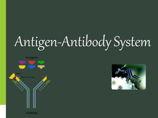
8. • An antibody has a Y-shaped structure, made up of four
polypeptide subunits.
• Each subunit has two identical light and heavy chains
• N-terminus of each heavy chain forms an antigen-
binding domain with a light chain.
• There are two antigen-binding domains forming the
arms of the “Y” shape. They are known as ‘fragment
antigen-binding’ (Fab) domains.
Antibody Structure
9. • The C-terminus of the heavy chains forms ‘fragment
crystallization’ (Fc) domain, which helps in the
interaction with the effector cells.
• All four polypeptide subunits are held together by
disulfide and non-covalent bonds.
• The heavy chains of the antibodies contain a variable
region and three constant regions.
• Each antibody has two identical antigen-binding sites
and they differ in the antibodies.
Antibody Structure
10. • Antibodies or immunoglobulins(Ig) are of five different
isotypes. This classification is on the basis of their H
chains.
1. IgM
2. IgG
3. IgA
4. IgD
5. IgE
Types Of Antibodies
11. 1. IgM
• IgM is the first antibody produced in response to a
microbial attack by B cells.
• Largest antibody and is found in a pentameric form.
• Circulates in the blood and lymph and constitutes 6% of
the total antibody content in the serum.
• Involved in agglutination and opsonization.
• It has a large number of antigenic sites on its surface
and therefore facilitates efficient activation of the
immune system.
Types Of Antibodies
12. 2. IgG
• Most abundant isotype in the plasma, and comprises
80% of the total antibody content in the serum
• It is transferred to the placenta through the fetus and
protects the infant until its birth.
• IgG is divided into four subclasses- IgG1, IgG2, IgG3,
and IgG4. Among these, only IgG3 and IgG4 possess
the ability to cross the placenta.
• It facilitates the process of phagocytosis and provides
immunity to the developing fetus.
Types Of Antibodies
13. 3. IgA
• Usually found in liquids such as breast milk, serum,
saliva, fluids of the intestine.
• IgA in breast milk protects an infant’s gastrointestinal
tract from microbial activity.
• It constitutes 13% of the total antibody content in the
serum and is divided into 2 sub-classes- IgA1 and IgA2.
• Among these, IgA1 is highly found in the secretions
and is also called the secretory immunoglobulin.
• Activates the complement pathway and participates in
the immune response.
Types Of Antibodies
14. 4. IgD
• It is involved in the production of the antibody by B
cells
• It comprises less than 1% of the total antibody content
in serum.
• It acts as a receptor on B cell surface and participates in
B cell activation and differentiation
Types Of Antibodies
15. 5. IgE
• IgE is present in the least amounts, around 0.02% of the
antibody content in the serum.
• These are present in the linings of the respiratory and
intestinal tracts and respond to allergic reactions.
Types Of Antibodies
18. Antigen Antibody Reactions
• The antigens and antibodies combine specifically with
each other. This interaction between them is called
Antigen –Antibody reaction.
• It may be abbreviated asAg –Ab reaction.
• The first correct description of the antigen-antibody
reaction was given by Richard J. Goldberg at the
University of Wisconsin in 1952.
19. • These reactions form the basis for detection of
infectious disease causing agents and also some non
specific antigens like enzymes.
• The reactions betweenAg and Ab occur in three stages.
In first or primary stage the reaction involves
formation of Ag-Ab complex.
The secondary stage leads to visible events like
precipitation, agglutination etc.
The tertiary stage includes destruction of Ag or its
neutralization.
20. Its USES are
1. In vivo
• Forms basis of immunity against infectious diseases.
2. In vitro
• For diagnosis of infections
• Helpful in epidemiological studies
• Detection and quantification of antigens or antibodies.
21. Characteristics
• Reaction is specific, an antigen combines only with its
homologous antibody and vice-versa. However, cross
reactions may occur due to antigenic similarity.
• Entire molecules of antigen and antibody react and not
the fragments.
• Only the surface antigens participate in the antigen
antibody reaction.
• The reaction is firm but reversible.
23. Precipitation reactions
When a soluble antigen reacts with its antibody in the
presence of electrolytes at an optimal temperature and
pH, antigen antibody complex forms an insoluble
precipitate that usually sediments at the bottom of the
tube and it is called precipitation.
Precipitation may occur in liquid media or in gels
such as agar, agarose etc.
24.
25. Applications
• Identification of bacteria.
• Detection of antibody for diagnostic purposes.
• Forensic application in identification of human blood
and seminal stains
• To standardize toxins and antitoxins.
28. Agglutination
It is an antigen antibody reaction in which a particulate
antigen combines with its antibody in the presence of
electrolytes at an optimal temperature and pH resulting
in visible clumping of particles.
It is more sensitive than precipitation for detection of
antibodies.
The reactions take place better with IgM antibody.
29. Types
SlideAgglutination test
Routine procedure to identify
bacterial stains. E.g., Salmonella species
Also used for blood grouping.
TubeAgglutination test
Standard quantitative method for determination of
antibodies.
Routinely employed in diagnosis of typhoid and
typhoid fever
30. Coombs Test
• Originally devised by Coombs, Mourant and Race (1945)
for detection of incomplete Rh antibodies.
• When sera containing incomplete anti-Rh antibodies are
mixed with corresponding Rh-positive erythrocytes but no
agglutination occurs.
• When such erythrocytes are treated with antiglobulin or
COOMBS serum (rabbit antiserum with human gamma
globulin), the cells are
agglutinated.
31. Neutralization Test
• Bacterial exotoxins are capable of producing neutralizing
antibodies (antitoxins) which play protective role in
diseases such as diphtheria and tetanus.
• Toxin – antitoxin neutralization can be measured in vivo
and in vitro.
32. In vivo tests:
Toxigenicity test. E.g., Diphtheriae
In vitro test:
Virus neutralization tests.
33. Immunofluorescence
• Fluorescence is the property of certain dyes which absorb rays of
one particular wavelength (ultraviolet light) and emit rays with a
different wavelength (visible light)
• Most commonly used dyes are:
1. Fluorescin isothiocyanate – blue green
2. Lissamine rhodamine – orange red
They are of two types:
1. Direct immunofluorescence
2. Indirect immunofluorescence
34.
35. Direct immunofluorescence
Uses:
• Commonly employed for detection of bacteria, viruses or other
antigens in blood, urine, tissues and other specimens.
• Sensitive method to diagnosis Rabies.
Disadvantage: Separate fluorescent labelled
prepared for each antigen to be tested.
antibody has to be
Indirect immunofluorescence
• A single antihuman globulin fluorescent conjugate
employed for detection of antibody to any antigen
can be
• This has overcome the disadvantage of direct immunofluorescence
36. ELISA
• Enzyme linked immunosorbent assay is a simple and a
sensitive test.
• Requires only microlitre quantities of test reagents.
• The principle of ELISA is almost same as that of
immunofluorescence, the only difference being, an
enzyme is used instead of fluorescent dye.
• It can be used for detection of Antigen or Antibody.
• Types: Sandwich, Indirect, Competitive ELISA
37.
38. Uses:
Detection of HIV antibodies in serum
Detection of mycobacterial antibodies in TB
Detection of Hepatitis B markers in serum
Detection of enterotoxin of E.coli in feces.
40. • Immuno electron microscopy: Viral particles are mixed
with specific antisera and are observed under electron
microscope. These are seen as clumps.
Used in detection of Hepatitis Avirus.
• Immuno ferritin test: Ferritin (electron dense substance)
conjugated antibody is used to react with an antigen.
Used in identification of different species.
• Immuno enzyme test: Tissue sections are treated with
peroxidase labelled antisera to detect the corresponding
antigen and in viewed under electron microscope.
41. • Therefore we see the application of antigen antibody
reactions in the diagnosis of diseases which can
help in developments of varieties of diagnostic tests.
• In clinical practice, they help in:
Preventing destructive diseases.
Preventing progression of the diseases.
Identifying high risk patients
Target treatment of specific diseases
Monitor the effects of the treatment.
Applications