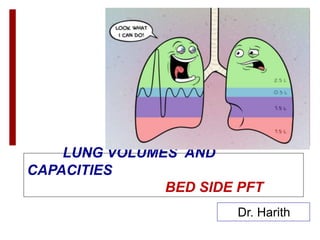
2. Lung volumes and lung capacities refer to
the volume of air associated with different phases
of the respiratory cycle.
Lung volumes are directly measured; Lung
capacities are inferred from lung volumes.
Instrument is spirometry
6. 1) TIDAL VOLUME (TV):
VOLUME OF AIR INHALED/EXHALED
IN EACH BREATH DURING QUIET
RESPIRATION.
N – 6-8 ml/kg.
TV FALLS WITH DECREASE IN
COMPLIANCE, DECREASED
VENTILATORY MUSCLE STRENGTH.
7. 2) INSPIRATORY RESERVE VOLUME (IRV):
MAX. VOL. OF AIR WHICH CAN BE
INSPIRED AFTER A NORMAL TIDAL
INSPIRATION
i.e. FROM END INSPIRATION
N- 1900 ml- 3300 ml.
8. 3) EXPIRATORY RESERVE VOLUME (ERV):
MAX. VOLUME OF AIR WHICH CAN BE
EXPIRED AFTER A NORMAL TIDAL
EXPIRATION
i.e. FROM END EXPIRATION PT.
N- 700 ml – 1000 ml
700-1000ml
9. Residual Volume (RV):
Volume of air remaining in lungs
after maximium exhalation (20-25
ml/kg) (1700-2100ml)
Indirectly measured (FRC-ERV)
10. CAPACITIES
Two or more Volumes combined together
Functional Residual Capacity (FRC):
Volume of air remaining in the lungs at
passive end expiration.
N- 2.3 -3.3 L OR 30-35 ml/kg
FRC = RV + ERV
11. FRC INCREASES WITH
Increased height
Erect position (30% more than in supine)
Decreased lung recoil (e.g. emphysema)
FRC DECREASES WITH
Obesity
Muscle paralysis (especially in supine)
Supine position
Restrictive lung disease (e.g. fibrosis, Pregnancy)
FRC does NOT change with age.
12. Total Lung Capacity (TLC):
Maximum volume of air
attained in lungs after maximal
inspiration.
N- 4-6 l or 80-100 ml/kg
TLC= VC + RV
13. Inspiratory Capacity (IC):
MAX. VOL. OF AIR WHICH CAN BE
INSPIRED AFTER A NORMAL TIDAL
EXPIRATION.
IC = IRV + TV
N-2400 ml – 3800 ml.
22. • Patient breathes in and out of a spirometer filled with 10%
helium and 90% o2, till conc. In spirometer and lung
becomes same (equilibirium).
As no helium is lost; (as he is insoluble in blood)
C1 X V1 = C2 ( V1 + V2)
V2 = V1 ( C1 – C2)
C2
• V1= VOL. OF SPIROMETER
• V2= FRC
• C1= Conc.of He in the spirometer before equilibrium
• C2 = Conc, of He in the spirometer after equilibrium
2(10-5)
5
23. Volume above residual
where airway closure begins
Total volume in the lung
where airway closure begins
CLOSING VOLUME AND CAPACITY
24. BED SIDE PFT
Sabrasez breath holding test:
• Ask the patient to take a full but not too deep breath &
hold it as long as possible.
>25 SEC.-NORMAL Cardiopulmonary Reserve
(CPR)
15-25 SEC- LIMITED CPR
<15 SEC- VERY POOR CPR (Contraindication for
elective surgery)
25 - 30 SEC - 3500 ml VC
20 - 25 SEC - 3000 ml VC
15 - 20 SEC - 2500 ml VC
10 - 15 SEC - 2000 ml VC
5 - 10 SEC - 1500 ml VC
25. Single breath count:
After deep breath, hold it and start counting till the next breath.
N- 30-40 COUNT
Indicates vital capacity
SCHNEIDER’S MATCH BLOWING TEST: MEASURES Maximum Breathing
Capacity.
Ask to blow a match stick from a distance of 6” (15 cms) with-
Mouth wide open
Chin rested/supported
No purse lipping
No head movement
No air movement in the room
Mouth and match at the same level
26. Can not blow out a match
MBC < 60 L/min
FEV1 < 1.6L
Able to blow out a match
MBC > 60 L/min
FEV1 > 1.6L
MODIFIED MATCH TEST:
DISTANCE MBC
9” >150 L/MIN.
6” >60 L/MIN.
3” > 40 L/MIN.
27. COUGH TEST: DEEP BREATH F/BY COUGH
ABILITY TO COUGH
STRENGTH
EFFECTIVENESS
INADEQUATE COUGH IF: FVC<20 ML/KG
FEV1 < 15 ML/KG
PEFR < 200 L/MIN.
VC ~ 3 TIMES TV FOR EFFECTIVE COUGH.
A wet productive cough / self propagated
paraoxysms of coughing – patient susceptible for
pulmonary Complication.
28. DEBONO WHISTLE BLOWING TEST
MEASURES PEFR.
Patient blows down a wide bore tube at
the end of which is a whistle, on the
side is a hole with adjustable knob.
As subject blows → whistle blows,
leak hole is gradually increased till the
intensity of whistle disappears.
At the last position at which the
whistle can be blown , the PEFR can
be read off the scale.
29. FORCED EXPIRATORY TIME:
After deep breath, exhale maximally and forcefully & keep
stethoscope over trachea & listen.
Normal FET – 3-5 SECS.
Obstructive Lung Disease - > 6 SEC
Restrictive Lung Disease - < 3 SEC
MICROSPIROMETERS – MEASURE VC.