SlideShare a Scribd company logo
1 of 83
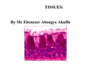
TISSUES:
By Mr Ebenezer Aboagye Akuffo
Learning outcomes
 describe the structure and functions of
these tissues: epithelial, connective,
muscle, nervous
 explain the capacity of different types of
tissue to regenerate
 outline the structure and functions of
membranes
 compare and contrast the structure and
functions of exocrine and endocrine
glands.
Tissues
 Definition: a group of closely associated
cells that perform related functions and are
similar in structure
 Between cells: nonliving extracellular
material
 Four basic types of tissue…function
Epithelium…covering
Connective tissue…support
Muscle tissue…movement
Nervous tissue…control
Epithelia (plural)
 Epithelium: sheet of cells that covers a body
surface or lines a body cavity; also form most of
the body’s glands
 Roles: as interfaces and as boundaries
 Functions:
Protection Absorption
Sensory reception Ion transport
Secretion Filtration
Formation of slippery surfaces for movement
Special characteristics of epithelia
 Cellularity(mostly composed of cells)
 Covers body surfaces
 Polarity(distinct cell surface)
 Free upper (apical) surface
 Lower (basal) surface contributing basal lamina to
basement membrane
 Cell and matrix connections
 Avascular but innervated
 Without vessels
 With nerve endings
 capable of Regeneration
Classification of epithelia
 According to thickness
“simple” - one cell layer
“stratified” – more than one layer of cells
(which are named according to the shape of
the cells in the apical layer)
Pseudostratified -false
 According to shape
“squamous” – wider than tall
“cuboidal” – as tall as wide
“columnar” - taller than wide
to protect
where diffusion is important
where tissues are involved
in secretion and absorption:
larger cells because of the
machinery of production,
packaging, and energy
requirements
“ciliated” literally =
eyelashes
(see next page)
STRATIFIED EPITHELIA
 several layers of cells of various shapes
 Basement membrane is usually absent
 function is to protect against wear and tear
 two types –stratified squamous epithelium
-transitional epithelium
stratified squamous epithelium
 Non-keratinised Stratified Epithelium- found
on wet surface subjected to wear & tear but are
protected from drying. eg Conjunctiva, lining of
the mouth, the pharynx, the oesophagus and the
vagina.
 Keratinised Stratified Epithelium- found in dry
surface subjected to wear and tear, contain the
protein keratin. Eg skin, hair and nails

Stratified: regenerate from below
Rare…
Rare…
TRANSITIONAL EPITHELIUM
 cells rounded and can spread out when
the organ expands
 found lining organs which must expand
and must be water proof eg. The bladder
Endothelium
A simple squamous
epithelium that lines
the interior of the
circulatory vessels
and heart
Mesothelium
Simple squamous epithelium that lines the
peritoneal, pleural and pericardial cavities and
covers the viscera
Glands
 Glands are cells or organs that secrete
something; that is, they produce a substance
that has a function either at that site or at a
more distant site.
 Production & secretion of needed
substances
 Are aqueous (water-based) products
 The protein product is made in rough ER,
packed into secretory granules by Golgi
apparatus, released from the cell by
exocytosis
Classification of glands
 By where they release their product
Exocrine: external secretion onto body
surfaces (skin) or into body cavities
Endocrine: secrete messenger molecules
(hormones) which are carried by blood to
target organs; “ductless” glands
 By whether they are unicellular or
multicellular
Exocrine glands
unicellular or multicellular
Unicellular: goblet cell
scattered within epithelial
lining of intestines and
respiratory tubes
Product: mucin
mucus is mucin & water
Multicellular exocrine glands
Examples of exocrine gland products
 Many types of mucus secreting glands
 Sweat glands of skin
 Oil glands of skin
 Salivary glands of mouth
 Liver (bile)
 Pancreas (digestive enzymes) The exocrine
portions secrete digestive enzymes that are
carried by ducts to the duodenum of the small
intestine, their site of action
 Mammary glands (milk)
Endocrine glands
 Ductless glands
 Release hormones into extracellular space
Hormones are messenger molecules
 Hormones enter blood and travel to
specific target organs
Examples of endocrine glands
 thyroid gland, adrenal glands, and pituitary
gland.
 The pancreas is an organ that is both an
exocrine and an endocrine gland
 The endocrine portions of the pancreas,
called pancreatic islets or islets of
Langerhans, secrete the hormones insulin
and glucagon directly into the blood.
Epithelial surface features
 Lateral surface
Adhesion proteins
Cell junctions: see next slide
 Basal surface
Basal lamina: noncellular sheet of protein
together with reticular fibers form basement
membrane
 Apical surface
Cell Junctions
 Tight junctions
 So close that are
sometimes impermeable
 Adherens junctions
 Transmembrane linker
proteins
 Desmosomes
 Anchoring junctions
 Filaments anchor to the
opposite side
 Gap junctions
 Allow small molecules to
move between cells
Apical surface features
 Microvilli – maximize surface area
Fingerlike extensions of the plasma
membrane of apical epithelial cells
On moist and mucus secreting epithelium
Longest on epithelia that absorb nutrients
(small intestine) or transport ions (kidneys)
(continued)
 Cilia
Whiplike motile
extentions of the
apical surface
membranes
 Flagellum
Long isolated
cilium
Only found as
sperm in human
 Four basic types of tissue
Epithelium
Connective tissue
 Connective tissue proper (examples: fat
tissue, fibrous tissue of ligaments)
 Cartilage
 Bone
 Blood
Muscle tissue
Nervous tissue
Connective Tissue
 Connective tissue is distinguished by its
extracellular matrix.
 Functions of Connective Tissue
Connective tissues enclose and separate
organs and tissue; connect tissues to one
another; play a role in support for
movement; store high-energy molecules;
cushion; insulate; transport; and protect.
Cells of Connective Tissue
 The extracellular matrix results from the activity
of specialized connective tissue cells; in general,
blast cells form the matrix, cyte cells maintain it,
and clast cells break it down. Fibroblasts form
protein fibers of many connective tissues,
osteoblasts form bone, and chondroblasts form
cartilage.
Adipose (fat) cells, mast cells, white blood cells,
macrophages, and mesenchymal cells (stem cells)
are commonly found in connective tissue.
Connective Tissue
 Originate from embryonic tissue called
mesenchyme
 Most diverse and abundant type of tissue
 Many subclasses
 Function: to protect, support and bind together
other tissues
 Bones, ligaments, tendons
 Areolar cushions; adipose insulates and is food
source
 Blood cells replenished; body tissues repaired
 Cells separated from one another by large
amount of nonliving extracellular matrix
Extracellular Matrix explained
 Nonliving material between cells
 Produced by the cells and then extruded
 Responsible for the strength
 Two components
1. Ground substance
 Of fluid, adhesion proteins, proteoglycans
 Liquid, semisolid, gel-like or very hard
2. Fibers: collagen, elastic or reticular
Basic functions of connective tissue
reviewed
 Support and binding of other tissues
 Connecting tissues to one another
 Defending the body against infection
macrophages, plasma cells, mast cells, WBCs
 Storing nutrients as fat
 Cushioning and insulating
MAJOR CELLS OF THE CONNECTIVE
TISSUE
 Fibroblast- star-shape, produce fibres (collagen)
by secreting protein into the matrix of the
connective tissue.
 Macrophages- specialized to carry on
phagocytosis, defend against infections agents
-monocytes in blood
- Phagocytes in the alveoli of the lungs
-kupffer cells in liver
-fibroblasts in lymph nodes and spleen
-microglial cells in the brain
MAJOR CELLS cont.
 Mast Cells- found in loose connective
tissue
- under the fibrous capsule of some
organs
 release heparin- that prevent blood
clotting
 Histamine- that promotes some of the
reactions associated with inflammation
and allergies
41
Connective Tissue
Fiber Types Present
• Collagenous fibers
• Thick
• Composed of collagen
• Great tensile strength
• Abundant in dense CT
• Hold structures
together
• Tendons, ligaments
• Elastic fibers
• Bundles of
microfibrils embedded
in elastin
• Fibers branch
• Elastic
• Vocal cords, air
passages
• Reticular fibers
• Very thin collagenous fibers
• Highly branched
• Form supportive networks
Classes of Connective Tissue
*
1. Loose connective tissue
■ Loose (areolar) connective tissue has many
different cell types and a random arrangement of
protein fibers with space between the fibers. This
tissue fills spaces around the organs and attaches
the skin to underlying tissues.
2.Dense connective tissue
■ Dense regular connective tissue is composed of
fibers arranged in one direction, which provides
strength in a direction parallel to the fiber
orientation. Two types of dense regular connective
tissue exist: collagenous (tendons and most
ligaments) and elastic (ligaments of vertebrae).
 Dense irregular connective tissue has
fibers organized in many directions, which
produces strength in different directions.
 Two types of dense irregular connective
tissue exist: collagenous (capsules of
organs and dermis of skin) and elastic
(large arteries).
 Connective tissue with special properties
•
Adipose tissue has fat cells (adipocytes) filled with
lipid and very little extracellular matrix (a few
reticular fibers).
Adipose tissue functions as energy storage,
insulation, and protection. Adipose tissue can be
yellow (white) or brown. Brown fat is specialized
for generating heat.
Reticular tissue is a network of reticular fibers and
forms the framework of lymphoid tissue, bone
marrow, and the liver.
Hemopoietic tissue, or red bone marrow, is the site
of blood cell formation, and yellow bone marrow is
a site of fat storage.
*
Classes of Connective Tissue
*
Classes of Connective Tissue
4. Cartilage
Cartilage has a relatively rigid matrix composed of
protein fibers and proteoglycan aggregates. The
major cell type is the chondrocyte, which is located
within lacunae.
Hyaline cartilage has evenly dispersed collagen
fibers that provide rigidity with some flexibility.
Examples include the costal cartilage, the covering
over the ends of bones in joints, the growing
portion of long bones, and the embryonic skeleton.
Fibrocartilage has collagen fibers arranged
in thick bundles; it can withstand great
pressure, and it is found between vertebrae,
in the jaw, and in the knee.
Elastic cartilage is similar to hyaline
cartilage, but it has elastin fibers. It is more
flexible than hyaline cartilage. It is found in
the external ear.
Classes of Connective Tissue
*
Bone
Bone cells, or osteocytes, are located in
lacunae that are surrounded by a
mineralized matrix (hydroxyapatite) that
makes bone very hard. Cancellous bone has
spaces between bony trabeculae, and
compact bone is more solid.
Classes of Connective Tissue
*
Blood and hemopoietic tissue
■ Blood cells are suspended in a fluid
matrix.
■ Hemopoietic tissue forms blood cells.
 Four basic types of tissue
Epithelium
Connective tissue
Muscle tissue
 Skeletal
 Cardiac
 Smooth
Nervous tissue
66
5.5: Muscle Tissues
• General characteristics:
• Muscle cells also called
muscle fibers
• Contractile
• Three (3) types:
• Skeletal muscle
• Smooth muscle
• Cardiac muscle
• Skeletal muscle
• Attached to bones
• Striated
• Voluntary
• Smooth muscle
• Walls of organs
• Skin
• Walls of blood vessels
• Involuntary
• Non-striated
• Cardiac muscle
• Heart wall
• Involuntary
• Striated
• Intercalated discs
Muscle Tissue
 1. Muscle tissue has the ability to contract, or shortens
2. Skeletal (striated(banded) voluntary) muscle attaches to
bone and is responsible for body movement. Skeletal
muscle cells are long, cylindrically shaped cells with many
peripherally located nuclei.
3. Cardiac (striated involuntary) muscle cells are cylindrical,
branching cells with a single, central nucleus. Cardiac
muscle is found in the heart and is responsible for pumping
blood through the circulatory system.
4. Smooth (nonstriated involuntary) muscle forms the walls
of hollow organs, the iris of the eye, and other structures.
Its cells are spindle shaped with a single, central nucleus.
 Four basic types of tissue
Epithelium
Connective tissue
Muscle tissue
Nervous tissue
Neurons
Supporting cells
 It is found in the brain, spinal cord, and
nerves and is characterized by the ability
to conduct electric signals called action
potentials
 It consists of neurons, which are
responsible for this conductive ability, and
support cells called neuroglia.
 Neurons , or nerve cells, are the
conducting cells of nervous tissue.
 They are composed of three major parts:
cell body, dendrites, and axon.
 Neuron- basic
structural unit of the
nervous system
 Dendrites- carry
impulses towards the
cell
 Axon-carry impulses
away from the cell
 Myelin sheath
 Synaptic terminal
 Epinephrine
 Norepinephrine
 Acetylcholine
 Neurons that possess several dendrites
and one axon are called multipolar
neurons
 Neurons that possess a single dendrite
and an axon are called bipolar neurons
 unipolar neurons have only one axon
and no dendrites
Membranes that combine epithelial
sheets plus underlying connective tissue
proper (see next slide)
 Cutaneous membranes
 Skin: epidermis and dermis
 Mucous membranes, or mucosa
 Lines every hollow internal organ that opens to the
outside of the body
 Serous membranes, or serosa
 Slippery membranes lining the pleural, pericardial and
peritoneal cavities
 The fluid formed on the surfaces is called a transudate
 Synovial membranes
 Line joints
(a) Cutaneous membrane
(b) Mucous membrane
(c) Serous membrane
Tissue response to injury
 Immune: takes longer and is highly specific
 Inflammation
 Nonspecific, local, rapid
 Signs: heat, swelling, redness, pain
 Repair – two ways
 Regeneration
 Fibrosis and scarring
 Severe injuries
 Cardiac and nervous tissue
Inflammation
 The inflammatory response occurs when
tissues are damaged or associated with an
immune response
 Agents that cause injury, microorganisms,
cold, heat, radiant energy, chemicals,
electricity, and mechanical trauma
 Infla. removes foreign materials and
damaged cells so that tissue repair can
proceed
 After a person is injured, chemical
substances called mediators of
inflammation are released in the tissues
and the adjacent blood vessels.
 The mediators include histamine, kinins,
prostaglandins, leukotrienes, and others
 Some mediators induce dilation of blood
vessels and produce redness and heat
 some also stimulate pain receptors and
increase the permeability of blood vessels
 which leads edema
Tissue Repair
1. Tissue repair is the substitution of viable cells for dead ones. Tissue
repair occurs by regeneration or replacement.
■ Labile cells divide throughout life and can undergo regeneration.eg skin,
mucous membranes, and hemopoietic and lymphatic tissues,
■ Stable cells do not ordinarily divide after growth is complete but can
regenerate if necessary.eg liver, pancreas, and endocrine glands
■ Permanent cells cannot replicate. If killed, permanent tissue is repaired by
replacement. Eg. Neurons
2. Tissue repair by primary union occurs when the edges of the wound
are close together.
Secondary union occurs when the edges are far apart.
Tumors (neoplasms): abnormal growth
of cells
 Adenoma – neoplasm of glandular
epithelium, benign or malignant
 Carcinoma – cancer arising in an
epithelium (90% of all human cancers)
 Sarcoma – cancer arising in mesenchyme-
derived tissue (connective tissues and
muscle)
Thanks
Any question

More Related Content

What's hot (20)

Lecture 11 the lymphatic system and immunity
Lecture 11 the lymphatic system and immunityLecture 11 the lymphatic system and immunity
Lecture 11 the lymphatic system and immunity
 
Chapter 1 organization of the human body
Chapter 1  organization of the human bodyChapter 1  organization of the human body
Chapter 1 organization of the human body
 
Muscle system
Muscle systemMuscle system
Muscle system
 
Epithelium, cells,tissues & histology
Epithelium, cells,tissues & histologyEpithelium, cells,tissues & histology
Epithelium, cells,tissues & histology
 
Histology: Tissues
Histology: TissuesHistology: Tissues
Histology: Tissues
 
CELL ANATOMY
CELL ANATOMYCELL ANATOMY
CELL ANATOMY
 
A&P2 Cell Tissue
A&P2 Cell TissueA&P2 Cell Tissue
A&P2 Cell Tissue
 
Histology of bone
Histology of boneHistology of bone
Histology of bone
 
Blood Physiology
Blood PhysiologyBlood Physiology
Blood Physiology
 
Animal tissue and organization
Animal tissue and organizationAnimal tissue and organization
Animal tissue and organization
 
Histology- Cell structure
Histology- Cell structure Histology- Cell structure
Histology- Cell structure
 
Histology of Muscle tissue
Histology of Muscle tissueHistology of Muscle tissue
Histology of Muscle tissue
 
Muscular tissue
Muscular tissueMuscular tissue
Muscular tissue
 
Tissue
TissueTissue
Tissue
 
Nervous tissue histology
Nervous tissue  histology Nervous tissue  histology
Nervous tissue histology
 
Connective tissue
Connective tissueConnective tissue
Connective tissue
 
Connective tissues
Connective tissuesConnective tissues
Connective tissues
 
Plants and Animals Tissues
Plants and Animals TissuesPlants and Animals Tissues
Plants and Animals Tissues
 
Ana212 histology of integumentary system
Ana212 histology of integumentary systemAna212 histology of integumentary system
Ana212 histology of integumentary system
 
Histology various type of epithelium
Histology   various type of epitheliumHistology   various type of epithelium
Histology various type of epithelium
 

Similar to Structure of a Tissue,Functions and types of Tissue

Similar to Structure of a Tissue,Functions and types of Tissue (20)

4. Tissues: Living Communities
4. Tissues: Living Communities4. Tissues: Living Communities
4. Tissues: Living Communities
 
Animal Tissues And Homeostasis
Animal Tissues And HomeostasisAnimal Tissues And Homeostasis
Animal Tissues And Homeostasis
 
Tissues
TissuesTissues
Tissues
 
Biology
BiologyBiology
Biology
 
Histology 1 fro medicine and science student
Histology 1 fro medicine and science student Histology 1 fro medicine and science student
Histology 1 fro medicine and science student
 
Architectural pattern of an animal
Architectural pattern of an animalArchitectural pattern of an animal
Architectural pattern of an animal
 
3.tissues of body
3.tissues of body3.tissues of body
3.tissues of body
 
Chapter 4 - Tissues
Chapter 4 - TissuesChapter 4 - Tissues
Chapter 4 - Tissues
 
Tissues
TissuesTissues
Tissues
 
Animal tissues epithelial & connective
Animal tissues epithelial & connectiveAnimal tissues epithelial & connective
Animal tissues epithelial & connective
 
Animal tissues epithelial & connective
Animal tissues epithelial & connectiveAnimal tissues epithelial & connective
Animal tissues epithelial & connective
 
A and P Mod. #2 Tissues
A and P Mod. #2 TissuesA and P Mod. #2 Tissues
A and P Mod. #2 Tissues
 
Histology
HistologyHistology
Histology
 
tissue and histology.pptx
tissue and histology.pptxtissue and histology.pptx
tissue and histology.pptx
 
Connective Tissue in Animals
Connective Tissue in AnimalsConnective Tissue in Animals
Connective Tissue in Animals
 
Connective tissue
Connective tissueConnective tissue
Connective tissue
 
The Tissue level of organization .pptx
The Tissue level of organization   .pptxThe Tissue level of organization   .pptx
The Tissue level of organization .pptx
 
Anatomy :O
Anatomy :OAnatomy :O
Anatomy :O
 
Connective tissue
Connective tissueConnective tissue
Connective tissue
 
Connective tissues
Connective tissuesConnective tissues
Connective tissues
 

Recently uploaded

Scheme-of-Work-Science-Stage-4 cambridge science.docx
Scheme-of-Work-Science-Stage-4 cambridge science.docxScheme-of-Work-Science-Stage-4 cambridge science.docx
Scheme-of-Work-Science-Stage-4 cambridge science.docxyaramohamed343013
 
Pests of soyabean_Binomics_IdentificationDr.UPR.pdf
Pests of soyabean_Binomics_IdentificationDr.UPR.pdfPests of soyabean_Binomics_IdentificationDr.UPR.pdf
Pests of soyabean_Binomics_IdentificationDr.UPR.pdfPirithiRaju
 
User Guide: Capricorn FLX™ Weather Station
User Guide: Capricorn FLX™ Weather StationUser Guide: Capricorn FLX™ Weather Station
User Guide: Capricorn FLX™ Weather StationColumbia Weather Systems
 
Pests of Bengal gram_Identification_Dr.UPR.pdf
Pests of Bengal gram_Identification_Dr.UPR.pdfPests of Bengal gram_Identification_Dr.UPR.pdf
Pests of Bengal gram_Identification_Dr.UPR.pdfPirithiRaju
 
Base editing, prime editing, Cas13 & RNA editing and organelle base editing
Base editing, prime editing, Cas13 & RNA editing and organelle base editingBase editing, prime editing, Cas13 & RNA editing and organelle base editing
Base editing, prime editing, Cas13 & RNA editing and organelle base editingNetHelix
 
Analytical Profile of Coleus Forskohlii | Forskolin .pptx
Analytical Profile of Coleus Forskohlii | Forskolin .pptxAnalytical Profile of Coleus Forskohlii | Forskolin .pptx
Analytical Profile of Coleus Forskohlii | Forskolin .pptxSwapnil Therkar
 
Pests of castor_Binomics_Identification_Dr.UPR.pdf
Pests of castor_Binomics_Identification_Dr.UPR.pdfPests of castor_Binomics_Identification_Dr.UPR.pdf
Pests of castor_Binomics_Identification_Dr.UPR.pdfPirithiRaju
 
Pests of Blackgram, greengram, cowpea_Dr.UPR.pdf
Pests of Blackgram, greengram, cowpea_Dr.UPR.pdfPests of Blackgram, greengram, cowpea_Dr.UPR.pdf
Pests of Blackgram, greengram, cowpea_Dr.UPR.pdfPirithiRaju
 
Davis plaque method.pptx recombinant DNA technology
Davis plaque method.pptx recombinant DNA technologyDavis plaque method.pptx recombinant DNA technology
Davis plaque method.pptx recombinant DNA technologycaarthichand2003
 
Microteaching on terms used in filtration .Pharmaceutical Engineering
Microteaching on terms used in filtration .Pharmaceutical EngineeringMicroteaching on terms used in filtration .Pharmaceutical Engineering
Microteaching on terms used in filtration .Pharmaceutical EngineeringPrajakta Shinde
 
Sulphur & Phosphrus Cycle PowerPoint Presentation (2) [Autosaved]-3-1.pptx
Sulphur & Phosphrus Cycle PowerPoint Presentation (2) [Autosaved]-3-1.pptxSulphur & Phosphrus Cycle PowerPoint Presentation (2) [Autosaved]-3-1.pptx
Sulphur & Phosphrus Cycle PowerPoint Presentation (2) [Autosaved]-3-1.pptxnoordubaliya2003
 
Environmental Biotechnology Topic:- Microbial Biosensor
Environmental Biotechnology Topic:- Microbial BiosensorEnvironmental Biotechnology Topic:- Microbial Biosensor
Environmental Biotechnology Topic:- Microbial Biosensorsonawaneprad
 
User Guide: Pulsar™ Weather Station (Columbia Weather Systems)
User Guide: Pulsar™ Weather Station (Columbia Weather Systems)User Guide: Pulsar™ Weather Station (Columbia Weather Systems)
User Guide: Pulsar™ Weather Station (Columbia Weather Systems)Columbia Weather Systems
 
Behavioral Disorder: Schizophrenia & it's Case Study.pdf
Behavioral Disorder: Schizophrenia & it's Case Study.pdfBehavioral Disorder: Schizophrenia & it's Case Study.pdf
Behavioral Disorder: Schizophrenia & it's Case Study.pdfSELF-EXPLANATORY
 
STOPPED FLOW METHOD & APPLICATION MURUGAVENI B.pptx
STOPPED FLOW METHOD & APPLICATION MURUGAVENI B.pptxSTOPPED FLOW METHOD & APPLICATION MURUGAVENI B.pptx
STOPPED FLOW METHOD & APPLICATION MURUGAVENI B.pptxMurugaveni B
 
Citronella presentation SlideShare mani upadhyay
Citronella presentation SlideShare mani upadhyayCitronella presentation SlideShare mani upadhyay
Citronella presentation SlideShare mani upadhyayupadhyaymani499
 
RESPIRATORY ADAPTATIONS TO HYPOXIA IN HUMNAS.pptx
RESPIRATORY ADAPTATIONS TO HYPOXIA IN HUMNAS.pptxRESPIRATORY ADAPTATIONS TO HYPOXIA IN HUMNAS.pptx
RESPIRATORY ADAPTATIONS TO HYPOXIA IN HUMNAS.pptxFarihaAbdulRasheed
 
THE ROLE OF PHARMACOGNOSY IN TRADITIONAL AND MODERN SYSTEM OF MEDICINE.pptx
THE ROLE OF PHARMACOGNOSY IN TRADITIONAL AND MODERN SYSTEM OF MEDICINE.pptxTHE ROLE OF PHARMACOGNOSY IN TRADITIONAL AND MODERN SYSTEM OF MEDICINE.pptx
THE ROLE OF PHARMACOGNOSY IN TRADITIONAL AND MODERN SYSTEM OF MEDICINE.pptxNandakishor Bhaurao Deshmukh
 
Call Girls In Nihal Vihar Delhi ❤️8860477959 Looking Escorts In 24/7 Delhi NCR
Call Girls In Nihal Vihar Delhi ❤️8860477959 Looking Escorts In 24/7 Delhi NCRCall Girls In Nihal Vihar Delhi ❤️8860477959 Looking Escorts In 24/7 Delhi NCR
Call Girls In Nihal Vihar Delhi ❤️8860477959 Looking Escorts In 24/7 Delhi NCRlizamodels9
 
The dark energy paradox leads to a new structure of spacetime.pptx
The dark energy paradox leads to a new structure of spacetime.pptxThe dark energy paradox leads to a new structure of spacetime.pptx
The dark energy paradox leads to a new structure of spacetime.pptxEran Akiva Sinbar
 

Recently uploaded (20)

Scheme-of-Work-Science-Stage-4 cambridge science.docx
Scheme-of-Work-Science-Stage-4 cambridge science.docxScheme-of-Work-Science-Stage-4 cambridge science.docx
Scheme-of-Work-Science-Stage-4 cambridge science.docx
 
Pests of soyabean_Binomics_IdentificationDr.UPR.pdf
Pests of soyabean_Binomics_IdentificationDr.UPR.pdfPests of soyabean_Binomics_IdentificationDr.UPR.pdf
Pests of soyabean_Binomics_IdentificationDr.UPR.pdf
 
User Guide: Capricorn FLX™ Weather Station
User Guide: Capricorn FLX™ Weather StationUser Guide: Capricorn FLX™ Weather Station
User Guide: Capricorn FLX™ Weather Station
 
Pests of Bengal gram_Identification_Dr.UPR.pdf
Pests of Bengal gram_Identification_Dr.UPR.pdfPests of Bengal gram_Identification_Dr.UPR.pdf
Pests of Bengal gram_Identification_Dr.UPR.pdf
 
Base editing, prime editing, Cas13 & RNA editing and organelle base editing
Base editing, prime editing, Cas13 & RNA editing and organelle base editingBase editing, prime editing, Cas13 & RNA editing and organelle base editing
Base editing, prime editing, Cas13 & RNA editing and organelle base editing
 
Analytical Profile of Coleus Forskohlii | Forskolin .pptx
Analytical Profile of Coleus Forskohlii | Forskolin .pptxAnalytical Profile of Coleus Forskohlii | Forskolin .pptx
Analytical Profile of Coleus Forskohlii | Forskolin .pptx
 
Pests of castor_Binomics_Identification_Dr.UPR.pdf
Pests of castor_Binomics_Identification_Dr.UPR.pdfPests of castor_Binomics_Identification_Dr.UPR.pdf
Pests of castor_Binomics_Identification_Dr.UPR.pdf
 
Pests of Blackgram, greengram, cowpea_Dr.UPR.pdf
Pests of Blackgram, greengram, cowpea_Dr.UPR.pdfPests of Blackgram, greengram, cowpea_Dr.UPR.pdf
Pests of Blackgram, greengram, cowpea_Dr.UPR.pdf
 
Davis plaque method.pptx recombinant DNA technology
Davis plaque method.pptx recombinant DNA technologyDavis plaque method.pptx recombinant DNA technology
Davis plaque method.pptx recombinant DNA technology
 
Microteaching on terms used in filtration .Pharmaceutical Engineering
Microteaching on terms used in filtration .Pharmaceutical EngineeringMicroteaching on terms used in filtration .Pharmaceutical Engineering
Microteaching on terms used in filtration .Pharmaceutical Engineering
 
Sulphur & Phosphrus Cycle PowerPoint Presentation (2) [Autosaved]-3-1.pptx
Sulphur & Phosphrus Cycle PowerPoint Presentation (2) [Autosaved]-3-1.pptxSulphur & Phosphrus Cycle PowerPoint Presentation (2) [Autosaved]-3-1.pptx
Sulphur & Phosphrus Cycle PowerPoint Presentation (2) [Autosaved]-3-1.pptx
 
Environmental Biotechnology Topic:- Microbial Biosensor
Environmental Biotechnology Topic:- Microbial BiosensorEnvironmental Biotechnology Topic:- Microbial Biosensor
Environmental Biotechnology Topic:- Microbial Biosensor
 
User Guide: Pulsar™ Weather Station (Columbia Weather Systems)
User Guide: Pulsar™ Weather Station (Columbia Weather Systems)User Guide: Pulsar™ Weather Station (Columbia Weather Systems)
User Guide: Pulsar™ Weather Station (Columbia Weather Systems)
 
Behavioral Disorder: Schizophrenia & it's Case Study.pdf
Behavioral Disorder: Schizophrenia & it's Case Study.pdfBehavioral Disorder: Schizophrenia & it's Case Study.pdf
Behavioral Disorder: Schizophrenia & it's Case Study.pdf
 
STOPPED FLOW METHOD & APPLICATION MURUGAVENI B.pptx
STOPPED FLOW METHOD & APPLICATION MURUGAVENI B.pptxSTOPPED FLOW METHOD & APPLICATION MURUGAVENI B.pptx
STOPPED FLOW METHOD & APPLICATION MURUGAVENI B.pptx
 
Citronella presentation SlideShare mani upadhyay
Citronella presentation SlideShare mani upadhyayCitronella presentation SlideShare mani upadhyay
Citronella presentation SlideShare mani upadhyay
 
RESPIRATORY ADAPTATIONS TO HYPOXIA IN HUMNAS.pptx
RESPIRATORY ADAPTATIONS TO HYPOXIA IN HUMNAS.pptxRESPIRATORY ADAPTATIONS TO HYPOXIA IN HUMNAS.pptx
RESPIRATORY ADAPTATIONS TO HYPOXIA IN HUMNAS.pptx
 
THE ROLE OF PHARMACOGNOSY IN TRADITIONAL AND MODERN SYSTEM OF MEDICINE.pptx
THE ROLE OF PHARMACOGNOSY IN TRADITIONAL AND MODERN SYSTEM OF MEDICINE.pptxTHE ROLE OF PHARMACOGNOSY IN TRADITIONAL AND MODERN SYSTEM OF MEDICINE.pptx
THE ROLE OF PHARMACOGNOSY IN TRADITIONAL AND MODERN SYSTEM OF MEDICINE.pptx
 
Call Girls In Nihal Vihar Delhi ❤️8860477959 Looking Escorts In 24/7 Delhi NCR
Call Girls In Nihal Vihar Delhi ❤️8860477959 Looking Escorts In 24/7 Delhi NCRCall Girls In Nihal Vihar Delhi ❤️8860477959 Looking Escorts In 24/7 Delhi NCR
Call Girls In Nihal Vihar Delhi ❤️8860477959 Looking Escorts In 24/7 Delhi NCR
 
The dark energy paradox leads to a new structure of spacetime.pptx
The dark energy paradox leads to a new structure of spacetime.pptxThe dark energy paradox leads to a new structure of spacetime.pptx
The dark energy paradox leads to a new structure of spacetime.pptx
 

Structure of a Tissue,Functions and types of Tissue

  • 1. TISSUES: By Mr Ebenezer Aboagye Akuffo
  • 2. Learning outcomes  describe the structure and functions of these tissues: epithelial, connective, muscle, nervous  explain the capacity of different types of tissue to regenerate  outline the structure and functions of membranes  compare and contrast the structure and functions of exocrine and endocrine glands.
  • 3. Tissues  Definition: a group of closely associated cells that perform related functions and are similar in structure  Between cells: nonliving extracellular material  Four basic types of tissue…function Epithelium…covering Connective tissue…support Muscle tissue…movement Nervous tissue…control
  • 4. Epithelia (plural)  Epithelium: sheet of cells that covers a body surface or lines a body cavity; also form most of the body’s glands  Roles: as interfaces and as boundaries  Functions: Protection Absorption Sensory reception Ion transport Secretion Filtration Formation of slippery surfaces for movement
  • 5. Special characteristics of epithelia  Cellularity(mostly composed of cells)  Covers body surfaces  Polarity(distinct cell surface)  Free upper (apical) surface  Lower (basal) surface contributing basal lamina to basement membrane  Cell and matrix connections  Avascular but innervated  Without vessels  With nerve endings  capable of Regeneration
  • 6. Classification of epithelia  According to thickness “simple” - one cell layer “stratified” – more than one layer of cells (which are named according to the shape of the cells in the apical layer) Pseudostratified -false  According to shape “squamous” – wider than tall “cuboidal” – as tall as wide “columnar” - taller than wide
  • 8. where diffusion is important where tissues are involved in secretion and absorption: larger cells because of the machinery of production, packaging, and energy requirements
  • 9.
  • 10.
  • 12.
  • 13. STRATIFIED EPITHELIA  several layers of cells of various shapes  Basement membrane is usually absent  function is to protect against wear and tear  two types –stratified squamous epithelium -transitional epithelium
  • 14. stratified squamous epithelium  Non-keratinised Stratified Epithelium- found on wet surface subjected to wear & tear but are protected from drying. eg Conjunctiva, lining of the mouth, the pharynx, the oesophagus and the vagina.  Keratinised Stratified Epithelium- found in dry surface subjected to wear and tear, contain the protein keratin. Eg skin, hair and nails 
  • 18. TRANSITIONAL EPITHELIUM  cells rounded and can spread out when the organ expands  found lining organs which must expand and must be water proof eg. The bladder
  • 19.
  • 20. Endothelium A simple squamous epithelium that lines the interior of the circulatory vessels and heart Mesothelium Simple squamous epithelium that lines the peritoneal, pleural and pericardial cavities and covers the viscera
  • 21. Glands  Glands are cells or organs that secrete something; that is, they produce a substance that has a function either at that site or at a more distant site.  Production & secretion of needed substances  Are aqueous (water-based) products  The protein product is made in rough ER, packed into secretory granules by Golgi apparatus, released from the cell by exocytosis
  • 22. Classification of glands  By where they release their product Exocrine: external secretion onto body surfaces (skin) or into body cavities Endocrine: secrete messenger molecules (hormones) which are carried by blood to target organs; “ductless” glands  By whether they are unicellular or multicellular
  • 23. Exocrine glands unicellular or multicellular Unicellular: goblet cell scattered within epithelial lining of intestines and respiratory tubes Product: mucin mucus is mucin & water
  • 25. Examples of exocrine gland products  Many types of mucus secreting glands  Sweat glands of skin  Oil glands of skin  Salivary glands of mouth  Liver (bile)  Pancreas (digestive enzymes) The exocrine portions secrete digestive enzymes that are carried by ducts to the duodenum of the small intestine, their site of action  Mammary glands (milk)
  • 26. Endocrine glands  Ductless glands  Release hormones into extracellular space Hormones are messenger molecules  Hormones enter blood and travel to specific target organs
  • 27. Examples of endocrine glands  thyroid gland, adrenal glands, and pituitary gland.  The pancreas is an organ that is both an exocrine and an endocrine gland  The endocrine portions of the pancreas, called pancreatic islets or islets of Langerhans, secrete the hormones insulin and glucagon directly into the blood.
  • 28. Epithelial surface features  Lateral surface Adhesion proteins Cell junctions: see next slide  Basal surface Basal lamina: noncellular sheet of protein together with reticular fibers form basement membrane  Apical surface
  • 29. Cell Junctions  Tight junctions  So close that are sometimes impermeable  Adherens junctions  Transmembrane linker proteins  Desmosomes  Anchoring junctions  Filaments anchor to the opposite side  Gap junctions  Allow small molecules to move between cells
  • 30. Apical surface features  Microvilli – maximize surface area Fingerlike extensions of the plasma membrane of apical epithelial cells On moist and mucus secreting epithelium Longest on epithelia that absorb nutrients (small intestine) or transport ions (kidneys) (continued)
  • 31.  Cilia Whiplike motile extentions of the apical surface membranes  Flagellum Long isolated cilium Only found as sperm in human
  • 32.
  • 33.  Four basic types of tissue Epithelium Connective tissue  Connective tissue proper (examples: fat tissue, fibrous tissue of ligaments)  Cartilage  Bone  Blood Muscle tissue Nervous tissue
  • 34. Connective Tissue  Connective tissue is distinguished by its extracellular matrix.  Functions of Connective Tissue Connective tissues enclose and separate organs and tissue; connect tissues to one another; play a role in support for movement; store high-energy molecules; cushion; insulate; transport; and protect.
  • 35. Cells of Connective Tissue  The extracellular matrix results from the activity of specialized connective tissue cells; in general, blast cells form the matrix, cyte cells maintain it, and clast cells break it down. Fibroblasts form protein fibers of many connective tissues, osteoblasts form bone, and chondroblasts form cartilage. Adipose (fat) cells, mast cells, white blood cells, macrophages, and mesenchymal cells (stem cells) are commonly found in connective tissue.
  • 36. Connective Tissue  Originate from embryonic tissue called mesenchyme  Most diverse and abundant type of tissue  Many subclasses  Function: to protect, support and bind together other tissues  Bones, ligaments, tendons  Areolar cushions; adipose insulates and is food source  Blood cells replenished; body tissues repaired  Cells separated from one another by large amount of nonliving extracellular matrix
  • 37. Extracellular Matrix explained  Nonliving material between cells  Produced by the cells and then extruded  Responsible for the strength  Two components 1. Ground substance  Of fluid, adhesion proteins, proteoglycans  Liquid, semisolid, gel-like or very hard 2. Fibers: collagen, elastic or reticular
  • 38. Basic functions of connective tissue reviewed  Support and binding of other tissues  Connecting tissues to one another  Defending the body against infection macrophages, plasma cells, mast cells, WBCs  Storing nutrients as fat  Cushioning and insulating
  • 39. MAJOR CELLS OF THE CONNECTIVE TISSUE  Fibroblast- star-shape, produce fibres (collagen) by secreting protein into the matrix of the connective tissue.  Macrophages- specialized to carry on phagocytosis, defend against infections agents -monocytes in blood - Phagocytes in the alveoli of the lungs -kupffer cells in liver -fibroblasts in lymph nodes and spleen -microglial cells in the brain
  • 40. MAJOR CELLS cont.  Mast Cells- found in loose connective tissue - under the fibrous capsule of some organs  release heparin- that prevent blood clotting  Histamine- that promotes some of the reactions associated with inflammation and allergies
  • 41. 41 Connective Tissue Fiber Types Present • Collagenous fibers • Thick • Composed of collagen • Great tensile strength • Abundant in dense CT • Hold structures together • Tendons, ligaments • Elastic fibers • Bundles of microfibrils embedded in elastin • Fibers branch • Elastic • Vocal cords, air passages • Reticular fibers • Very thin collagenous fibers • Highly branched • Form supportive networks
  • 42.
  • 44. 1. Loose connective tissue ■ Loose (areolar) connective tissue has many different cell types and a random arrangement of protein fibers with space between the fibers. This tissue fills spaces around the organs and attaches the skin to underlying tissues. 2.Dense connective tissue ■ Dense regular connective tissue is composed of fibers arranged in one direction, which provides strength in a direction parallel to the fiber orientation. Two types of dense regular connective tissue exist: collagenous (tendons and most ligaments) and elastic (ligaments of vertebrae).
  • 45.  Dense irregular connective tissue has fibers organized in many directions, which produces strength in different directions.  Two types of dense irregular connective tissue exist: collagenous (capsules of organs and dermis of skin) and elastic (large arteries).
  • 46.  Connective tissue with special properties • Adipose tissue has fat cells (adipocytes) filled with lipid and very little extracellular matrix (a few reticular fibers). Adipose tissue functions as energy storage, insulation, and protection. Adipose tissue can be yellow (white) or brown. Brown fat is specialized for generating heat. Reticular tissue is a network of reticular fibers and forms the framework of lymphoid tissue, bone marrow, and the liver. Hemopoietic tissue, or red bone marrow, is the site of blood cell formation, and yellow bone marrow is a site of fat storage.
  • 47.
  • 48.
  • 49.
  • 51.
  • 52.
  • 54. 4. Cartilage Cartilage has a relatively rigid matrix composed of protein fibers and proteoglycan aggregates. The major cell type is the chondrocyte, which is located within lacunae. Hyaline cartilage has evenly dispersed collagen fibers that provide rigidity with some flexibility. Examples include the costal cartilage, the covering over the ends of bones in joints, the growing portion of long bones, and the embryonic skeleton.
  • 55.
  • 56.
  • 57. Fibrocartilage has collagen fibers arranged in thick bundles; it can withstand great pressure, and it is found between vertebrae, in the jaw, and in the knee. Elastic cartilage is similar to hyaline cartilage, but it has elastin fibers. It is more flexible than hyaline cartilage. It is found in the external ear.
  • 58.
  • 60. Bone Bone cells, or osteocytes, are located in lacunae that are surrounded by a mineralized matrix (hydroxyapatite) that makes bone very hard. Cancellous bone has spaces between bony trabeculae, and compact bone is more solid.
  • 61.
  • 63. Blood and hemopoietic tissue ■ Blood cells are suspended in a fluid matrix. ■ Hemopoietic tissue forms blood cells.
  • 64.
  • 65.  Four basic types of tissue Epithelium Connective tissue Muscle tissue  Skeletal  Cardiac  Smooth Nervous tissue
  • 66. 66 5.5: Muscle Tissues • General characteristics: • Muscle cells also called muscle fibers • Contractile • Three (3) types: • Skeletal muscle • Smooth muscle • Cardiac muscle • Skeletal muscle • Attached to bones • Striated • Voluntary • Smooth muscle • Walls of organs • Skin • Walls of blood vessels • Involuntary • Non-striated • Cardiac muscle • Heart wall • Involuntary • Striated • Intercalated discs
  • 67. Muscle Tissue  1. Muscle tissue has the ability to contract, or shortens 2. Skeletal (striated(banded) voluntary) muscle attaches to bone and is responsible for body movement. Skeletal muscle cells are long, cylindrically shaped cells with many peripherally located nuclei. 3. Cardiac (striated involuntary) muscle cells are cylindrical, branching cells with a single, central nucleus. Cardiac muscle is found in the heart and is responsible for pumping blood through the circulatory system. 4. Smooth (nonstriated involuntary) muscle forms the walls of hollow organs, the iris of the eye, and other structures. Its cells are spindle shaped with a single, central nucleus.
  • 68.
  • 69.
  • 70.
  • 71.  Four basic types of tissue Epithelium Connective tissue Muscle tissue Nervous tissue Neurons Supporting cells
  • 72.
  • 73.  It is found in the brain, spinal cord, and nerves and is characterized by the ability to conduct electric signals called action potentials  It consists of neurons, which are responsible for this conductive ability, and support cells called neuroglia.  Neurons , or nerve cells, are the conducting cells of nervous tissue.  They are composed of three major parts: cell body, dendrites, and axon.
  • 74.  Neuron- basic structural unit of the nervous system  Dendrites- carry impulses towards the cell  Axon-carry impulses away from the cell  Myelin sheath  Synaptic terminal  Epinephrine  Norepinephrine  Acetylcholine
  • 75.  Neurons that possess several dendrites and one axon are called multipolar neurons  Neurons that possess a single dendrite and an axon are called bipolar neurons  unipolar neurons have only one axon and no dendrites
  • 76. Membranes that combine epithelial sheets plus underlying connective tissue proper (see next slide)  Cutaneous membranes  Skin: epidermis and dermis  Mucous membranes, or mucosa  Lines every hollow internal organ that opens to the outside of the body  Serous membranes, or serosa  Slippery membranes lining the pleural, pericardial and peritoneal cavities  The fluid formed on the surfaces is called a transudate  Synovial membranes  Line joints
  • 77. (a) Cutaneous membrane (b) Mucous membrane (c) Serous membrane
  • 78. Tissue response to injury  Immune: takes longer and is highly specific  Inflammation  Nonspecific, local, rapid  Signs: heat, swelling, redness, pain  Repair – two ways  Regeneration  Fibrosis and scarring  Severe injuries  Cardiac and nervous tissue
  • 79. Inflammation  The inflammatory response occurs when tissues are damaged or associated with an immune response  Agents that cause injury, microorganisms, cold, heat, radiant energy, chemicals, electricity, and mechanical trauma  Infla. removes foreign materials and damaged cells so that tissue repair can proceed
  • 80.  After a person is injured, chemical substances called mediators of inflammation are released in the tissues and the adjacent blood vessels.  The mediators include histamine, kinins, prostaglandins, leukotrienes, and others  Some mediators induce dilation of blood vessels and produce redness and heat  some also stimulate pain receptors and increase the permeability of blood vessels  which leads edema
  • 81. Tissue Repair 1. Tissue repair is the substitution of viable cells for dead ones. Tissue repair occurs by regeneration or replacement. ■ Labile cells divide throughout life and can undergo regeneration.eg skin, mucous membranes, and hemopoietic and lymphatic tissues, ■ Stable cells do not ordinarily divide after growth is complete but can regenerate if necessary.eg liver, pancreas, and endocrine glands ■ Permanent cells cannot replicate. If killed, permanent tissue is repaired by replacement. Eg. Neurons 2. Tissue repair by primary union occurs when the edges of the wound are close together. Secondary union occurs when the edges are far apart.
  • 82. Tumors (neoplasms): abnormal growth of cells  Adenoma – neoplasm of glandular epithelium, benign or malignant  Carcinoma – cancer arising in an epithelium (90% of all human cancers)  Sarcoma – cancer arising in mesenchyme- derived tissue (connective tissues and muscle)