2. site (26). Furthermore, compound A-803467 showed an
unusual removal of resting block (“disinhibition”) for the
human NaV1.8 channel during repetitive stimulation.2
Whereas the role of the S6 transmembrane segments in volt-
age-gated Naϩ
channel functional properties and drug binding
is well established for other subtypes, and the key residues have
been identified (see above), the corresponding residues for the
NaV1.8 subtype are yet to be investigated. Here we have used
whole-cell patch clamp to study the functional properties of
mutant NaV1.8 channels containing alanine substitutions at the
corresponding key positions in the S6 segments. The effect of
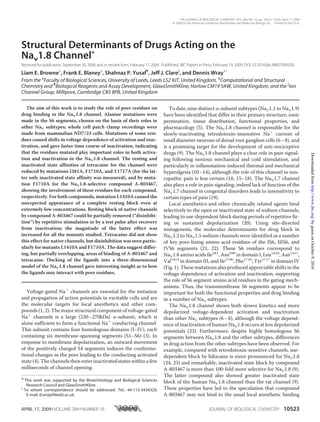
these mutations on the action of compound A-803467 (Fig. 1)
on the NaV1.8 channel was investigated, and compared with the
actions of tetracaine (Fig. 1), to further understand the action of
this compound, which selectively acts on this promising thera-
peutic target for anti-nociceptive drugs. In this study for the
first time the drug binding sites of the NaV1.8 channel have
been addressed, and furthermore, we have investigated the
human channel in an appropriate mammalian host background
more like the native situation, rather than in oocytes.
EXPERIMENTAL PROCEDURES
Mutagenesis of Human Nav1.8
Channels—The human NaV1.8
␣-subunit (Swiss-Prot accession
Q9Y5Y9, polymorph 1073V, Ref. 7)
in the pFastBacMam1 vector (27)
was used in this study. Mutant
channels I381A, N390A, L1410A,
V1414A, I1706A, F1710A, and
Y1717A were generated using the
QuikChange XL Mutagenesis Kit
(Stratagene) together with appro-
priate mutagenic primers. All muta-
tions were validated by restriction
mapping and sequencing of the
entire channel cDNA.
ND7/23 cells (ECACC, Salisbury,
UK) were cultured in Dulbecco’s
modified Eagle’s medium supple-
mented with 2 mM L-glutamine, 10%
heat-inactivated fetal bovine serum,
and 1ϫ non-essential amino acids.
Cells were seeded at 60% confluence
and co-transfected with 3.0 g of
wild type or mutant NaV1.8 ␣-sub-
unit cDNA and 0.3 g of EBO-pCD-
Leu2 cDNA (for CD8 marker) or
pEGFP-N1 cDNA (for fluorescence
marker) using Lipofectamine 2000
(Invitrogen). Transfection-positive
cells were identified 2–4 days after
transfection with Immunobeads
(anti-CD8 Dynabeads; Invitrogen)
or green fluorescence.
Electrophysiological Recording
and Data Analysis—Whole-cell patch clamp recordings were
performed at room temperature (20–22 °C) using patch
pipettes pulled from thin-walled borosilicate glass capillaries
and coated with Sigmacote (Sigma). Pipettes (tip resistances
1.5–2.5 M⍀) were filled with solution containing (mM): CsF,
120; HEPES, 10; EGTA, 10; and NaCl, 15 adjusted to pH 7.2
with CsOH. Cells were continuously perfused with an external
solution containing (mM): NaCl, 140; HEPES, 5; MgCl2, 1.3;
CaCl2, 1; glucose, 11; and KCl, 4.7, adjusted to pH 7.4 with
NaOH. Tetrodotoxin (200 nM, Tocris) was used to block endog-
enous Naϩ
channel currents.
Cells were allowed to equilibrate for 10 min in the whole-cell
configuration before currents were acquired using an Axo-
patch-1C or Axopatch 200B amplifier (Molecular Devices) fil-
tered at 5 kHz, and sampled at 10 kHz using pClamp8 or
pClamp9 software (Clampex; Molecular Devices). The online
P/4 subtraction procedure was used to subtract linear leak and
capacitance currents where appropriate. Clampfit versions 9 or
10 (Molecular Devices) and Origin version 5.0 (MicroCal Inc.)
were used for offline data analysis.
Conductance-voltage curves were determined from the peak
sodium current (INa), using the equation: GNa ϭ INa/(V Ϫ ENa),
2
Browne, L. E., Clare, J. J., and Wray, D. (2009) Neuropharmacology
10.1016/j.neuropharm.2009.01.018.
FIGURE 1. A, the structures of the compounds tetracaine and A-803467. B, alignments of the S6 segments of
different human NaV subtypes. The figure also summarizes results from this study. The residues indicated are
shown in this study to be important for the affinity of tetracaine (T) and A-803467 (A) using mutagenesis, or
tetracaine (t) and A-803467 (a) using computational modeling. The residues indicated (͉) did not appear to be
important for the binding of tetracaine or A-803467 by mutagenesis or by computational modeling.
Sites of Drug Action on NaV1.8
10524 JOURNAL OF BIOLOGICAL CHEMISTRY VOLUME 284•NUMBER 16•APRIL 17, 2009
byguestonOctober9,2016http://www.jbc.org/Downloadedfrom
3. where V is the membrane potential and ENa is the reversal
potential obtained from the I–V curve for each cell. These
curves were fit with the Boltzmann equation: G ϭ Gmax/(1 ϩ
exp((V1⁄2 Ϫ V)/k)), where Gmax is maximum conductance, V1⁄2 is
the potential for half-maximal activation, and k is the slope
factor. Voltage-dependence of inactivation curves were fit with
the Boltzmann equation: I ϭ A ϩ(B Ϫ A)/(1 ϩ exp((V Ϫ V1⁄2)/
k)), where B is the maximum current, A is the amplitude of a
non-inactivating component, V1⁄2 is the potential for half-max-
imal inactivation.
Inactivation time constants were obtained by fitting the
decay phase of individual currents with double exponential fits.
Recovery from inactivation was studied in twin pulse experi-
ments, and the time course of recovery was fit with double
exponentials; in the presence of some drugs used, a third expo-
nential was required.
All test compounds were prepared in dimethyl sulfoxide and
diluted to the desired concentration in the external solution
giving a final concentration of Յ1% dimethyl sulfoxide. Control
test currents were recorded before the application of com-
pounds. Compounds were applied during a 3–4-min incuba-
tion period after which currents were recorded in the continual
presence of test compound. The dissociation constants for rest-
ing channels (Kr) and inactivated channels (Ki) were deter-
mined using the model of Kuo and Bean (28) as described by Liu
et al. (29) and Yarov-Yarovoy et al. (30). The resting state drug
dissociation constant was calculated using a test pulse before
and after drug application from a resting potential of Ϫ120 mV,
at which channels are in the resting state. The dissociation con-
stants were determined using Kr ϭ [D]/((1/IDr) Ϫ 1), where D is
the concentration of test compound and IDr is the peak test
current amplitude in the presence of drug expressed as a frac-
tion of the peak test current amplitude in the absence of drug.
The protocol for obtaining inactivated state dissociation con-
stants is shown in Fig. 5C, where a 4-s depolarizing pulse was
used to inactivate currents, and a test pulse before and after
drug application was used to measure the extent of inactivated
current block. The inactivated state dissociation constants were
calculated using Ki ϭ [D](1 Ϫ h)/((1/IDi) Ϫ 1), where D is the
concentration of test compound, h is the fraction of non-inac-
tivated sodium current (in the absence of the compound) fol-
lowing the 4-s depolarization, and IDi is the peak test current
amplitude in the presence of drug expressed as a fraction of the
peak test current amplitude in the absence of drug (Fig. 5C).
The voltage of the 4-s depolarizing pulse was chosen to always
give approximately the same (60–80%) inactivation in wild
type and mutant channels; the measurement of the Ki value
should therefore only represent the effect of the mutation on
drug binding rather than any effect of the mutation on inacti-
vation itself. Indeed, in previous work the effects of mutations
on inactivation and on the Ki value were not found to correlate
(29, 30). Statistics are presented as mean Ϯ S.E., and the Stu-
dent’s t test was used to test significance.
Computational Modeling of the Human Nav1.8 Channel and
Docking of Drugs—Previous models of sodium channels
described in the literature have been based on bacterial potas-
sium channel structures. More recently, however, a structure of
the rat KV1.2 potassium channel has been solved (31). It was
considered that this would be closer in structure to human
NaV1.8, and our model was therefore based on its homology
with this structure (PDB code 2A79). Our model consisted of
the S5 helices, P-loop, and S6 helices (along with the short
linker between the P-loop and the S6 helix in the first domain).
Whereas sequence alignments between the rat KV1.2 channel
and NaV1.8 domains were unsuccessful; Kyte-Doolittle
hydropathy plots (32) were used to identify the S5-P-loop-S6
region of each domain. Alignment of the loops was made so as
to form a good filter geometry with the DEKA residues. Align-
ment of the S6 regions was straightforward and was based on
the conserved glycine (serine in domain IV). The alignment of
the S5 helices was not obvious so use was made of the conser-
vation moment method using the HELANAL program (33).
The starting structure was constructed manually using the
homology tools within the Quanta program (Wavefunction
Inc.). This was subsequently refined by energy minimization
with CHARMm (34). Initially the backbone was held fixed but
this was subsequently relaxed and the helices were maintained
by using distance (“nuclear Overhauser effect”) constraints
between the backbone amide bonds. 500 steps of Steepest
Descent followed by 5000 steps of Adopted Basis Newton-
Rhaphson were used for the minimization phases. The Karplus
rotamer library (35) was used to set the side chain dihedral
angles at standard values. The two ligands were constructed
with the Spartan program using HF-3–21G* charges (accelrys.
com). They were docked manually into the protein in multiple
poses, ensuring each time that initial side chain dihedral angles
were set at the rotamer library values. Each pose was minimized
as above and ranked according to interaction energy.
RESULTS
Effects of S6 Segment Mutations on the Functional Properties
of the NaV1.8 Channel—Mutations were chosen at positions in
the S6 segments of domains I, III, and IV of the NaV1.8 channel,
corresponding to the positions found in other NaV channel sub-
types where a number of drugs have been found to act (21, 22).
Example current traces for each human NaV1.8 channel muta-
tion are shown in Fig. 2. The voltage dependence of activation
(Fig. 3A) was measured at a holding potential of Ϫ120 mV.
Conductance-voltage relationships for some of the mutant
NaV1.8 channels showed shifts in the curves as compared with
wild type channels (Fig. 3A); a 9-mV shift to more positive
potentials was observed for mutations N390A and V1414A
(Fig. 3B), and negative shifts of ϳ6 mV were observed for muta-
tions I381A and F1710A. The V1⁄2 for activation was unaffected
by mutations L1410A, I1706A, and Y1717A. The k values were
not affected by most of the mutations, although there were
small but significant reductions for two of the mutants (Fig. 3B).
To study the effects of the mutations on the voltage depend-
ence of inactivation (Fig. 3C), a 4-s prepulse to various poten-
tials was used to inactivate channels followed by a test pulse to
0 mV (holding potential Ϫ120 mV). The steady-state inactiva-
tion curves show that all the mutations except F1710A showed
shifts to more negative potentials (Fig. 3, C and D). The k values
were not significantly affected as compared with wild type
(10.5 Ϯ 0.4 mV, n ϭ 84), except for mutations L1410A
(decrease in k to 6.4 Ϯ 0.4 mV, n ϭ 6) and Y1717A (increase in
Sites of Drug Action on NaV1.8
APRIL 17, 2009•VOLUME 284•NUMBER 16 JOURNAL OF BIOLOGICAL CHEMISTRY 10525
byguestonOctober9,2016http://www.jbc.org/Downloadedfrom
4. k to 15.6 Ϯ 1.2 mV, n ϭ 14). For wild type channels, inactivation
was not complete even at very positive inactivating potentials
(A/B parameter in Fig. 3D, see “Experimental Procedures”);
mutations I1706A, F1710A, and Y1717A (all located in the IVS6
segment) showed even less complete inactivation at positive
potentials.
During depolarizing pulses, inactivation kinetics were faster
than wild type for the mutations. An example current trace for
V1414A, and the voltage dependence of the inactivation time
constant are shown in Fig. 3E for all the mutants. It can be seen
that the time constants were generally reduced for the mutants,
particularly at the Ϫ10 mV test potential (Fig. 3F). Thus the
mutations studied here enter inactivated states from open
states faster than the wild type NaV1.8 channel.
Binding Sites for Tetracaine and A-803467 at Inactivated
Wild Type Channels—Tetracaine and A-803467 have been
shown to bind preferentially to inactivated rather than resting
wild type NaV1.8 channels (9, 36). Drugs that preferentially act
on the inactivated state rather than on the resting state show a
hyperpolarizing shift in the steady-state inactivation curve (⌬V)
without altering the slope (k) (37). The magnitude of the shift in
the inactivation curve may be used to determine whether tetra-
caine and A-803467 act on the same binding site. Inactivation
curves were obtained in the presence and absence of 10 M
tetracaine (Fig. 4A), 150 nM A-803467 (Fig. 4B). The values
obtained for these shifts agree with the predictions calculated
from a model with single binding sites (37) (Fig. 4D). For both
drugs applied together (at concentrations of 5 M tetracaine
and 75 nM A-803467, Fig. 4C), the shift clearly agrees with the
predicted value for overlapping binding sites, rather than sepa-
rate binding sites for each drug, again using the models in Ref.
37 (Fig. 4D). Details of the formula used in these models are
given in Fig. 4, legend.
Affinity of Tetracaine and A-803467 for Resting and Inacti-
vated Mutant NaV1.8 Channels—To determine the site of
action for tetracaine and A-803467 on the NaV1.8 channel, here
we have studied the effects of S6 mutations on both the resting
and inactivated state affinities.
In the resting state (holding potential Ϫ120 mV), the extent
of block of a test pulse current by the compounds was used to
measure the affinity of compounds for the resting state (more
precisely, dissociation constant, Kr, see “Experimental Proce-
dures”). For tetracaine, the resting state affinity was signifi-
cantly reduced (i.e. dissociation constants were increased) for
mutations I381A and F1710A as compared with wild type chan-
nels (Fig. 5A). For compound A-803467, an appreciable
decrease in the resting state affinity was only observed for
mutation F1710A (Fig. 5B). Thus residues Ile381
and Phe1710
are
involved in the resting state binding of tetracaine, whereas res-
idue Phe1710
is involved in resting state binding of compound
A-803467.
To calculate the inactivated state affinity of test compounds
for mutant NaV1.8 channels, a twin pulse protocol (Fig. 5C) was
used before and after application of tetracaine or A-803467.
Before compound application, the fractional amount of non-
inactivated current during the test pulse after a 4-s depolariza-
tion (h, see “Experimental Procedures”) was obtained from the
ratio of test to control pulse peak amplitude. After compound
application, the fraction of test current not blocked by the com-
pounds (IDi, see “Experimental Procedures”) was measured and
the inactivated state dissociation constant, Ki, calculated from h
and IDi. Example currents are shown in Fig. 5D. Mutations
I381A, F1710A, and Y1717A showed marked decreases in affin-
ity for tetracaine (Fig. 5E). For compound A-803467, only
mutation F1710A caused a decrease in the inactivated state
affinity (Fig. 5F). Thus residue Phe1710
is important in deter-
mining both resting and inactivated state affinities for both tet-
racaine and A-803467, whereas residues Ile381
and Tyr1717
are
also important for tetracaine binding. The effects of test com-
pounds on mutant L1410A channels are considered separately
in the next section.
Tetracaine and A-803467 Drug Block of NaV1.8 Mutation
L1410A—Data for the affinity of mutant L1410A channels
could not readily be obtained using the above protocols because
even concentrations 1000 times smaller than used in the previ-
ous section still gave almost complete resting block (Fig. 6A)
(indicating very high resting state affinities of Ͼ10 nM for tet-
racaine, and Ͼ100 pM for A-803467), and also because of
unusual behaviors under repetitive stimulation (Fig. 6B). As can
FIGURE 2. Example current traces for human NaV1.8 channels. Current
traces are shown for ND7/23 cells transfected with wild type or mutant
hNaV1.8 channel cDNA in the presence of 200 nM tetrodotoxin. Currents were
elicited for voltage steps to Ϫ100 to ϩ60 mV (in 10-mV increments) from a
holding potential of Ϫ120 mV. The peak current amplitudes were Ϫ79 Ϯ 7
pA/pF (n ϭ 79) for wild type NaV1.8, Ϫ53 Ϯ 5 pA/pF (n ϭ 50) for mutant I381A,
Ϫ112 Ϯ 21 pA/pF (n ϭ 52) for mutant N390A, Ϫ32 Ϯ 5 pA/pF (n ϭ 31) for
mutant L1410A, Ϫ64 Ϯ 6 pA/pF (n ϭ 58) for mutant V1414A, Ϫ35 Ϯ 4 pA/pF
(n ϭ 38) for mutant I1706A, Ϫ69 Ϯ 7 pA/pF (n ϭ 45) for mutant F1710A, and
Ϫ40 Ϯ 3 pA/pF (n ϭ 54) for mutant Y1717A.
Sites of Drug Action on NaV1.8
10526 JOURNAL OF BIOLOGICAL CHEMISTRY VOLUME 284•NUMBER 16•APRIL 17, 2009
byguestonOctober9,2016http://www.jbc.org/Downloadedfrom
5. be seen in the latter figure, in the presence of tetracaine or
A-803467, during repetitive stimulation at 10 Hz, currents sur-
prisingly increased from almost zero to values comparable with
currents before test compound, as if stimulation removed the
unusually high affinity resting block of the compounds
(disinhibition).
FIGURE 3. Effects of S6 mutations on Na؉
channel activation and inactivation. A, conductance-voltage curves are shown for wild type NaV1.8 (f, n ϭ 79),
and mutations I381A (F, n ϭ 50), N390A (Œ, n ϭ 52), L1410A (, n ϭ 31), V1414A (Ⅺ, n ϭ 58), I1706A (E, n ϭ 38), F1710A (⌬, n ϭ 45), and Y1717A (ƒ, n ϭ 54).
Curves were fit with the Boltzmann equation and normalized to maximum conductance. The pulse protocol is shown in the inset. B, bar diagrams show the
voltage for half-maximal activation (V1⁄2) and the slope factor (k) for wild type and mutant channels (same experiments as in A). C, curves are shown for the
voltage dependence of inactivation for wild type NaV1.8 (f, n ϭ 84), and mutations I381A (F, n ϭ 13), N390A (Œ, n ϭ 14), L1410A (, n ϭ 6), V1414A (Ⅺ, n ϭ
25), I1706A (E, n ϭ 12), F1710A (⌬, n ϭ 18), and Y1717A (ƒ, n ϭ 14). The curves were fit with the Boltzmann equation and normalized to the maximum current.
The pulse protocol is shown in the inset. D, bar diagrams (same experiments as C) showing the voltage for half-maximal inactivation (V1⁄2), and the amplitude
of the non-inactivated component normalized to the maximum current (A/B, see “Experimental Procedures”). *, p Ͻ 0.05. E, the inset shows example currents
for wild type and mutant (V1414A) NaV1.8 channels elicited by a voltage step to 0 mV from a holding potential of Ϫ120 mV. Current traces were normalized and
superimposed. The time constant, , obtained from the inactivation time course is shown for wild type NaV1.8 (f, n ϭ 56), and mutations I381A (F, n ϭ 35),
N390A (Œ, n ϭ 34), L1410A (, n ϭ 32), V1414A (Ⅺ, n ϭ 36), I1706A (E, n ϭ 32), F1710A (⌬, n ϭ 37), and Y1717A (ƒ, n ϭ 38), for the test potentials shown. F, bar
diagrams are shown for the mean values of the time constant of inactivation, , for each mutant at Ϫ10 mV test potential (same experiments as in E). *, p Ͻ 0.05.
Sites of Drug Action on NaV1.8
APRIL 17, 2009•VOLUME 284•NUMBER 16 JOURNAL OF BIOLOGICAL CHEMISTRY 10527
byguestonOctober9,2016http://www.jbc.org/Downloadedfrom
6. To determine whether sustained depolarization would show
a similar effect, a 600-ms conditioning depolarizing pulse was
first applied, followed 100 ms later (to allow for recovery from
inactivation) by a test pulse (Fig. 6C). The 600-ms pulse is com-
parable with the 60 10-ms pulses used during repetitive stimu-
lation. Although current during the conditioning pulse was
almost completely blocked by tetracaine or A-803467, the
test current was comparable with that in the absence of the
compounds (Fig. 6D). Thus it appears that depolarization
removes the high-affinity resting block of the compounds,
underlying the process of disinhibition.
The Effect of A-803467 and Tetracaine on the Recovery from
Inactivation of S6 Segment Mutations—The effects of
A-803467 and tetracaine on the recovery from inactivation (in
the continual presence of the compound) were investigated
using the protocol shown in the inset in Fig. 7C. Following the
application of A-803467, the time course for recovery involved
not simply the removal of inactivation, but also an additional
component with increased current above resting control values
(disinhibition), apparently due to partial removal of the resting
block by stimulation in the presence of the compound. This
effect, although small for wild type, can be seen from example
current traces (Fig. 7A). The effect can also be seen for mean
current values in Fig. 7C, where all currents have been normal-
ized to control pulse amplitude in the absence of the com-
pound. Recovery from disinhibition followed a very slow time
course (1.7 Ϯ 0.2 s, Fig. 7F).
For the mutants in the presence of compound A-803467 (100
nM), this disinhibitory component was larger than for wild type.
For instance, example current traces for V1414A clearly show
the effect (Fig. 7B), as do the mean current values for V1414A
and L1410A (Fig. 7, D and E). For the latter mutant (with almost
zero current in the resting state in the presence of the com-
pound at 100 pM), marked disinhibitory current was seen, cor-
responding to extensive removal of resting block during stim-
ulation (Fig. 7E). Data for all the mutants are summarized in Fig.
FIGURE 4. Binding sites for tetracaine and A-803467 on the wild type NaV1.8 channel. The figure shows steady-state inactivation curves for (A) tetracaine
(10 M, E, n ϭ 4), (B) A-803467 (150 nM, E, n ϭ 4), and (C) tetracaine (5 M) plus A-803467 (75 nM) (E, n ϭ 8). Control curves in the absence of drug are shown
(F, paired values in each case). The protocol used was as in Fig. 3C, and Boltzmann curves fit as before. The shifts in the inactivation curves (⌬V1⁄2) were
determined and are shown in D. Predicted values of the shifts are shown for separate binding sites and for overlapping binding sites using the model of Kuo
(37). Briefly, in this model, in the presence of a single drug of concentration ([D]), and affinity Ki, the shift is given by ⌬V ϭ k(ln(1 ϩ ([D]/Ki))). For both drugs
applied together, if the two drugs act on an overlapping site, the shift of the inactivation curve is given by⌬V ϭ k(ln(1 ϩ ([D1]/Ki1) ϩ ([D2]/Ki2))), where [D1] and
[D2] are the concentrations of each drug with the respective Ki values Ki1 and Ki2. In contrast, if the two drugs act on separate sites, then the shift in the
inactivation curve is given by ⌬V ϭ k(ln(1 ϩ ([D1]/Ki1) ϩ ([D2]/Ki2) ϩ ([D1]/Ki1)([D2]/Ki2))). For the predictions in D, values of Ki are as indicated in Fig. 5, and
values of k for each drug application were obtained from the above inactivation curves, taking mean values in each case.
Sites of Drug Action on NaV1.8
10528 JOURNAL OF BIOLOGICAL CHEMISTRY VOLUME 284•NUMBER 16•APRIL 17, 2009
byguestonOctober9,2016http://www.jbc.org/Downloadedfrom
7. 7F, where it can be seen that the disinhibitory component
(expressed as a fraction of the current blocked at rest by the
compound) is greater for all the mutants considered than for
wild type. For mutant Y1717A (not
shown in the figure), currents were
too small to be measured in the
presence of 100 nM A-803467; how-
ever, even at 10 nM, disinhibition
was much greater than for wild type
at 10 nM. All mutants (except for
I1706A) showed a similar time con-
stant for the recovery of this disin-
hibitory component (Fig. 7F).
For tetracaine (10 M), by con-
trast, the wild type channel showed
no component of disinhibition, as
can been seen from the example of
current traces (Fig. 8A), and the cur-
rents during recovery shown in Fig.
8C. On the other hand, mutant
L1410A showed a striking disinhibi-
tory component in the presence of
tetracaine (Fig. 8D). For mutant
F1710A, a clear disinhibitory com-
ponent was observed in the pres-
ence of tetracaine (Fig. 8, B and E);
this component was relatively large
when expressed as a fraction of the
resting block (Fig. 8F). For the other
mutants, disinhibitory components
were observed in detailed fits to the
time course but their amplitudes
relative to extent of resting block
were small (Fig. 8F). Time constants
of recovery were in the range 0.8 to
1.8 s (Fig. 8F). Taken together the
data show that for tetracaine, muta-
tions L1410A and F1710A clearly
induced the disinhibitory com-
ponent, whereas for compound
A-803467, all the mutations consid-
ered led to more pronounced disin-
hibitory components. The time
course of recovery of this compo-
nent appears to be simply a reflec-
tion of the time course of reinstate-
ment of the resting block. It is also
noteworthy that the extent of disin-
hibition (Figs. 7F and 8F) for each
mutant did not correlate with the
effects of the mutations themselves
on inactivation (Fig. 3), suggesting
that disinhibition is not the result of
altered channel function per se.
Molecular Modeling—A model
was constructed for the S6 and P
loop regions of the NaV1.8 channel,
using the alignment shown in Fig. 9
(see “Experimental Procedures”) with the rat KV1.2 channel in
the open state. The model suggests that, unlike the other S6
residues mutated in this study, Asn390
and Val1414
do not face
FIGURE 5. Dissociation constants for resting and inactivated states of mutant NaV1.8 channels. A, bar
diagrams are shown representing mutant NaV1.8 channel resting state dissociation constants (Kr) for tetra-
caine. Values were calculated for tetracaine at 10 M for wild type NaV1.8 (n ϭ 6) and mutations I381A (n ϭ 7),
N390A (n ϭ 8), V1414A (n ϭ 5), I1706A (n ϭ 6), and F1710A (n ϭ 4). B, bar diagrams are shown representing
resting state dissociation constants (Kr) for compound A-803467. Values were calculated for A-803467 at 100
nM for wild type NaV1.8 (n ϭ 5) and mutations I381A (n ϭ 4), N390A (n ϭ 9), V1414A (n ϭ 6), I1706A (n ϭ 2), and
F1710A (n ϭ 7). It was not possible to determine the resting state dissociation constant for mutation Y1717A
under the conditions used here, because a large proportion (23%) of channels are inactivated at a holding
potential of Ϫ120 mV as a consequence of the strong negative shift in the inactivation curve. C, the figure
shows the twin pulse protocol used to determine the inactivated state affinities (Ki). This was used before and
after test compound application; the depicted 4-s depolarizing pulse was to potentials such that 60–80%
inactivation was observed. In the case of mutant Y1717A, the resting level of inactivation at Ϫ120 mV (as
above) was taken into account in obtaining the parameter h. D, the figure shows example current traces
elicited by the pulse protocol in C, where currents in bold are before test compound application and fine traces
are after test compound application; the currents larger in magnitude correspond to the control pulses and
currents smaller in magnitude correspond to the test pulse following a 4-s depolarization. E, bar diagrams are
shown representing mutant NaV1.8 channel-inactivated state dissociation constants (Ki) for tetracaine. Values
were calculated for tetracaine at 1–10 M for wild type NaV1.8 (n ϭ 7) and mutations I381A (n ϭ 6), N390A (n ϭ
6), V1414A (n ϭ 6), I1706A (n ϭ 5), F1710A (n ϭ 7), and Y1717A (n ϭ 7). F, bar diagrams are shown representing
mutant NaV1.8 channel-inactivated state dissociation constants (Ki) for compound A-803467. Values were
calculated for A-803467 at 10–100 nM for wild type NaV1.8 (n ϭ 7) and mutations I381A (n ϭ 6), N390A (n ϭ 7),
V1414A (n ϭ 5), I1706A (n ϭ 6), F1710A (n ϭ 7), and Y1717A (n ϭ 7). *, p Ͻ 0.05.
Sites of Drug Action on NaV1.8
APRIL 17, 2009•VOLUME 284•NUMBER 16 JOURNAL OF BIOLOGICAL CHEMISTRY 10529
byguestonOctober9,2016http://www.jbc.org/Downloadedfrom
8. into the pore but rather form interhelical interactions with
adjacent helices. Thus Asn390
in domain I forms hydrogen
bonds with Asn1724
in S6 of domain IV and Thr250
in S5 of
domain I, whereas Val1414
in domain III makes hydrophobic
contacts with Leu889
and Phe893
in S6 of domain II (Fig. 10, A
and B). These residues are all located in the cytoplasmic side of
the transmembrane bundle.
Using the model, docking studies were performed with tet-
racaine and compound A-803467 (Fig. 10, C–F). The lack of any
acidic residues in the S6 helices suggested that binding of the
protonated nitrogen of tetracaine and the amide NH of
A-803467 might occur with the aspartate, Asp356
in the P loop
of domain I (i.e. the Asp of the DEKA motif). This allowed the
ligands to adopt poses where all the observed mutation results
could be explained. Residue Phe1710
forms - stacking with an
aromatic ring in both ligands, whereas Leu1410
forms good
hydrophobic interactions with both drugs, consistent with the
experimental finding that its mutation altered the drug action.
However, compound A-803467 extends further into the P loop
of the channel where the two methoxy groups of the compound
can form hydrogen bonds with Thr354
and Ser1660
in P loops.
These hydrogen bonds effectively lock A-803467 in this posi-
tion so that the substituted ring cannot form a favorable hydro-
phobic interaction with Ile381
in S6. The dimethyl amino group
of tetracaine, however, is able to interact favorably with Ile381
.
The tyrosine residue, Tyr1717
in S6, can form a good hydrogen
bond to the ester carbonyl of tetracaine, but it is relatively far
from the biaryl rings of A-803467, so cannot interact favorably
with it. The experimental results for affinities of the com-
pounds found in our mutational study support the model.
DISCUSSION
Functional Properties of NaV1.8 S6 Mutants—Mutations in
Asn390
and Val1414
gave positive shifts in steady-state activa-
tion, suggesting that the corresponding native residues stabilize
open states relative to closed states. This seems to be consistent
with our molecular model in the open state, because these two
residues and those that interact with
them are all located in the cytoplas-
mic side of the transmembrane bun-
dle, and interactions are probably
more likely to be found in the open
state, so that these residues may
indeed stabilize open states. At res-
idues corresponding to these posi-
tions in rNaV1.2 and rNaV1.4, simi-
lar positive shifts were observed in
previous work (30, 38, 39).
Other mutations in NaV1.8 gave
negative shifts in activation (I381A
and F1710A), indicating relative sta-
bilization of corresponding native
closed states. However, for residues
corresponding to these, negative
shifts were not observed for muta-
tions in the rNaV1.2 channel (30,
40). Furthermore, in contrast to our
results showing lack of shift for
mutations L1410A and I1706A in NaV1.8, homologous rNaV1.2
mutations showed positive shifts (41). Thus, the S6 segment
residues play an important role in voltage dependence of acti-
vation but this role in activation appears to be different for the
NaV1.8 channel from other subtypes.
For the voltage dependence of steady-state inactivation in
NaV1.8, all mutations studied here (except F1710A) caused
strong negative shifts. One possibility might be that shifts in
inactivation curves might simply be the result of shifts in the
activation curves. However, because NaV1.8 activation showed
both negative and positive shifts depending on the mutation
studied, this suggests that inactivation gating is not simply
linked to activation, as already noted for NaV1.4 (42). These
curves represent mainly inactivation from closed states (41).
Thus, for NaV1.8, closed-state inactivation is less favored for
the native channel than for the mutants. We also showed that
the time course of inactivation of NaV1.8 was faster for all the
mutants considered here. Because this represents open-state
inactivation (41), the data show that open-state inactivation is
also less favorable for the native channel than for the mutants.
The mutational data shows that all the S6 residues studied here
are involved in inactivation processes in the native channel.
As for activation, the effects of mutations on inactivation
appear to be subtype-specific. For closed-state inactivation,
shifts for N390A, V1414A, and I381A in NaV1.8 were in the
opposite direction to those for corresponding mutations in
NaV1.2; there was no shift for F1710A in NaV1.8, whereas cor-
responding mutations in NaV1.2 and NaV1.5 gave positive shifts
(30, 40, 41, 43). For open state inactivation (observed from the
time course of decay of currents), all the mutations in NaV1.8
caused a faster time course, whereas the corresponding muta-
tions in NaV1.2 did not affect it (nor did mutations in NaV1.4
corresponding to I1706A and Y1717A in NaV1.8). Also the
mutation corresponding to F1710A in NaV1.8 was slower for
NaV1.4 (30, 40–42). The underlying reason at the molecular
level for these differences is not known, but may be related to
FIGURE 6. Disinhibition of resting block for mutation L1410A. A, the figure shows example L1410A mutant
currents in the absence and presence of very low concentrations (indicated) of tetracaine and A-803467. The
current traces were elicited by a test pulse to 0 mV from a holding potential of Ϫ120 mV. B, currents, Inorm, for
mutant L1410A NaV1.8 channels are shown during a 10-Hz train of pulses (10-ms duration to 0 mV from a
holdingpotentialofϪ120mV),plottedagainstpulsenumberandnormalizedtothefirstpulseoftheuntreated
cell. The currents are shown before the application of tetracaine (f, n ϭ 6) or A-803467 (F, n ϭ 8) and after
tetracaine (10 nM, Ⅺ) or A-803467 (100 pM, E) in paired cells. C, as shown in the protocol, current amplitude was
measured at a test pulse following a 600-ms depolarizing pulse to 0 mV and a 100-ms recovery period. D, the
bar diagrams show the mean currents using the protocol in C, before (filled bars) and after (unfilled bars) the
application of tetracaine (10 nM, n ϭ 5) or compound A-803467 (100 pM, n ϭ 6) in paired cells.
Sites of Drug Action on NaV1.8
10530 JOURNAL OF BIOLOGICAL CHEMISTRY VOLUME 284•NUMBER 16•APRIL 17, 2009
byguestonOctober9,2016http://www.jbc.org/Downloadedfrom
9. FIGURE 7. The effect of A-803467 on the recovery from inactivation. Example NaV1.8 channel currents are shown for wild type (A) and V1414A mutant (B) in the
presence or absence of A-803467 (100 nM). Currents were elicited by an initial control pulse (to 0 mV), followed by test pulses (to 0 mV) at the indicated times during
recovery (protocol shown in the inset of C). The graphs show the test pulse amplitude normalized to control pulse during the recovery from inactivation for wild type
(C), and example mutations, V1414A (D) and L1410A (E), using the protocol shown in the inset. Time courses of recovery from inactivation are shown before (f) and
after(F)theapplicationofA-803467(100nM,except100pM forL1410A)inpairedcells.F,bardiagramsareshownfortheamplitude(Idis,normalizedtoextentofresting
currentblock)andtimecourse()oftheslowestcomponentofthethree-exponentialfittothetimecourseofrecoveryofinactivationforwildtype(nϭ5),andmutants
I381A (n ϭ 7), N390A (n ϭ 7), L1410A (n ϭ 5), V1414A (n ϭ 6), I1706A (n ϭ 2), and F1710A (n ϭ 7). The dotted line in C–E represents the level of resting block.
Sites of Drug Action on NaV1.8
APRIL 17, 2009•VOLUME 284•NUMBER 16 JOURNAL OF BIOLOGICAL CHEMISTRY 10531
byguestonOctober9,2016http://www.jbc.org/Downloadedfrom
10. FIGURE 8. The effect of tetracaine on the recovery from inactivation. Example NaV1.8 channel currents are shown for wild type (A) and F1710A mutant (B) in the
presence or absence of tetracaine (10 M). Currents were elicited by an initial control pulse (to 0 mV), followed by test pulses (to 0 mV) at the indicated times during
recovery (protocol shown in the inset of C). The graphs show the test pulse amplitude normalized to control pulse during the recovery from inactivation for wild type
(C), and example mutations, L1410A (D) and F1710A (E), using the protocol shown in the inset. Time courses of recovery from inactivation are shown before (f) and
after (F) the application of tetracaine (10 M, except 10 nM for L1410A) in paired cells. F, bar diagrams are shown for the amplitude (Idis, which is the disinihibitory
componentI3 expressedasafractionofrestingcurrentblock)andtimecourse()oftheslowestcomponentofthethree-exponentialfittothetimecourseofrecovery
of inactivation for mutants N390A (n ϭ 4), L1410A (n ϭ 7), V1414A (n ϭ 3), I1706A (n ϭ 3), F1710A (n ϭ 4), and Y1717A (n ϭ 6), whereas wild type (n ϭ 6) and mutant
I381A (n ϭ 6) did not show disinhibition. The dotted line in C–E represents the level of resting block.
Sites of Drug Action on NaV1.8
10532 JOURNAL OF BIOLOGICAL CHEMISTRY VOLUME 284•NUMBER 16•APRIL 17, 2009
byguestonOctober9,2016http://www.jbc.org/Downloadedfrom
11. the property of slower inactivation observed in native NaV1.8
than in other NaV subtypes.
For the IVS6 segment mutations I1706A, F1710A, and
Y1717A, but not for the other mutations considered here, there
was incomplete inactivation, even at very positive potentials. A
similar phenomenon of incomplete inactivation has also been
reported for NaV1.2 for the mutations homologous to F1710A
and Y1717A in NaV1.8, and for NaV1.4 mutation homologous
to N390A in NaV1.8 (40, 44). Thus, as for the other subtypes, for
NaV1.8 these residues play an important role in inactivation.
Mutation Y1717A gave the largest effect in NaV1.8; as this
mutation is located at the intracellular mouth of the pore where
the inactivation domain III–IV linker acts, it may be that this
residue is somehow involved in the receptor site for fast inacti-
vation (40).
Effects of S6 Segment Mutations on Drug Action on the NaV1.8
Channel—Tetracaine and A-803467 have been previously
shown to preferentially bind to inactivated rather than resting
wild type NaV1.8 channels (9, 36). Here we have analyzed the
actions of tetracaine and compound A-803467 on the human
channel using an appropriate mammalian expression system.
Similar magnitude shifts in the steady-state inactivation curve
were observed following the application of either tetracaine (10
M) or compound A-803467 (150 nM) separately, or following
the application of both drugs together (at half the above con-
centrations). This finding indicates that one drug precludes the
binding of the other. The observed shifts were all of a similar
magnitude to the value predicted by a model with a single bind-
ing site, rather than a model with separate binding sites for each
compound. Thus, tetracaine and A-803467 appear to bind to
overlapping, or partially overlapping, binding sites on the
NaV1.8 channel.
To determine more precisely the site of action of tetracaine
and A-803467 on the NaV1.8 channel, residues were chosen for
mutation in the S6 regions guided by their role in drug binding
for other NaV subtypes (21, 22). We have shown that residues
Ile381
, Leu1410
, Phe1710
, and Tyr1717
are involved for tetracaine
affinity, but only residues Leu1410
and Phe1710
for A-803467.
The roles of these residues in binding of the respective com-
pounds was fully supported in our docking study using the
molecular model for NaV1.8 (Fig. 10, C–F). In addition, the
model implicates binding of other residues in the pore region of
the channel. The S6 residues implicated in binding to NaV1.8 by
molecular modeling and by our mutational studies are summa-
rized in Fig. 1B.
The increased resting affinity observed with the L1410A
mutant is difficult to explain in our molecular model; one pos-
sibility would be that the compounds are being trapped in the
bound state, although it is difficult to understand how the ala-
nine mutation would enhance trapping. As the mutation did
not show unusual functional properties in the absence of drugs
(see above), it is unlikely that the mutation causes severe distor-
tion of the molecular structure.
In previous studies with a range of drugs and NaV subtypes,
the residue corresponding to Phe1710
in NaV1.8 has generally
been found to be most important in determining inactivated
state affinity (36). We have indeed found this residue to be
important in the present study for NaV1.8, both for binding of
tetracaine and for A-803467. For tetracaine the F1710A muta-
tion reduced the inactivated state affinity far more than the
resting affinity, and so this residue has a key role in contributing
to the preferential inactivated state block of tetracaine. A sim-
ilar result for tetracaine was found for NaV1.3 channels (36). For
compound A-803467, no previous mutagenesis work has been
carried out, and as mentioned in the Introduction, it was spec-
ulated that this drug does not bind to the local anesthetic bind-
ing site (26). However, our results for mutation of the local
anesthetic binding site Phe1710
indeed further suggests binding
of this compound to at least part of the established local anes-
thetic binding site. Although this residue is indeed important
for binding of compound A-803467, by contrast with tetracaine
other residues not mutated here must also be important for
binding. The reason for this is that mutation of this residue in
NaV1.8 affected both resting and inactivated state affinities for
A-803467 by similar proportional amounts, and because affin-
ity of this compound for inactivated native channels is much
greater than for resting native channels, other residues must
also be involved. Our modeling leads us to suggest that other
residues in S6 regions of all four domains and the P-loop may
also be important for A-803467 binding (Fig. 10, C–F), and it
would be interesting to investigate the role of these residues in
future mutational studies.
Although the residues considered here have been shown to
be involved for a range of NaV subtypes and compounds, tetra-
caine has so far only been examined for NaV1.3 where residues
corresponding to Phe1710
(as above) and Tyr1717
are involved,
although the interaction with the latter residue was suggested
to be indirect (36). For NaV1.2 channels, the local anesthetic
etidocaine also has important interactions with these residues
(45). The key role for residue Phe1710
in drug binding has also
been generally shown for NaV1.3, NaV1.4, and NaV1.5 (22). This
residue is proposed to directly bind to local anesthetics by a
cation- interaction (46), although our model for NaV1.8 sug-
gests a - stacking interaction. Overall it is surprising that
compound A-803467 is selective for NaV1.8, whereas the
sequence alignments with other human NaV subtypes (Fig. 1B)
show that the residues that we have implicated in drug binding
(whether by the mutational study or by modeling) are almost
FIGURE 9. Sequence alignments used in the model. The S5 helices, P-loops,
and S6 helices are aligned with the rat KV1.2 channel (31). The DEKA motif in
the filter and the glycines in S6 are shown shaded.
Sites of Drug Action on NaV1.8
APRIL 17, 2009•VOLUME 284•NUMBER 16 JOURNAL OF BIOLOGICAL CHEMISTRY 10533
byguestonOctober9,2016http://www.jbc.org/Downloadedfrom
12. Sites of Drug Action on NaV1.8
10534 JOURNAL OF BIOLOGICAL CHEMISTRY VOLUME 284•NUMBER 16•APRIL 17, 2009
byguestonOctober9,2016http://www.jbc.org/Downloadedfrom
13. completely identical between NaV subtypes. Of the residues
mutated here, only the Val1414
residue is different in other sub-
types; the isoleucine present in other subtypes at this position
may contribute to subtype differences, although it is remote to
the ligand binding site. However, the other regions of the chan-
nel may indirectly involve drug binding; indeed slow inactiva-
tion processes and their conformational implications for the
whole channel have been suggested to play a role in selectivity
of compounds (24). Another possible explanation of the
reported selectivity may lie in the presence of a double glycine
motif in the S6 helix of domain III. The glycine at 1406 is a
serine in other NaV channels. This double glycine greatly
increases the flexibility in the helix and indeed a brief molecular
dynamics simulation of 200 ps3
showed that Leu1410
moved
closer to the chlorophenyl ring of A-803467, thus further stabi-
lizing the interaction. A more extensive mutational study of
other residues coupled with more extensive molecular dynam-
ics simulations in other NaV channel models would be required
in the future to establish the reason for the selectivity of
A-803467 for the NaV1.8 channel.
For native NaV1.8 channels, in the continual presence of
compound A-803467, depolarization and subsequent repolar-
ization leads not only to inactivation and recovery but also to
partial removal of resting block by the compound. The latter
mechanism involves a component with an increase in current
(disinhibition), an entirely distinct process from removal of
inactivation during recovery. All the mutations considered here
increased this disinhibitory current for A-803467. An
extremely interesting example of this was for mutant L1410A,
where resting block by the compound was almost complete. For
this mutant, stimulation in the presence of the compound gave
an appreciable current corresponding to marked removal of the
resting block. This phenomenon was observed whether stimu-
lation was applied repetitively (Fig. 6B), or after long (600 ms,
Fig. 6D) or short (50 ms, Fig. 7E) depolarizations. For the other
mutations considered here, whereas resting block was generally
not substantially affected, all mutations showed greater disin-
hibition than for the wild type channel. Thus all the residues
mutated here appear to be involved in this disinhibitory effect
of A-803467.
Tetracaine did not show the disinhibitory effect for native
NaV1.8 channels. However, mutations caused the appearance
of disinhibitory components after stimulation in the presence
of tetracaine. As for compound A-803467, tetracaine almost
completely blocked resting channels with high affinity for
mutant L1410A, and yet stimulation in the presence of tetra-
caine again showed a disinhibitory component, whether for
repetitive stimulation or for long or short pulses (Figs. 6 and 8).
Of the remaining mutants considered here, clear disinhibitory
currents were seen for F1710A, although smaller effects were
seen for most of the other mutants. Taken together, the data
suggest that for tetracaine, residues Leu1410
and Phe1710
con-
tribute to the mechanism underlying the main disinhibitory
component on the NaV1.8 channel.
The phenomenon of disinhibition is not easy to understand
by the molecular model. One may speculate that disinhibition
may be the result of weakened ligand binding to the pore region
(and the DEKA filter) caused by channel opening and the com-
pound moving away from the pore loop region, so allowing
increased channel currents. With tetracaine, the salt bridge
with Asp356
of the DEKA filter is stronger than the hydrogen
bonds formed with A-803467, and so less disinhibition would
occur for tetracaine. The hydrogen bonds from the P-loop res-
idues Thr354
and Ser1660
to A-803467 hold the ligand to the side
of the P loop and would then perhaps allow partial flow of ions
through the channel, perhaps allowing disinhibition for the
native channel in the case of A-803467, although not for
tetracaine.
Although it seems reasonable to interpret the disinhibitory
component as due to removal of resting block by stimulation in
the presence of the compounds, another possible mechanism
may be the induction of a new single channel state with a higher
conductance. Thus it would be interesting to test this in the
future using single channel recordings. Either way, during
recovery from disinhibition there is reinstatement of the resting
equilibrium state in the presence of the drug. Although the
detailed mechanism for this process is not understood, because
the time course of removal of the disinhibitory component is
very slow (order of seconds), that would imply that the process
does not involve simply unbinding and rebinding of the drug
(which would be much quicker). One may perhaps hypothesize
that depolarization in the presence of the drug induces a new
type of state with slow recovery back to equilibrium.
In summary, our mutational study has indicated that various
residues in the S6 region play important roles in the activation
and inactivation properties of the NaV1.8 channel. We have
identified residues in S6 involved in the resting and inactivating
state block by tetracaine and A-803467. We also identified res-
idues involved in the phenomenon of removal of resting block
(disinhibition) by stimulation in the presence of these com-3
F. E. Blaney, unpublished data.
FIGURE 10. Modeling of the pore region of NaV1.8. A, local environment around Asn390
, with hydrogen bonds between this residue and Asn1724
on S6 of
domain IV and Thr250
on S5 of domain I. B, local environment around Val1414
, with hydrophobic interactions with Leu889
and Phe893
on S6 of domain II. These
interactions occur at the cytoplasmic side of the bundle in the open state model. C, the figure shows docking of tetracaine to IIS6, IVS6, and P-loops, with -
stacking Phe1710
(in IVS6) and Tyr1717
(in IVS6), whereas the latter residue also forms a hydrogen bond with the ester carbonyl group of tetracaine. Hydrophobic
interactions are also observed between tetracaine and Leu882
(in IIS6) and Leu1711
(IVS6). D, docking is shown for tetracaine to IS6, IIIS6, and P-loops, with the
protonated amino group of the compound forming a salt bridge with Asp356
(in the P-loop), whereas the amino-methyl groups have hydrophobic interactions
with Ile381
(in IS6) and Phe382
(in IS6). The butyl group of tetracaine also forms hydrophobic interactions with Leu1410
(in IIIS6) and Phe1413
(in IIIS6). E, docking
of A-803467 is shown to IIS6, IVS6, and P-loops, with P-loop residues Thr354
and Ser1660
hydrogen bonding to the two methoxy groups of the compound. There
is - stacking between the ligand and Phe1710
(in IVS6) and additional hydrophobic interaction with Leu882
(in IIS6) and Val1714
(in IVS6). F, the figure shows
docking of A-803467 to IS6, IIIS6, and P-loops, with primary interaction with Asp356
(in the P-loop) and Leu1410
(in IIIS6) in the proximity of the chlorophenyl ring
of the compound. There is also a hydrogen bond between the amide carbonyl group of the compound and Ser385
(in IS6). Residues Asn390
(in IS6) and Val1414
(in IIIS6) do not interact with the ligand. The figures in C–F show P-loops for domains I and IV and S6 helices for all four domains. The ␣-carbon ribbons are
color-coded as follows: gray, pore loops; blue, IS6; yellow, IIS6; orange, IIIS6; red, IVS6. The underlined residues were mutated.
Sites of Drug Action on NaV1.8
APRIL 17, 2009•VOLUME 284•NUMBER 16 JOURNAL OF BIOLOGICAL CHEMISTRY 10535
byguestonOctober9,2016http://www.jbc.org/Downloadedfrom
14. pounds. The data suggest differing but partially overlapping
areas of binding for tetracaine and compound A-803467.
Acknowledgments—We thank Dr. Andrew Powell and Tim Dale for
continuing expert technical assistance and advice. We are grateful to
Dr. Lin-Hua Jiang for help with initial experiments in his laboratory
and for comments on the manuscript.
REFERENCES
1. Catterall, W. A., Goldin, A. L., and Waxman, S. G. (2005) Pharmacol. Rev.
57, 397–409
2. Hille, B. (2001) Ionic Channels of Excitable Membranes, Sinauer, Sunder-
land, MA
3. Catterall, W. A. (2000) Neuron 26, 13–25
4. Yang, N., George, A. L., Jr., and Horn, R. (1996) Neuron 16, 113–122
5. Goldin, A. L., Barchi, R. L., Caldwell, J. H., Hofmann, F., Howe, J. R.,
Hunter, J. C., Kallen, R. G., Mandel, G., Meisler, M. H., Netter, Y. B., Noda,
M., Tamkun, M. M., Waxman, S. G., Wood, J. N., and Catterall, W. A.
(2000) Neuron 28, 365–368
6. Akopian, A. N., Sivilotti, L., and Wood, J. N. (1996) Nature 379, 257–262
7. Rabert, D. K., Koch, B. D., Ilnicka, M., Obernolte, R. A., Naylor, S. L.,
Herman, R. C., Eglen, R. M., Hunter, J. C., and Sangameswaran, L. (1998)
Pain 78, 107–114
8. Sangameswaran, L., Delgado, S. G., Fish, L. M., Koch, B. D., Jakeman, L. B.,
Stewart, G. R., Sze, P., Hunter, J. C., Eglen, R. M., and Herman, R. C. (1996)
J. Biol. Chem. 271, 5953–5956
9. Jarvis, M. F., Honore, P., Shieh, C. C., Chapman, M., Joshi, S., Zhang, X. F.,
Kort, M., Carroll, W., Marron, B., Atkinson, R., Thomas, J., Liu, D., Kram-
bis, M., Liu, Y., McGaraughty, S., Chu, K., Roeloffs, R., Zhong, C., Mikusa,
J. P., Hernandez, G., Gauvin, D., Wade, C., Zhu, C., Pai, M., Scanio, M., Shi,
L., Drizin, I., Gregg, R., Matulenko, M., Hakeem, A., Gross, M., Johnson,
M., Marsh, K., Wagoner, P. K., Sullivan, J. P., Faltynek, C. R., and Krafte,
D. S. (2007) Proc. Natl. Acad. Sci. U. S. A. 104, 8520–8525
10. Abrahamsen, B., Zhao, J., Asante, C. O., Cendan, C. M., Marsh, S., Mar-
tinez-Barbera, J. P., Nassar, M. A., Dickenson, A. H., and Wood, J. N.
(2008) Science 321, 702–705
11. Akopian, A. N., Souslova, V., England, S., Okuse, K., Ogata, N., Ure, J.,
Smith, A., Kerr, B. J., McMahon, S. B., Boyce, S., Hill, R., Stanfa, L. C.,
Dickenson, A. H., and Wood, J. N. (1999) Nat. Neurosci. 2, 541–548
12. Kerr, B. J., Souslova, V., McMahon, S. B., and Wood, J. N. (2001) Neuro-
report 12, 3077–3080
13. Laird, J. M., Souslova, V., Wood, J. N., and Cervero, F. (2002) J. Neurosci.
22, 8352–8356
14. Nassar, M. A., Levato, A., Stirling, L. C., and Wood, J. N. (2005) Mol. Pain
1, 24
15. Joshi, S. K., Mikusa, J. P., Hernandez, G., Baker, S., Shieh, C. C., Neelands,
T., Zhang, X. F., Niforatos, W., Kage, K., Han, P., Krafte, D., Faltynek, C.,
Sullivan, J. P., Jarvis, M. F., and Honore, P. (2006) Pain 123, 75–82
16. Lai, J., Gold, M. S., Kim, C. S., Bian, D., Ossipov, M. H., Hunter, J. C., and
Porreca, F. (2002) Pain 95, 143–152
17. Porreca, F., Lai, J., Bian, D., Wegert, S., Ossipov, M. H., Eglen, R. M.,
Kassotakis, L., Novakovic, S., Rabert, D. K., Sangameswaran, L., and
Hunter, J. C. (1999) Proc. Natl. Acad. Sci. U. S. A. 96, 7640–7644
18. Dong, X. W., Goregoaker, S., Engler, H., Zhou, X., Mark, L., Crona, J.,
Terry, R., Hunter, J., and Priestley, T. (2007) Neuroscience 146, 812–821
19. Cox, J. J., Reimann, F., Nicholas, A. K., Thornton, G., Roberts, E., Springell,
K., Karbani, G., Jafri, H., Mannan, J., Raashid, Y., Al-Gazali, L., Hamamy,
H., Valente, E. M., Gorman, S., Williams, R., McHale, D. P., Wood, J. N.,
Gribble, F. M., and Woods, C. G. (2006) Nature 444, 894–898
20. Hille, B. (1977) J. Gen. Physiol. 69, 497–515
21. Catterall, W. A. (2002) Novartis Found. Symp. 241, 206–218; Discussion
218–232
22. Nau, C., and Wang, G. K. (2004) J. Membr. Biol. 201, 1–8
23. Akiba, I., Seki, T., Mori, M., Iizuka, M., Nishimura, S., Sasaki, S., Imoto, K.,
and Barsoumian, E. L. (2003) Receptors Channels 9, 291–299
24. Leffler, A., Reiprich, A., Mohapatra, D. P., and Nau, C. (2007) J. Pharmacol.
Exp. Ther. 320, 354–364
25. Roy, M. L., and Narahashi, T. (1992) J. Neurosci. 12, 2104–2111
26. Cummins, T. R., and Rush, A. M. (2007) Exp. Rev. Neurother. 7,
1597–1612
27. Condreay, J. P., Witherspoon, S. M., Clay, W. C., and Kost, T. A. (1999)
Proc. Natl. Acad. Sci. U. S. A. 96, 127–132
28. Kuo, C. C., and Bean, B. P. (1994) Mol. Pharmacol. 46, 716–725
29. Liu, G., Yarov-Yarovoy, V., Nobbs, M., Clare, J. J., Scheuer, T., and Catter-
all, W. A. (2003) Neuropharmacology 44, 413–422
30. Yarov-Yarovoy, V., McPhee, J. C., Idsvoog, D., Pate, C., Scheuer, T., and
Catterall, W. A. (2002) J. Biol. Chem. 277, 35393–35401
31. Long, S. B., Campbell, E. B., and Mackinnon, R. (2005) Science 309,
897–903
32. Kyte, J., and Doolittle, R. F. (1982) J. Mol. Biol. 157, 105–132
33. Blaney, F. E., and Tennant, M. (1996) in Membrane Protein Models (Find-
lay, J. B. C., ed) pp. 161–176, Bios Scientific Publishers Ltd., Oxford
34. Brooks, B. R., Bruccoleri, R. E., Olafson, B. D., States, D. J., Swaminathan,
S., and Karplus, M. (1983) J. Comput. Chem. 4, 187–217
35. Dunbrack, R. L., Jr., and Karplus, M. (1993) J. Mol. Biol. 230, 543–574
36. Li, H. L., Galue, A., Meadows, L., and Ragsdale, D. S. (1999) Mol. Pharma-
col. 55, 134–141
37. Kuo, C. C. (1998) Mol. Pharmacol. 54, 712–721
38. Nau, C., Wang, S. Y., Strichartz, G. R., and Wang, G. K. (1999) Mol. Phar-
macol. 56, 404–413
39. Wang, S. Y., and Wang, G. K. (1997) Biophys. J. 72, 1633–1640
40. McPhee, J. C., Ragsdale, D. S., Scheuer, T., and Catterall, W. A. (1995)
J. Biol. Chem. 270, 12025–12034
41. Yarov-Yarovoy, V., Brown, J., Sharp, E. M., Clare, J. J., Scheuer, T., and
Catterall, W. A. (2001) J. Biol. Chem. 276, 20–27
42. Kondratiev, A., and Tomaselli, G. F. (2003) Mol. Pharmacol. 64, 741–752
43. Carboni, M., Zhang, Z. S., Neplioueva, V., Starmer, C. F., and Grant, A. O.
(2005) J. Membr. Biol. 207, 107–117
44. Nau, C., Wang, S. Y., and Wang, G. K. (2003) Mol. Pharmacol. 63,
1398–1406
45. Ragsdale, D. S., McPhee, J. C., Scheuer, T., and Catterall, W. A. (1996) Proc.
Natl. Acad. Sci. U. S. A. 93, 9270–9275
46. Ahern, C. A., Eastwood, A. L., Dougherty, D. A., and Horn, R. (2008) Circ.
Res. 102, 86–94
Sites of Drug Action on NaV1.8
10536 JOURNAL OF BIOLOGICAL CHEMISTRY VOLUME 284•NUMBER 16•APRIL 17, 2009
byguestonOctober9,2016http://www.jbc.org/Downloadedfrom
15. Liam E. Browne, Frank E. Blaney, Shahnaz P. Yusaf, Jeff J. Clare and Dennis Wray
1.8 ChannelvStructural Determinants of Drugs Acting on the Na
doi: 10.1074/jbc.M807569200 originally published online February 19, 2009
2009, 284:10523-10536.J. Biol. Chem.
10.1074/jbc.M807569200Access the most updated version of this article at doi:
Alerts:
When a correction for this article is posted•
When this article is cited•
to choose from all of JBC's e-mail alertsClick here
http://www.jbc.org/content/284/16/10523.full.html#ref-list-1
This article cites 44 references, 22 of which can be accessed free at
byguestonOctober9,2016http://www.jbc.org/Downloadedfrom