UNIT 2__DDP__DrSushilNeupane.pdf
•
0 likes•15 views

Animal Product Technology I is a fundamental course that aims to provide you with a comprehensive understanding of various aspects related to the processing and preservation of animal-derived products. As we explore this subject, we will delve into the techniques, technologies, and principles involved in transforming raw animal products into valuable commodities that meet the needs of consumers.

Recommended
More Related Content
Similar to UNIT 2__DDP__DrSushilNeupane.pdf
Similar to UNIT 2__DDP__DrSushilNeupane.pdf (20)
More from Dr. Sushil Neupane
More from Dr. Sushil Neupane (20)
Recently uploaded
Recently uploaded (20)
UNIT 2__DDP__DrSushilNeupane.pdf
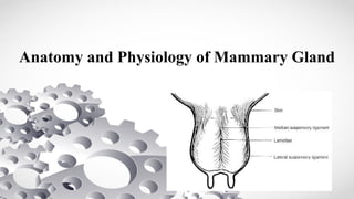
- 1. Anatomy and Physiology of Mammary Gland
- 3. Development of Mammary Gland Ø The development of the mammary gland starts early in the fetal life. Ø Already in the second month of gestation teat formation starts and the development continues up to the sixth month of gestation. Ø When the calf fetus is six months, the udder is almost fully developed with four separate glands and a median ligament, teat and gland cisterns. Ø The developments of milk ducts and the milk secreting tissue takeplace between puberty and parturition. Ø The udder continues to increase in cell size and cell numbers throughout the first five lactations, and the milk producing capacity increases correspondingly. Ø This is not always fully utilized, since the productive life time of many cows today is as short as 2.5 lactations.
- 4. Anatomy of Mammary Gland v The mammary gland of the dairy cow consists of four separate glands each with a teat. v Milk which is synthesized in one gland cannot pass over to any of the other glands. v The right and left side of the udder are also separated by a median ligament, while the front and the hind quarters are more diffusely separated. v The udder is a very big organ weighing, around 50 kg (including milk and blood). However, weights up to 100 kg have been reported. Therefore, the udder has to be very well attached to the skeleton and muscles. v The median ligaments are composed of elastic fibrous tissue, while the lateral ligaments are composed of connective tissue with less elasticity.
- 5. Anatomy of Mammary Gland v The mammary gland consists of secreting tissue and connective tissue. v The amount of secreting tissue, or the number of secreting cells is the limiting factor for the milk producing capacity of the udder. v It is a common belief that a big udder is related to a high milk production capacity. This is, however, not true in general, since a big udder might include a lot of connective and adipose tissue. v The milk is synthesized in the secretory cells, which are arranged as a single layer on a basal membrane in a spherical structure called alveoli. v Several alveoli together form a lobule. The structure of this area is very similar to the structure of the lung. v The milk which is continuously synthesized in the alveolar area, is stored in the alveoli, milk ducts, udder and teat cistern between milkings.
- 6. Anatomy of Mammary Gland Ø 60-80% of the milk is stored in the alveoli and small milk ducts, while the cistern only contains 20-40%. Ø However, there are relatively big differences between dairy cows when it comes to the cistern capacity. Ø The teat consists of a teat cistern and a teat canal. Where the teat cistern and teat Ø canal meet, 6-10 longitudinal folds form the so called Fürstenberg' s rosette, which is Ø involved in the local defense against mastitis. Ø The teat canal is surrounded by bundles of smooth muscle fibers, longitudinal as well as circular. Ø Between milkings the smooth muscles function to keep the teat canal closed. The teat canal is also provided with keratin or keratin like substances which between milkings act as a barrier for the pathogenic bacteria.
- 9. Physiology of Mammary Gland Ø The mammary gland is densely innervated especially in the teat. Ø The skin of the teat is provided with sensory nerves which are sensitive to the suckling performed by the calf, and thus influenced by pressure, heat and frequency of suckling. Ø The mammary gland is very well supported with blood vessels,arteries and veins. Ø Right and left udder halves generally have their own arterial supply. Ø The primary function of the arterial system is to provide a continuous supply of nutrients to the milk synthesizing cells. Ø To produce 1 liter of milk 500 ltr. of blood have to pass through the udder. Ø The udder also contains a lymphatic system. It carries waste products away from the udder. Ø The lymph nodes serve as a filter that destroy foreign substances but also provide a source of lymphocytes to fight infections.
- 10. Milk Secretion and Milk Components Ø Milk synthesis takes place in the alveoli where the milk secretingcells in the mammary gland are provided with a continuous supply of nutrients. Ø Milk fat consists mainly of triglycerides, which are synthesized from glyceroles and fatty acids. Ø Long chained fatty acids are absorbed from the blood. Short chained fatty acids are synthesized in the mammary gland from the components acetate and beta hydroxybutyrate which have their origins in the blood. Ø Milk protein is synthesized from amino acids also with origin from the blood, and consists mainly of caseins and to a smaller extent whey proteins. Ø Lactose is synthesized from glucose and galactose within the milk secreting cell. Ø Vitamins, minerals, salts and antibodies are transformed from the blood across the cell cytoplasm into the alveolar lumen.
- 11. Induction of Milk Letdown Ø Physical stimulation of the teats, either by the calf’s suckling or the milkers hands, excite receptors from which nerve impulses are sending to the posterior pituitary gland causing secretion of the hormone oxytocin. Ø The hormone is transported via the blood to the mammary gland. Because both hormones and nerve impulses are involved in the milk ejection reflex, it is called a neuro-hormonal reflex. Ø Oxytocin stimulates the contraction of the alveoli and small ducts thereby emptying the milk into the larger ducts and the cistern. Hereafter the milk can be evacuated from the udder. Ø The contraction of the alveoli may, to some extent, be enhanced by tactile stimuli of the udder (massaging, squeezing) the so called tap reflex. Ø When calves suckle, they butt at the udder in order increase milk secretion. Manual massage of the udder during milking imitates this reflex.
- 12. Milk Letdown Ø Milk is initially secreted into small sacs within the mammary gland called alveoli, from which it must be ejected for consumption or harvesting. Ø Mammary alveoli are surrounded by smooth muscle (myoepithelial) cells which are a prominent target cell for oxytocin. Ø Oxytocin stimulates contraction of myoepithelial cells, causing milk to be ejected into the ducts and cisterns above the teat. Ø Oxytocin is released after the cow receives an appropriate stimulus, this can be visual, aural or physical, and should be predictable and consistent at every milking. Ø Handling/massage of the teats for at least 15 seconds is a strong stimulus, but cows can also learn to let down through the association of the dairy environment to the milking process. Ø The pressure of milk being forced into the ducts/cistern and down towards the teat causes the teat to swell with milk and become ‘plump’.
- 15. Inhibition of Milk Letdwon v Cows/Buffaloes are sensitive to changes in the environment. They may withhold the milk if they are uncomfortable with the situation. v If the animals are stressed, scared or in pain, the hormone adrenaline is secreted. This hormone causes constriction of the blood vessels, thereby hindering the supply of sufficient amount of oxytocin to the udder. v Adrenaline also directly acts on the myoepithelial cells in the alveoli by blocking the oxytocin receptors. v The inhibition if milk letdown will result in the leaving of milk in the secretary parts of the udder. v Continuous exposure of stress to the buffaloes will affect the milk production negatively. v Change of milker or milking routine, application of wrong milking technique or milking machines in bad conditions are some reasons for the buffaloes to with hold the milk..
- 16. Milk Letdown Ø It takes 60 – 90 seconds for teats to become plump after let down has been initiated. Ø Cows with well-filled udders require a shorter period of stimulation to elicit milk let Ø down response than cows with less-filled udders. Ø The action of oxytocin is essential for emptying of the udder during milking. As much as 80% of a cow’s milk is unavailable if this oxytocin release is insufficient or does not occur. Ø Its let down action lasts for about 5 minutes and is strongest for the first 3 minutes of milking. Ø It is important to get the cups attached / hand milking quickly after let down has started to make full use of the increased udder pressure that occurs.