dentin.pptx
•Download as PPTX, PDF•
1 like•35 views

The document discusses dentin, the bony tissue that makes up the bulk of the tooth below the enamel. It describes dentin's physical properties, including its composition of hydroxyapatite and collagen, and that it is harder than bone but softer than enamel. It also discusses the histology of dentin, including dentinal tubules that contain vital contents and the different types of dentin like primary, secondary and tertiary dentin.

Recommended
More Related Content
Similar to dentin.pptx
Similar to dentin.pptx (20)
More from Dr. Shubhangi Mishra
More from Dr. Shubhangi Mishra (6)
Recently uploaded
Recently uploaded (20)
dentin.pptx
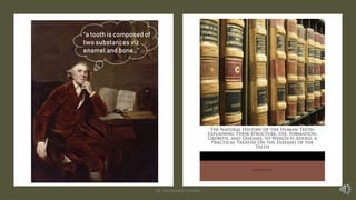
- 1. “a tooth is composed of two substances viz enamel and bone…” DR. SHUBHANGI MISHRA
- 2. ‘a tooth is composed of two substances viz enamel and bone’ and ‘the other substance of which a tooth is composed is bony; but much harder than the most compact part of bones in general’ DR. SHUBHANGI MISHRA
- 6. Sir Richard Owen It’s DENTINE… DR. SHUBHANGI MISHRA
- 8. Sir Richard Owen It’s DENTINE… DR. SHUBHANGI MISHRA
- 10. DENTIN ivory, tooth bone, zahnsubstanz, substantia dentis DR. SHUBHANGI MISHRA
- 14. Physical Properties • YELLOW (effect of age?) • Located both on crown and root • Second hardest tissue of the body • 66%-70% mineralized (calcium hydroxyapatite) • 20%-25% organic (collagen and ground substance) • 10% bound water • Highly elastic and resilient • LIVING tissue (why is enamel not living?) • Harder than bone, softer than enamel! DR. SHUBHANGI MISHRA
- 15. Reminder ONE: You’re a Doctor! DR. SHUBHANGI MISHRA
- 16. Colloquy How is DENTIN different from ENAMEL and BONE DR. SHUBHANGI MISHRA
- 17. Histology of DENTIN • Dentinal tubules • Peritubular dentin • Intertubular dentin DR. SHUBHANGI MISHRA
- 18. Dentinal Tubules • Tubules with VITAL contents! • Diameter more towards pulp than enamel • Density more towards pulp than enamel (4 times) DR. SHUBHANGI MISHRA
- 20. Dentinal Tubules DR. SHUBHANGI MISHRA
- 22. Any possible reason behind the S-shaped curvatures? Movement of odontoblasts during tooth development DR. SHUBHANGI MISHRA
- 24. Peritubular Dentin • Found throughout dentin except nearer to pulp (present in minimal amount) • Thicker in outer dentin than towards pulp • 9% more mineralized than intertubular dentin • On the inner side, thin organic membrane called the lamina limitans DR. SHUBHANGI MISHRA
- 25. Intertubular dentin • Forms the main body of dentin • Less mineralized than peritubular dentin • About one half is organic (Collagen fibrils) • Hydroxyapatite crystals arranged parallel to the fibres DR. SHUBHANGI MISHRA
- 26. Reminder TWO: You’re a Doctor! DR. SHUBHANGI MISHRA
- 32. TYPES OF DENTIN • Mantle dentin • Circumpulpal dentin Primary dentin Secondary dentin Tertiary dentin DR. SHUBHANGI MISHRA
- 33. Primary dentin - Mantle Dentin (20 µ) • First-formed dentin • Just below DEJ • Soft (to provide cushioning effect) • Organic part has larger collagen fibrils (von Korff’s fibres) as compared to rest of the primary dentin • Less mineralised than circumpulpal dentin • Fewer defects than circumpulpal dentin • Matrix vesicles are involved in the mineralisation of mantle dentin DR. SHUBHANGI MISHRA
- 34. Primary dentin - Circumpulpal dentin • Remaining primary dentin • Bulk of the tooth • All of the dentin that forms BEFORE completion of root • Smaller and more closely packed collagen fibrils • Slightly more mineral than mantle dentin DR. SHUBHANGI MISHRA
- 35. Homework Differentiate between Mantle and Circumpulpal dentin DR. SHUBHANGI MISHRA
- 36. Secondary dentin • Narrow band around the pulp • Dentin formed AFTER root completion • Fewer tubules than primary dentin • A bend in the tubules at the primary and secondary dentin interface • Formation: Slow, uneven, and more on the floor and roof of pulp chamber • NOT in response to any stimuli DR. SHUBHANGI MISHRA
- 37. Tertiary dentin • Reparative/Response/Reactive • Localised on pulp-dentin border in reaction to trauma/caries/restorative procedures DR. SHUBHANGI MISHRA
- 38. Homework Differentiate between primary, secondary, and tertiary dentin DR. SHUBHANGI MISHRA
- 39. Incremental Lines • Of von Ebner • At 4-8 µ in crown and much lesser in root • Accentuated ones – Contour lines (of Owen) • Neonatal line only in ? teeth DR. SHUBHANGI MISHRA
- 40. Interglobular dentin • Sometimes, mineralisation begins in small globular areas that FAIL to coalesce into a homogenous mass • Thus, zones of HYPOminearalisation between the globules • Just below mantle dentin DR. SHUBHANGI MISHRA
- 41. Granular layer (Tomes’) DR. SHUBHANGI MISHRA
- 42. Think upon! The first-formed dentin is MANTLE DENTIN When dentin first forms, it is called PREDENTIN So, Logically tell the locations of mantle dentin and predentin! DR. SHUBHANGI MISHRA
- 44. Theories of DENTIN SENSITIVITY 1. Direct Neural Transmission Theory Unacceptance? - Nerves do not generally extend beyond the inner dentin - Topical application of local anaesthetics does not abolish sensitivity 2. Hydrodynamic Theory 3. Transduction Theory DR. SHUBHANGI MISHRA