
Recommended
More Related Content
What's hot
Similar to Tooth Development & Molecular aspect
Similar to Tooth Development & Molecular aspect (20)
More from Dr Monika Negi
More from Dr Monika Negi (18)
Recently uploaded
Recently uploaded (20)
Tooth Development & Molecular aspect
- 1. DR MONIKA NEGI MDS ORAL PATHOLOGY ,MICROBIOLOGY AND FORENSIC ODONTOLOGY
- 2. CONTENTS INTRODUCTION PRIMARY EPITHELIAL BAND DENTAL LAMINA DEVELOPMENTAL STAGES BUD STAGE CAP STAGE EARLY BELL STAGE ADVANCED BELL STAGE VASCULAR & NERVE SUPPLY DURING TOOTH DEVELOPMENT TIME LINE OF HUMAN TOOTH DEVELOPMENT ROOT FORMATION HISTOPHYSIOLOGICAL & CLINICAL CONSIDERATIONS MOLECULAR INSIGHTS IN TOOTH MORPHOGENESIS REFERENCES
- 3. INTRODUCTION The stomodeum is lined by stratified squamous epithelium called the oral ectoderm The oral ectoderm contacts the endoderm of the foregut to form the buccopharyngeal membrane At about 27th day of gestation, this membrane ruptures and the primitive oral cavity establishes a connection with the foregut. Most of the connective tissue cells underlying the oral ectoderm are of neural crest or ectomesenchymal in origin.
- 4. PRIMARY EPITHELIAL BAND Certain areas of basal cells of oral ectoderm proliferate more rapidly than do the cells of adjacent areas, leading to formation of PRIMARY EPITHELIAL BAND. AT 6TH WEEK
- 7. PRIMARY EPITHELIAL BAND DENTAL LAMINA(inne r lingual process) Serves as primordium for the ectodermal portion of deciduous teeth. VESTIBULR LAMINA(outer buccal process) Labial and buccal to dental lamina in each arch, epithelial thickening develops later independently. Also called as LIP FURROW BAND AT 7TH WEEK
- 9. DENTAL LAMINA Serves as primordium for ectodermal portion of deciduous teeth At certain points along dental lamina(10 maxillary & 10 mandibular), the ectodermal cells multiply more rapidly and form little knobs that grow into underlying mesenchyme. This represents the beginning of enamel organ of tooth bud of a deciduous tooth. As cell proliferation continues, each enamel organ increases in size.
- 10. Later during the development of the jaws, the permanent molars arise directly from distal extension of dental lamina Development of 1st permanent molar- initiated at 4th month in utero 2nd molar- first year after birth 3rd molar-4th or 5th years Activity of DL- 5 yrs.
- 11. The lingual extension of free end of dental lamina, opposite to enamel organ of each deciduous tooth from which the permanent teeth arise is known as successional lamina. From 5th month in utero (Perm. CI) to 10th month of age (2nd PM).
- 13. FATE OF DENTAL LAMINA 5 years As the teeth continue to develop, they lose their connection with dental lamina. They later break up by mesenchymal invasion, which is at first incomplete and doesn’t perforate the total thickness of lamina. Remnants of dental lamina persist as epithelial pearls or islands within the jaw as well as in gingiva, referred to as CELL RESTS OF SERRES.
- 14. .
- 16. .
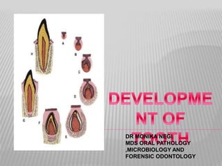
- 17. DEVELOPMENTAL STAGES Although the tooth development is a continuous process, yet it can be divided into three stages for descriptive purposes:- 1. BUD STAGE 2. CAP STAGE 3. Early BELL STAGE 4. Late/Advance BELL STAGE (named after the shape of the enamel organ)
- 19. BUD STAGE Bud stage is characterized by rounded, localized growth of epithelium surrounded by proliferating mesenchymal cells, which are packed closely beneath and around the epithelial buds
- 20. The epithelium of the dental lamina is separated from underlying ectomesenchyme by a basement membrane. The enamel organ consists of peripherally located low columnar cells and centrally located polygonal cells.
- 22. DENTAL PAPILLA- The area of ectomesenchymal condensation immediately subjacent to enamel organ DENTAL SAC- The condensed ectomesenchyme that surrounds the tooth bud and the dental papilla Both the dental papilla and dental sac become more well defined as the enamel organ grows into cap and bell shapes
- 23. TOOTH GERM ECTODERMAL COMPONENT ENAMEL ORGAN ECTOMESENCHYMAL COMPONENTS DENTAL PAPILLA DENTAL SAC (enamel) (dentin &pulp) (cementum, PDL & alveolar bone)
- 24. CAP STAGE The unequal growth in different parts of the tooth bud leads to the cap stage. At this stage , tooth germ consists of: Outer Enamel epithelium Inner enamel epithelium Stellate Reticulum Dental papilla Dental sac
- 27. TRANSITORY STRUCTURES ENAMEL KNOT (Ahrens knot)- The cells in the center of the enamel organ are densely packed and form the enamel knot. ENAMEL CORD- The vertical extension of the enamel knot towards the underlying dental papilla is known as enamel cord. The function of the enamel knot and cord may be to act as a reservoir of dividing cells for growing enamel organ. (Disappear before enamel formation begins)
- 30. ENAMEL SEPTUM- When enamel cord extends to meet the outer enamel epithelium, it is termed as enamel septum as it divides the stellate reticulum into two parts. ENAMEL NAVEL- A small depression that is formed at the point of meeting of enamel septum and outer enamel epithelium is known as enamel navel as it resembles umbilicus.
- 31. ENAMEL NICHE The enamel organ may seem to have a double attachment of dental lamina to the overlying oral epithelium enclosing ectomesenchyme called enamel niche between them.
- 33. BELL STAGE As the invagination of epithelium deepens & its margins continue to grow ,enamel organ assumes bell shape . Early Bell Stage Advanced Bell Stage
- 34. EARLY BELL STAGE Inner enamel epithelium Outer enamel epithelium Stratum Intermedium Stellate reticulum Cervical loop or zone of reflexion Dental Papilla Dental Sac
- 37. ADVANCED BELL STAGE Separation of tooth germ from Dental Lamina. Hard tissue formation
- 38. Separation of tooth germ from Dental Lamina: Dental lamina joining tooth germ to oral epithelium breaks into discrete islands of epithelial cells, thus separating developing tooth germ from oral epithelium.
- 41. Hard tissue formation
- 42. VASCULAR & NERVE SUPPLY DURING TOOTH DEVELOPMENT Vascular Supply: Clusters of blood vessels in dental follicle and papilla Clustering of vessels in papilla coincide with position of root formation Enamel organ is avascular, however vessels seen in close association in the follicle Nerve Supply: Initially noted in the dental follicle during bud to cap stage However after start of dentinogenesis, seen in dental papilla Nerve fibers do not enter enamel organ
- 43. TIME LINE OF HUMAN TOOTH DEVELOPMENT Age Developmental Characteristics 42 to 48 days Dental lamina formation 55 to 56 days Bud stage for deciduous teeth 14 weeks Bell stage for deciduous teeth; Bud stage for permanent teeth 18 weeks Dentin & functional ameloblasts in deciduous teeth 32 weeks Dentin & functional ameloblasts in permanent teeth.
- 44. ROOT FORMATION It begins after enamel & dentin formation has reached cemento enamel junction. The enamel organ plays important role by forming Hertwig’s epithelial root sheath. It is formed by proliferation of cervical loop cells . It consists of only inner & outer enamel epithelium. It molds the shape of root & initiate radicular dentin formation.
- 45. When dentin is formed ,it looses its structural integrity & its close relation with root surface. This loss of structural integrity is as a result of invasion of surrounding connective tissue of dental sac. The epithelium is moved away from surface of dentin so that connective tissue cells come into contact with outer surface dentin & differentiate into cementblasts that deposite a layer of cementum onto surface of dentin.
- 46. Remnants of Hertwig’s epithelial root sheath are found in periodontal ligament & are called rests of Malassez .
- 47. Prior to the beginning of root formation ,epithelial root sheath forms epithelial diaphragm by bending at future cemento enamel junction into horizontal plane ,narrowing the wide cervical opening of tooth. Proliferation of cells of epithelial diaphragm is accompanied by ectomesenchymal cell proliferation, adjacent to diaphragm.
- 51. HISTOPHYSIOLOGICAL & CLINICAL CONSIDERATIONS 1. Initiation 2. Proliferation 3. Histodifferentiation 4. Morphodifferentiation 5. Apposition
- 52. MOLECULAR INSIGHTS IN TOOTH MORPHOGENESIS The study of tooth development used manipulation of tooth germ explants from wild type and mutant mice. Studies on mammalian development are carried out with mice due to its suitability for both genetic and embryological manipulations.
- 53. TOOTH INITIATION POTENTIAL Experiments conducted combining first arch epithelium with neural crest in anterior chamber of eye resulted in formation of tooth, while epithelium from other sites like that of limb or second arch doesn’t produce same results. When dental epithelial organ is combined with skin, the tooth organ loses its dental characteristics and takes up the features of epidermis.
- 56. ESTABLISHMENT OF ORAL-ABORAL AXIS LIM-homeobox(Lhx) genes are the earliest mesenchymal markers for tooth formation. The expression of Fgf-8 establishes the anteroposterior axis of the first branchial arch and was shown restricted to the first arch. Fgf-8 has been attributed to be regulating the expression of Lhx-6 & Lhx-7 genes. Expression of Goosecoid in aboral mesenchyme
- 57. CONTROL OF TOOTH GERM POSITION The Pax-9 gene is one of the earliest mesenchymal genes that define the localization of the tooth germs. Pax-9 gene expression co-localizes with exact sites where tooth germs appear. Pax-9 gene is induced by Fgf-8 and is repressed by bone morphogenetic proteins(BMP-2 and BMP-4). Activin-βA also has role in control of tooth germ position but it is not regulated by Fgf-8 & BMP-4 interactions.
- 58. FUNCTIONAL REDUNDANCY & THEIR COMPLEXITIES Dlx genes Shh Lef-1
- 59. PATTERNING OF DENTITION The determination of specific tooth types at their correct positions in the jaws is referred to as patterning of the dentition. Two models have been proposed:- 1. Field model 2. Clone model
- 60. FIELD MODEL
- 61. CLONE MODEL
- 62. REGULATION OF ECTODERMAL BOUNDARIES Interactions between Wnt and Shh maintain boundaries between oral and dental ectoderm Shh specifies the sites where tooth buds will form in future Expression sites of Wnt-7b reciprocal to that of Shh Wnt-7b is expressed throughout oral ectoderm except for presumptive dental ectoderm where Shh is expressed Misexpression of Wnt-7b in presumptive dental ectoderm results in loss of Shh expression and failure of tooth bud formation.
- 63. STOMODEAL THICKENING STAGE- DENTAL LAMINA STAGE BUD STAGE
- 64. ENAMEL KNOT- SIGNALING CENTER FOR TOOTH MORPHOGENESIS Enamel knot cells express several signaling molecules like sonic hedgehog, bone morphogenetic proteins Bmp-2, Bmp-4, Bmp-7, Wnt signaling molecules as well as Fgf-4. As the same signaling molecules are expressed by well studied vertebrate signaling centres such as notochord, it was put forward that the enamel knot represents a signaling center for tooth morphogenesis.
- 65. REFERENCES ORBANS Oral Histology and embryology, 13th edition TENCATES Oral Histology Development, Structure and Function. Antonio Nanci. Seventh edition Oral Cells and Tissues. Philias R.Garant Oral Development nd Histology..Avery. Third edition The Genetic Control Of Early Tooth Development. R Mass and M Bei. Critical Reviews In Oral Biology And Medicine 1997 8:4