
Recommended
Recommended
More Related Content
What's hot
What's hot (20)
Similar to Dental Anatomy Guide for Implant Surgery
Similar to Dental Anatomy Guide for Implant Surgery (20)
More from DrBindu Kumari
Recently uploaded
Recently uploaded (20)
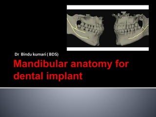
Dental Anatomy Guide for Implant Surgery
- 1. Dr Bindu kumari ( BDS)
- 2. A proficient knowledge of oral anatomy is needed to provide effective implant dentistry. The study of anatomy familiarizes the implant surgeon with normal and atypical oral structures. Knowledge of oral structures and ordinary anatomic variations, which usually differ with respect to size and shape, enhance patient evaluations and facilitate precise surgical procedures.
- 3. Mandibular foramen Inferior alveolar canal Mental foramen and nerve Mandibular incisive canal Lingual foramen and lateral canal Submental and sublingual arteries
- 4. Submandibular and sublingual fossae Lingual and mylohyoid nerve Long buccal nerve Muscles attached to mandible
- 5. Mandibular foramen The location of the mandibular foramen may vary based on race and ethnicity, and this can affect the success of block injections. It is advisable to inject patients 6 to 10 mm superior to the occlusal plane, which usually accounts for anatomic variations.
- 7. Inferior alveolar canal The trigeminal nerve, the fifth cranial nerve, has three main branches: ophthalmic, maxillary, and mandibular. The mandibular nerve gives rise to the inferior alveolar nerve (IAN).
- 9. Thickness of IAN canal= 3.4mm wide 2.2mm thick Within the canal there is a nerve, an artery, a vein, and lymphatic vessels.
- 10. When developing an osteotomy over the mandibular canal, cortical bone is penetrated first, and the preparation terminates within softer cancellous bone. The mandibular canal usually has cortical bone around it, which may provide some resistance to drilling.
- 13. The IAN may present in different anatomic configurations. The nerve may lower gently as it proceeds anteriorly, or there can be a sharp decline or the nerve can drape downward in catenary fashion (curled as hanging between two points).
- 14. The IAN crosses from the lingual to the buccal side of the mandible and often ,by the first molar, it is located midway between the buccal and lingual cortical plates of bone.
- 16. The IAN divides into the mental and incisive nerves in the premolar molar region. The mental nerve emerges from the mental canal, and anterior to the mental foramen the mandibular canal is referred to as the incisive canal.
- 18. Mental foramen and nerve Commonly, three nerve branches of the mental nerve emerge from the mental foramen They supply innervation to the skin of the mental foraminal area, the lower lip, chin, mucous membranes, and the gingiva until the second premolar
- 20. The anterior loop of the mental foramen refers to the IAN when it courses inferiorly and anteriorly to the foramen and then loops back to emerge from the foramen.
- 21. Several methods and techniques for identifying the extent of the AL( anterior loop) of the neurovascular bundle have been proposed using panoramic radiographs, computed tomography, and determination of the AL during surgery using a curved explorer.
- 23. If placement of the probe into the mental foramen on the distal side reveals that the mental canal is patent, then the anterior loop is not present. If placement of a probe into the mental foramen on the distal side reveals that the mental canal is not patent, then an anterior loop of the mental nerve exists. The nerve must have traversed inferiorly and looped back to the foramen creating an anterior loop.
- 24. Mandibular incisive canal Numerous investigations reported that there is a ‘‘true’’ incisive canal mesial to the mental foramen, which is a continuation of the mandibular canal. The incisive nerve supplies innervation to teeth (first bicuspid, canine, and lateral and central incisors).
- 25. The incisive canal is typically found in the middle third of the mandible (in 86% of cases). It usually narrows as it approaches the midline and only reaches the midline 18% of the time. The nerve usually terminates apical to the lateral incisor and sometimes apical to the central incisor.
- 27. Lingual foramen and lateral canal Vascular canals are often present in the midline and lateral to the midline of the mandible. The lingual foramen was detected in 99% of the mandibles when evaluating skull dissections.
- 28. The lingual foramen harbors an artery that corresponds to an anastomosis of the right and left sublingual arteries.
- 30. Submental and sublingual arteries The submental artery (2-mm average diameter) is derived from the facial artery, And the sublingual artery (2-mm average diameter) is a branch of the lingual artery.
- 31. The sublingual artery is found above the mylohyoid muscle and is the major nutrient vessel in the floor of the mouth. The submental artery frequently traverses inferiorly to the mylohyoid muscle
- 32. Hofschneider et al also reported that the sublingual and submental arteries may course anteriorly in close proximity to the lingual plate, and branches of these blood vessels enter accessory foramina along the lingual cortex.
- 33. Inadvertent penetration through the lingual cortical plate into the floor of the mouth while preparing an osteotomy can cause arterial trauma, there by resulting in development of a sublingual or submandibular hematoma.
- 34. Submandibular and sublingual fossae The submandibular fossa is a depression on the medial surface of the mandible inferior to the mylohyoid line, and it contains the submandibular gland. The sublingual gland is found in the sublingual fossa.
- 35. This fossa is a shallow depression on the medial surface of the mandible on both sides of the mental spine, superior to the mylohyoid line.
- 37. The submandibular and sublingual fossae must be palpated prior to osteotomy development; if there is a large undercut, the lingual bony plate can be perforated inadvertently, resulting in hemorrhaging.
- 38. If there is a large undercut, an instrument can be placed into and parallel to the undercut to visualize and measure the extent of the depression.
- 39. Lingual and mylohyoid nerve The mandibular branch of the trigeminal nerve gives rise to the lingual nerve. This nerve provides sensory innervation to the mucous membranes of the anterior two- thirds of the tongue and the lingual tissues.
- 41. At the time of implant surgery in the posterior mandible, the lingual nerve can be injured if the lingual flap is not reflected cautiously. The lingual nerve is usually located 3 mm apical to the osseous crest and 2 mm horizontally from the lingual cortical plate in the flap
- 42. However, in 15% to 20% of cases, the nerve may be situated at or above the crest of bone, lingual to the mandibular third molars.
- 43. The mylohyoid nerve is a branch of the inferior alveolar nerve.
- 44. It arises just prior to where the IAN enters the mandibular foramen. On the deep surface of the ramus, it moves down in a groove to reach and innervate the mylohyoid muscle and the anterior belly of the digastric muscle.
- 45. Long buccal nerve The buccal nerve is a branch of the mandibular nerve that is derived from the trigeminal nerve, and it begins high in the infratemporal fossa.
- 46. It transmits sensory innervation to the buccal gingiva and mucosa of the cheek from the retromolar area to the second premolar.
- 47. Muscles attached to mandible There are 26 muscles attached to the mandible. There are two single muscles (orbicularis oris and platysma) and 12 pairs of bilateral muscles.
- 48. Which are listed in alphabetic order: anteriorbelly of digastric, buccinator, depressor anguli oris, depressor labii inferioris, genioglossus, geniohyoid, masseter, mentalis, mylohyoid, lateral pterygoid, medial pterygoid, and temporalis.
- 49. 1. Mentalis Muscle 2. Mylohyoid Muscle 3. GenialTubercles (genioglossus and geniohyoid muscles) 4. Depressor Anguli Oris and Depressor Labii Inferiorus 5. Buccinator and Orbicularis Oris Muscles 6. Masseter Muscle
- 50. Mentalis Muscle The mentalis muscle is a paired small muscle that originates in the incisive fossa of the mandible and inserts into the integument of the chin.
- 51. When a flap is raised in this region, the entire mentalis muscle should not be released off from the mental protuberances, because the muscle may fail to reattach well. This can result in an appearance referred to as a witch’s chin (double chin).
- 52. Mylohyoid Muscle Two flat mylohyoid muscles form a sling inferior to the tongue, supporting the floor of the mouth. Their origin is the mylohyoid line on the medial aspect of the mandible, which extends from the symphysis to the last molar.
- 53. This muscle is an important anatomic barrier separating the sublingual and submandibular spaces.The submandibular fossa is below the mylohyoid muscle, and the sublingual fossa is superior to the muscle.
- 54. GenialTubercles (genioglossus and geniohyoid muscles) The genial tubercles are small, bony elevations located on the lingual surface of the mandible. They are found on either side of the midline close to the inferior border of the mandible .
- 55. There are two superior and two inferior tubercles. The genioglossus originates from the superior genial tubercles. And the geniohyoid originates from the inferior genial tubercles.
- 56. Depressor Anguli Oris and Depressor Labii Inferiorus TheseTwo muscles that overlie the mental foramen need to be displaced when exposing the roof of the foramen.
- 57. Once the flap is elevated past the mucogingival junction, these muscles can be released by using wet gauze to push back the flap.The wet gauze is used to protect the mental nerve.
- 58. Buccinator and Orbicularis Oris Muscles The submucosa is strongly attached to the buccinator muscle in the cheek region and the orbicularis oris in the lip area.
- 59. Masseter Muscle The masseter muscle consists of two portions: superficial and deep When the mandibular ramus area is used as a donor site for bone grafting(i.e., block graft), part of the masseter muscle is released from the ramus when the periosteum is elevated in this region.
- 60. Familiarity with the anatomic structures pertaining to dental implantology is critically important. Preplanning and review of anatomy before surgical procedures can help to avoid problems.