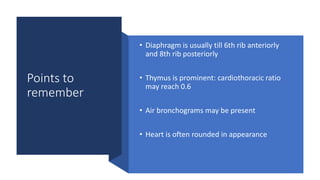
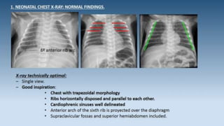
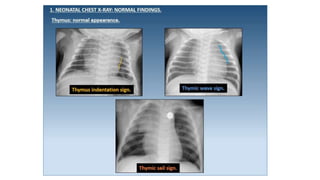
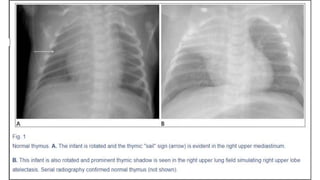
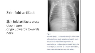
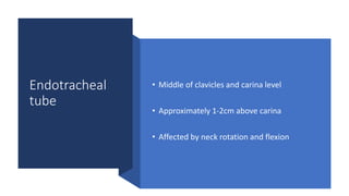
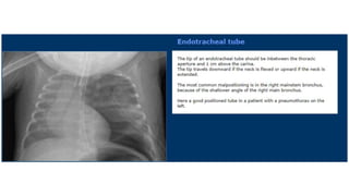
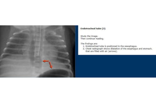
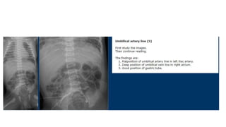
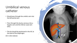
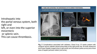
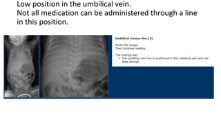
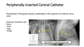
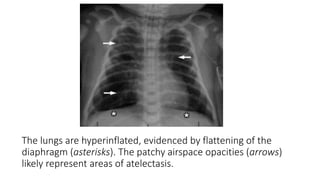
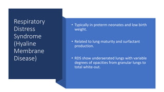
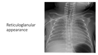
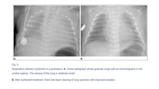
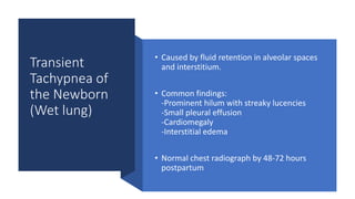
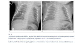
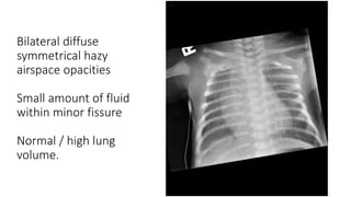
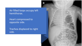
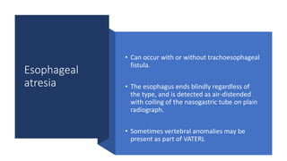
2. Learning objectives
1. When and when not to do a neonatal chest x-ray?
2. What does a normal chest x-ray look like?
3. Position of tubes and catheters?
4. Common causes of neonatal respiratory distress.
5. Surgical diagnoses.
6. References.
3. When to do a Chest X-ray?
For initial
diagnosis
Detioration
causes
Catheter/ tube
placements
4. When not to do a chest x-ray?
Daily x-ray for
ventilated babies
Repeated x-ray
after every
reajustment of
catheter / tube
Routine pre- and
post-intubation
Without clear
indication
Not routine to
cover abdomen
5. The normal
chest x-ray
technique
• In AP with patient supine
• Lateral films useful for:
1. Pneumothorax
2. Localisation of tube/catheter
6. Before commenting on a Chest X-ray: Review
History and indication
Is the film rotated
Is there good inspiration
Exposure
7. Fig. 2: Rotated chest X-ray may simulate cardiomegaly or hyperdense hemithorax (image on the left).
New X-ray study in the same patient reveals no cardiopulmonary disease (image on the right).
8. Term or
preterm?
In preterm babies:
Lack of subcutaneous fat
Lack of humeral head ossification
Look for umbilical clip:
Will remain until about 1-2 weeks after baby is
born
9. Points to
remember
• Diaphragm is usually till 6th rib anteriorly
and 8th rib posteriorly
• Thymus is prominent: cardiothoracic ratio
may reach 0.6
• Air bronchograms may be present
• Heart is often rounded in appearance
10.
11. The thymus
• The thymus is relatively prominent in infants
• Draped around the heart constituting the
cardiothymic silhouette.
• Sometimes it shows triangular projection
laterally, which is known as a "sail" sign
16. Endotracheal
tube
• Middle of clavicles and carina level
• Approximately 1-2cm above carina
• Affected by neck rotation and flexion
17.
18.
19.
20. Umbilical arterial
catheter
• Goes downwardly first and
then upwardly via the internal iliac
artery and common iliac artery before
entering the aorta
• Should demonstrate the typical loop
from the umbilicus inferiorly into the
internal iliac artery.
• Term used: hairpin turn
21. • Can be placed in
-high position (above the celiac,
mesenteric and renal arteries)
-high position: T6-T9
-low position (below the inferior
mesenteric artery)
-low position: L3-L5
• The high position is advisable since it
leads to less vascular complications.
22.
23.
24.
25. Umbilical venous
catheter
• Should pass through the umbilic vein into
the left portal vein.
• Then through the ductus venosus into a
hepatic vein and the inferior caval vein
(IVC).
• The tip should be positioned in the IVC at
the level of the diaphragm.
(0.5-1.0cm above)
26. This can lead to cardiac arrhythmias or
perforation.
27. Intrahepatic into
the portal venous system, both
right and
left, or even into the superior
mesenteric
or splenic vein.
This can cause thrombosis.
28. Low position in the umbilical vein.
Not all medication can be administered through a line
in this position.
29. Peripherally Inserted Central Catheter
Positioned in the great vessels, preferably in the superior or inferior vena
cava.
Find out insertion site
from:
-Arm
-Leg
-Scalp
31. The lungs are hyperinflated, evidenced by flattening of the
diaphragm (asterisks). The patchy airspace opacities (arrows)
likely represent areas of atelectasis.
34. • Classic radiographic findings of RDS:
-Small lung volume
-Ground-glass or hazy lung
-Air bronchogram
• Lung volumes are small since its pathophysiology is essentially
underinflated alveoli.
• Air bronchograms commonly seen in the central regions, more
pronounced when the endotracheal tube is placed.
38. Transient
Tachypnea of
the Newborn
(Wet lung)
• Caused by fluid retention in alveolar spaces
and interstitium.
• Common findings:
-Prominent hilum with streaky lucencies
-Small pleural effusion
-Cardiomegaly
-Interstitial edema
• Normal chest radiograph by 48-72 hours
postpartum
41. Meconium
Aspiration
Syndrome
• Contaminated meconium may be aspirated into the
baby's airway in utero or during delivery.
• More commonly found in term or near-term babies.
• Aspirated meconium particles mechanically occlude
the bronchioles.
• Air leak including pneumothorax is common and
pulmonary hypertension is a major prognostic
determinant.
44. Neonatal
pneumonia
• Most common pathogen for neonatal
pneumonia is group B beta hemolytic
streptococcus (GBS), which is known to mimick
RDS on radiographs.
• Unilateral lung involvement and presence of
pleural effusion favor GBS pneumonia.
• Radiographic features of neonatal pneumonia
are also nonspecific with a wide spectrum of
chest findings mimicking RDS, TTN, or meconium
aspiration.
45. Can be early onset (caused
by GBS) or late onset
(caused by gram positive
bacteria).
The most frequent and
characteristic alveolar
pattern is dense
bilateral air space filling
process with numerous air
bronchograms.
46. Right upper zone
and left lower zone
opacities consistent
with inflammatory /
infectious process.
47.
48. Differential diagnosis of diffuse pulmonary
disease in the newborn
High lung volumes, streaky perihilar densities Low lung volumes, granular opacities
1. Meconium aspiration syndrome
2. Transient tachypnea of the newborn
3. Neonatal pneumonia
1. Surfactant deficiency
2. Beta hemolytic streptococcal pneumonia
50. Diaphragmatic
hernia
• More common on the left side (Bochdalek
hernia)
• Air-filled bowel loops are seen in the
hemithorax and the heart and mediastinum
are shifted to the opposite side.
• Stomach gas can be seen either in the
thorax or in the abdomen
51. Air filled loops occupy left
hemithorax.
Heart compressed to
opposite side.
Trachea displaced to right
side.
52. Esophageal
atresia
• Can occur with or without trachoesophageal
fistula.
• The esophagus ends blindly regardless of
the type, and is detected as air-distended
with coiling of the nasogastric tube on plain
radiograph.
• Sometimes vertebral anomalies may be
present as part of VATERL
53.
54. References
• Survival Radiology: neonatal chest X-ray for
residents.
https://dx.doi.org/10.1594/ecr2015/C-2351
• Interpretation of Neonatal Chest
Radiography
https://doi.org/10.3348/jksr.2016.74.5.279
• Neonatal chest xray interpretation-Dr
Praveen Kumar
• The Neonatal and Paediatric Chest-
Radiology Key
Editor's Notes
Rotated might stimulate disease
meconium is rarely found in amniotic fluid before 34 gestational weeks of age.