Insulin
•Download as PPSX, PDF•
18 likes•7,312 views

Clinical pharmacology: endogenous insulin structure and secretion

Recommended
More Related Content
What's hot
What's hot (20)
Similar to Insulin
Similar to Insulin (20)
More from Domina Petric
More from Domina Petric (20)
Recently uploaded
Recently uploaded (20)
Insulin
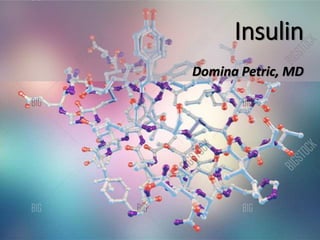
- 2. Structure of insulin Insulin is a protein comprising of 2 polypeptide chains: • chain A with 21 amino acid residues • chain B with 30 amino acid residues
- 3. Structure of insulin • Chains A and B are linked by disulphide bridges. • A-chain contains an intra-chain disulphide bridge linking residue 6 and 11. • C-chain connects A and B chains. • C-chain is liberated along with insulin after breakdown of proinsulin.
- 4. Structure of insulin Insulin monomers aggregate to form dimers and hexamers. Zn hexamer is composed of three insulin dimmers associated in threefold symmetrical pattern.
- 6. Biosynthesis of insulin Insulin is synthesized in the beta cells of pancreas in the form of preproinsulin. Preproinsulin is the ultimate precursor. Gene for preproinsulin is located on chromosome 11 close to that for insulin like growth factor-2 (IGF-2).
- 7. Biosynthesis of insulin • Within a minute after synthesis preproinsulin is discharged into cisternal space of rough endoplasmic reticulum. • In the rER it is cleaved into proinsulin by proteolytic enzymes. • Proinsulin has a C (connecting chain) linking A and B chains. • Proinsulin is transported by microvesicles to the Golgi apparatus.
- 8. Biosynthesis of insulin • Proinsulin is released in vesicles. • Conversion of proinsulin to insulin continues in maturing granules through the action of prohormone convertase 2 and 3 and carboxy peptidase H. • Maturing granules are translocated with the help of microtubules and microfilaments.
- 9. Biosynthesis of insulin Insulin Preproinsulin Removal of the signal peptide: N signal peptide Proinsulin Disulfide bridge formation Insulin Insulin with A and B chain and disulfide bridges Proinsulin Removal of the C chain
- 10. Insulin secretion • Insulin is secreted from the beta cells in response to various stimuli like glucose, arginine, sulphonylureas... • Glucose is the major determinant. • Glucose is taken up by beta cells through GLUT-2 receptors. • After entering the beta cell, glucose is oxidized by glucokinase. • Glucokinase acts as a glucose sensor.
- 11. Insulin secretion • Glucose concentration below 90 mg/dL does not cause any insulin release. • At such substimulatory glucose concentrations, K+ efflux through open KATP channels keeps the β cell membrane at a negative potential at which voltage- gated Ca2+ channels are closed. • If there is increase in plasma glucose, glucose uptake and metabolism by the β cell is enhanced.
- 12. Insulin secretion Rise in ATP concentration results in closure of KATP channels, leading to: • membrane depolarization • opening of voltage-gated Ca2+ channels • Ca2+ influx • rise in intracellular calcium concentration • exocytosis of insulin granules
- 13. KATP channels The pancreatic KATP channel consists of two unrelated subunits: • sulfonylurea receptor (SUR1 isoform) • potassium channel subunit (Kir6.2) that forms the central ion-conducting pathway
- 14. KATP channels • The mature KATP channel exists as an octamer of Kir6.2 and SUR1 subunits in a 4:4 stoichiometry. • A subunit specific site specific to pancreatic KATP channel, confers glimepiride an advantage over the other sulfonylurea secretagogues.
- 15. KATP channels • Sulfonylurea and non-sulphonylurea drugs act as insulin secretagogues by closing KATP channels bypassing the β cell metabolism. • Diazoxide is a K channel opener and inhibits insulin secretion, independent of blood glucose levels.
- 16. Literature • Joshi SR, Parikh RM, Das AK. Insulin-History, Biochemistry, Physiology and Pharmacology. Supplement of JAPI. 2007;55:19-25. • Biology.kenyon.edu