
Recommended
More Related Content
What's hot
What's hot (20)
Similar to PIVD Prolapsed Intervertebral Disc
Similar to PIVD Prolapsed Intervertebral Disc (20)
Recently uploaded
Recently uploaded (20)
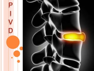
PIVD Prolapsed Intervertebral Disc
- 2. By Dr Dharma Patel (DPT)
- 5. THE IV DISC CONSISTS OF THREE DISTINCT COMPONENTS : 1.THE CARTILAGE END PLATES 2.NUCLEUS PULPOSUS 3.ANNULUS FIBROSUS. THE CARTILAGE RING ARE THIN LAYERS OF HYALINE CARTILAGE BETWEEN TWO ADJACENT VETEBRAL BODIES AND DISC PROPER.
- 17. CONGENTIAL DEVELOPMENTAL •POOR NUTRITION •ACCUMATED TRAUMA : SPORTS,AUTOMOBILE INJURY LEAD TO WEAKING OF I.V.DISC •BIOMECHANICAL FACTORS •POSTURE •REPITITIVE MICROTRAUMA , FLEXION (COMPRESSION INJURIES) , TENSIONAL INJURIES
- 20. IT IS SEQUENCE OF CHANGES OCURRING IN THE DISC WHICH LEAD TO PROTRUSION OR EXTRUSION OF NP THROUGH A RENT IN THE AF. THESE CHANGES CONSISTS OF THE FOLLOWING : A) NUCLEUSDEGENERATION : degenerative changes occur in the disc before displacement of the nuclear material. these changes are: (i) Softening of the nucleus and its fragmentation (ii) Weakening and disintegration of the posterior part of the annulus
- 21. B) NUCLEUS DISPLACEMENT : the nucleus is under positive pressure at all the times. When annulus become weak ,either because a small area of its entire thickness has been disintegrated spontaneously or because of the injury , the nucleus tends to bilge through the defect. This is known as disc protrusion. This tendency is greatly increased if the nucleus is degenerated and fragmented. Finally the nucleus comes out of the annulus , and the underthe post” longitudinal ligament though it has not lost contact between with parent disc. This is called as disc extrusion. Once the disc is extruded it cannot go back and PLL is not strong enoughto prevent the nucleus protruding further. So, the extruded disc, may loose its contact with parent disc. The sequestrated disc may come to lie behind the PLL or may
- 23. C) STAGE OF FIBROSIS : this the stage of repair. the begins alongside of degeneration . The residual nucleus pulposus becomes flattened , fibrosed , and finally undergoes calcification. At the same time, new bone formation occurs at the point where the PLL has been stripped from the vertebral body and spurformation occurs.
- 43. IN CASE OF Cx PIVD : a) Foraminal compression / spurling test b) Distraction test c) ULTT(upper limb tension test) d) Shoulder abduction test e) Tinel’s sign (a)
- 44. IN CASE OF Lx PIVD: a) SLR b) Lasegue’s test c) Bowstring test d) Prone knee bending (PKB) test e) Sensory impairment (over dermatomes motor weakness over myotomal distraction)
- 45. (d) (e)
- 46. (b) (c) (e) (d)
- 47. ASSESSMENT TREATMENT ASSESSSMENT : •POSTURE (rigid posture , loss of curve spine( i.e flattened back in lumbar spine , posterior pelvic tilt , sciatic scoliosis may present .) •MOVEMENT : trunk flexion (may be painful , flexion may produce pain .) •TENDERNESS : along midline or lateral to spinous processes.
- 49. Self Shift correction The patient stands with their shoulder against the wall. The bend their elbow and place their upper arm against their side. They then need to have their feet approximately 12-18 inches away from the wall, this allows more motion of the hips to the wall. They then glide their hips toward the wall.
- 53. Surgeries • Discectomy • Microdiscectomy • Laminectomy • Nucleoplasty • Artificial disc replacement • Foraminotomy