
Recommended
More Related Content
Similar to Structure and Function of the Inner Ear
Similar to Structure and Function of the Inner Ear (20)
More from DanjaarDasan
More from DanjaarDasan (17)
Recently uploaded
Recently uploaded (20)
Structure and Function of the Inner Ear
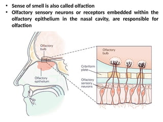
- 1. • Sense of smell is also called olfaction • Olfactory sensory neurons or receptors embedded within the olfactory epithelium in the nasal cavity, are responsible for olfaction
- 2. • There are three cell types at the base of the epithelium. 1. Olfactory Sensory Neurons: • 10 to 20 million bipolar olfactory sensory neurons are located in a specialized portion of the nasal mucosa. • Each neuron has a short, thick dendrite that projects into the nasal cavity where it terminates in a knob containing 10 to 20 cilia • The cilia are unmyelinated processes about 2 m long and 0.1 m in diameter and contain specific receptors for odorants (odorant receptors). • A single axon projects from each neuron to the olfactory bulb. • Odorants bind to specific odorant receptors on the cilia and initiate a cascade of events leading to generation of action potentials in the sensory axon. 2. Supporting Cells 3. Basal Stem Cells
- 3. Structure of the olfactory epithelium.
- 4. The axons of the olfactory sensory neurons pass through the cribriform plate of the ethmoid bone and enter the olfactory bulbs. In the olfactory bulbs, the axons of the olfactory nerve contact the primary dendrites of the mitral cells and tufted cells to form anatomically discrete synaptic units called olfactory glomeruli. The tufted cells are smaller than the mitral cells and have thinner axons, but both types send axons into the olfactory cortex. In addition to mitral and tufted cells, the olfactory bulbs contain • Periglomerular cells, which are inhibitory neurons connecting one glomerulus to another, • Granule cells, which have no axons and make reciprocal synapses with the lateral dendrites of the mitral and tufted cells.
- 5. The axons of the mitral and tufted cells pass posteriorly through the lateral olfactory stria to terminate on apical dendrites of pyramidal cells in the olfactory cortex: From these regions, information travels directly to the frontal cortex or via the thalamus to the orbitofrontal cortex.
- 6. Anosmia (inability to smell) and hyposmia or hypesthesia (diminished olfactory sensitivity) • can result from simple nasal congestion or be a sign of a more serious problem including damage to the olfactory nerves due to fractures of the cribriform plate, tumours such as neuroblastomas or meningioma, or infections (such as abscesses). • Alzheimer disease can also damage the olfactory nerves. • Aging is also associated with abnormalities in smell sensation Dysosmia (distorted sense of smell) can be caused by several disorders including sinus infections, partial damage to the olfactory nerves, and poor dental hygiene Hyperosmia (enhanced olfactory sensitivity) is less common than loss of smell, but pregnant women commonly become oversensitive to smell. Abnormalities in Odour Detection
- 7. • Inner ear is also called labyrinth consists of two structures: bony labyrinth and membranous labyrinth. • Membranous labyrinth is situated inside bony labyrinth. • The space between two labyrinths are filled with a fluid called perilymph. • The membranous labyrinth is filled with a fluid called endolymph and it consists of two structures: • Cochlea which is concerned with sense of hearing • Vestibular apparatus which is concerned with balance and equilibrium.
- 9. COCHLEA • It is a coiled structure like a snails shell. • It consists of a bony axis called modiolus and a bony spiral canal which winds around the modiolus. • Two membranous partitions called basilar membrane and vestibular membrane divide the spiral canal of cochlea in to three compartments; • Scala vestibuli lies above scala media. It arises from oval window. At the apex, it communicates with the scala tympani through a small canal called helicotremma. • Scala tympani lies below scala media. It lies parallel to scala vestibuli and end at the round window. • Scala media also called cochlear duct. It ends blindly at apex and at the base of cochlea. • Scala vestibuli and scala tympani contain perilymph. The scala media is filled with endolymph.
- 10. ORGAN OF CORTI
- 11. • The sensory part of cochlea is called Organ Of Corti which is situated on the upper surface of basilar membrane. • It is the receptor organ of hearing. It rest upon the lip of spiral lamina and basilar membrane. • The roof is formed by tectorial membrane. • It is made up of sensory elements called hair cells and various supporting cells. • The hair cells in organ of Corti are the receptor of auditory sensation. • They are of two types; outer hair cells and inner hair cells. • The surface of hair cells bear a cuticular plate and a number of short stiff hairs which are called stereocilia. • Each hair cell has about 100 stereocilia. • One of them is larger and it is called kinocilium. Sensory nerve fibres are distributed around hair cells.
- 12. • Sound transduction is a type of sensory transduction in the hair cell by which the energy caused by sound is converted in to action potentials in the auditory nerve fibre.
- 14. AUDITORY PATHWAY RECEPTORS • The outer and inner hair cells in organ or Corti are the receptors of the auditory sensation. FIRST ORDER NEURONS • The first order neurons of the auditory pathway are the dendrites of the bipolar cells are distributed around the hair cells of organ of Corti. • Their axons leave ear as cochlear nerve fibres and enter medulla oblongata. • The fibres are then divided in to two groups and end in dorsal and ventral cochlear nuclei of medulla oblongata
- 15. SECOND ORDER NEURONS • Second order neurons arises from the dorsal and ventral cochlear nuclei of medulla oblongata. The axons of second order neurons run in different directions: 1. Some of the fibres cross the midline and terminate in superior olivary nucleus and lateral lemniscus of opposite side. 2. Some fibres terminate in superior olivary nucleus and lateral lemniscus of the same side.
- 16. CORTICAL AUDITORY CENTRES • Are situated in temporal lobe. • The auditory areas are area 41, 42 and Wernicke's area. • Area 41 and 42 are primary auditory area and is concerned with perception of auditory impulses. • Wernicke's area is responsible for analysis and interpretation of sound with the help of auditorypshychic area. THIRD ORDER NEURONS • Third order neurons arises from the superior olivary nucleus and nucleus of lateral lemniscus and terminate in medial geniculate body which forms the subcortical auditory centre. • From medial geniculate body fibres reaches the temporal cortex via auditory radiation.
- 17. DEAFNESS Hearing loss is the most common sensory defect in humans Deafness can be divided into two major categories: conductive (or conduction) and sensorineural hearing loss. CONDUCTIVE DEAFNESS • It refers to impaired sound transmission in the external or middle ear and impacts all sound frequencies. • Among the causes of conduction deafness are • Obstruction of the external auditory canals with wax (cerumen) or foreign bodies, • Otitis externa (inflammation of the outer ear, "swimmer's ear") and otitis media (inflammation of the middle ear) causing fluid accumulation • Perforation of the eardrum, and • Osteosclerosis in which bone is resorbed and replaced with sclerotic bone that grows over the oval window.
- 18. SENSORINEURAL DEAFNESS OR NERVE DEAFNESS • It is caused by damage of any structure in cochlea. Most commonly the result of loss of cochlear hair cells but can also be due to damage in basilar membrane or cochlear duct or the lesion in auditory pathways • It often impairs the ability to hear certain pitches while others are unaffected. • Causes are • Antibiotics such as streptomycin and gentamicin obstruct the mechanosensitive channels in the stereocilia of hair cells. • Damage to the outer hair cells by prolonged exposure to noise is associated with hearing loss. • Other causes include tumours of the eighth cranial nerve and vascular damage in the medulla.
- 19. VESTIBULAR APPARATUS • It is formed by three semicircular canals and otolith organ or vestibule. • The semicircular canals are named as anterior, posterior and lateral semicircular canals. • Each semicircular canal has one narrow end and an enlarged end, which is called ampulla. Ampulla contains the receptor organ crista ampullaris. All the semicircular canals open in to utricle, which then opens in to saccule. • The receptors of semicircular canals give response to rotatory movements or angular acceleration of the head. • Otolith organ or vestibule is formed by utricle and saccule. • The receptor organ in otolith organ is called macula and it contain proprioreceptors. • The receptors of vestibule give response to linear acceleration of head.
- 20. CRISTAE AMPULLARIS • It is the receptor organ situated inside the semicircular canals • The receptors of semicircular canals give response to rotatory movements of the head. • The crest is formed by a receptor epithelium which consists of hair cells and supporting cells. • Hair cells are the proprioreceptor cells of cristae ampullaris. • They are of two types; type I and type II hair cells. • Type I are flask shaped. • The type II are cylindrical in shape. • The apex of each hair cell has a cuticular plate from which 40-60 cilia called stereocilia arise. one of them is taller and called kinocilium. • From the cristae ampullaris, a dome shaped gelatinous structure extends up to the roof of the ampulla. It is known as cupula and it encloses the cilia of hair cells.
- 22. MACULA • It is also formed by neuroepithelium and supporting cells. • The neuroepithelium made up of two type of cells; type I and type II. • Macula is also covered by a gelatinous membrane called otolith membrane. It is a flat structure and not dome shaped like cupula. • The stereocilium and kinocilium of each hair cell are embedded in otolith membrane. • It also contain some crystals called otoconia or statoconia or ear stones. The otoconia are usually calcium carbonate crystals.
- 23. Mechanotransduction • It is a type of sensory transduction in the hair cell by which mechanical energy caused by stimulus is converted to action potentials in vestibular nerve fibre. • The RMP of hair cell is about -60 mv. • The movement of stereocilia towards kinocilium causes development of mild depolarization in hair cells up to -50 mv which is called receptor potential. • The receptor potential causes the generation of action potentials in nerve fibre. • Movement of kinocilium in opposite direction causes hyperpolarization of hair cells and cessation of generation of action potential occurs.