
Recommended
More Related Content
Similar to CLASS 7 - CABG.pptx
Similar to CLASS 7 - CABG.pptx (20)
Recently uploaded
Recently uploaded (20)
CLASS 7 - CABG.pptx
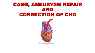
- 1. CABG, ANEURYSM REPAIR AND CORRECTION OF CHD
- 2. CONTENTS Anatomy of heart CABG oDefinition oPurpose oIndications & Contraindications oDiagnostic evaluation oTypes of CABG oTypes of Graft oProcedure oComplications Aortic Aneurysm oIndications of surgery oSurgical management Correction of CHD oClassification of CHD oSurgical management oNursing management
- 3. ANATOMY OF THE HEART • The heart contains 4 chambers oRight atrium oRight ventricle oLeft atrium oLeft ventricle
- 4. CABG • A form of bypass surgery that can create new routes around narrowed and blocked coronary arteries, permitting increased blood flow to deliver oxygen and nutrients to the heart muscle. • CABG surgery is one of the most commonly performed major operations.
- 5. PURPOSE Restore blood flow to the heart Relieve chest pain and ischemia Improves the patient’s quality of life Enables the patient to resume a normal life cycle Lower the risk of a heart attack
- 6. INDICATIONS & CONTRAINDICATIONS INDICATIONS Left main artery disease or equivalent Triple vessel disease Abnormal left ventricular function Failed PTCA Immediately after MI Life threatening arrhythmias caused by a previous MI Occlusion of grafts from previous CABG CONTRAINDICATIONS Aortic valve insufficiency Abdominal aortic aneurysm Hemorrhagic diseases Congenital heart diseases Cardiomyopathy Severe hypertension Uncontrolled arrhythmias Pregnancy
- 7. TYPES OF CABG Traditional/on pump Coronary Artery Bypass Grafting: It consists of the placement of arterial or venous grafts to provide blood between aorta or other blocked coronary arteries. Off-pump Coronary Artery Bypass Grafting (OPCAB): It uses a median sternotomy to access all coronary vessels. It is performed on a beating heart (no CPB) using mechanical stabilizers. Minimally Invasive Direct Coronary Artery Bypass (MIDCAB): It offers patients with disease of the LAD & RCA. It requires several small incisions between ribs or a mini-thoracotomy Robotic or Totally Endoscopic Coronary Artery Bypass(TECAB): It uses a robot in performing CABG surgery. It is done with or without use of CPB.
- 8. TYPES OF GRAFT Internal mammary artery graft – LIMA & RIMA Radial artery graft Saphenous vein graft Right gastro epiploic graft
- 10. PROCEDURE • Pt is brought to OT • Put IV & arterial lines • Administer analgesics & induction agents • Et tube is inserted and mechanical ventilation started • GA is maintained with anesthetic agent such as isoflurane • Chest is opened via a median sternotomy • Bypass grafts are harvested • When harvesting is done, the patient is given heparin to inhibit blood clotting
- 11. PROCEDURE (cont….) • In case of “on-pump” surgery, the surgeon sutures cannulae into the heart and instructs the perfusionist to start CPB • Protamine is given to reverse the effects of heparin • Chest tubes are placed in the mediastinal and pleural space to drain blood from around the heart and lungs • Sternum is wired together and incision is sutured • Dressing applied • Pt is transferred to ICU • Provide hemodynamic support & ventilator support
- 14. COMPLICATIONS Wound infection Bleeding Delay wound healing Reactions to anesthesia Fever Pain Stroke Heart attack Injury to arteries when graft harvesting time
- 15. AORTIC ANEURYSM • Aortic Aneurysm are out pouching or dilation of the arterial wall and are common problems involving aorta • TYPES oThoracic Aortic Aneurysm: Ballooning of the upper aspect of aorta, above the diaphragm oAbdominal Aortic Aneurysm: Enlargement in the area in the lower part of the aorta
- 16. INDICATION OF SURGERY FOR AORTIC ANEURYSM Persistent pain Aortic valve involvement Coronary artery involvement Diameter greater than 5.5 or rapidly expanding Symptomatic patients Acute rupture
- 17. SURGICAL MANAGEMENT Open Aneurysm Repair (OARs) Endo Vascular Graft Procedure (EVAR)
- 18. OPEN ANEURYSM REPAIR It involves large abdominal incision and cut into diseased aortic segment Remove any thrombus or plaque Sutures a synthetic graft to the aorta Sutures the native aortic wall around the graft to act as a protective cover It requires aortic cross clamping After which clamp is removed and blood is restored
- 20. ENDO VASCULAR GRAFT PROCEDURE • It is an alternative to conventional surgical repair of AAA • This technique involves the placement of a suture less aortic graft into the abdominal aorta inside the aneurysm via femoral artery cut down • After the graft is delivered to the predetermined point, the graft is pressed or implanted against the vessel wall by balloon inflation • Blood then flows through the vascular graft, thus preventing the expansion of the aneurysm due to pressure, and the aneurysm wall begin to shrink over time
- 22. OPEN Vs EVAR OAR EVAR • Longer recovery time • Longer hospital stay • 90% long term success • Younger patients typically • Shorter length of stay • Reduction in blood loss • ICU utilization reduced • Reduce morbidity/mortality rate • Needs long term follow up • May need secondary procedures for end leaks
- 23. CONGENITAL HEART DEFECTS • According to arterial oxygen saturation, congenital heart diseases are classified into: • ACYANOTIC CHD- Normal arterial oxygen saturation oASD, VSD, PDA, Coarctation of aorta, Pulmonary stenosis • CYANOTIC CHD- Reduced arterial oxygen saturation oTOF, TGA, Tricuspid atresia, Truncus arteriosus,Total anomalies pulmonary venous drainage
- 24. BASIS OF SURGERY FOR CHD • Two main categories: PALLIATIVE PROCEDURES: Aiming to increase pulmonary blood flow • Aortopulmonary shunt (Eg: TOF, Pulmonary atresia) • Pulmonary artery banding COMPLETE REPAIR: • Repair of extra cardiac anomalies (Eg: PDA, and Coarctation of Aorta) • Repair of intra cardiac anomalies (Eg:VSD, ASD and TOF)
- 25. AORTOPULMONARY SHUNT CLASSIC BLALOCK-TAUSSIG SHUNT (BT shunt) : • Classically, it consists of anastomosing the subclavian artery to the Pulmonary artery on the side opposite the aortic arch • However, with some technical modifications the subclavian artery can be anastomosed to the pulmonary artery on the same side of aortic arch (Rarely used) MODIFIED BLALOCK-TAUSSIG SHUNT (MBT shunt) : • It consists of interposition of a polytetraflurothelene (GORE-TEX) tube graft between the subclavian or innominate artery and the right or left pulmonary artery
- 26. REPAIR OF EXTRA CARDIAC ANOMALIES PDA: • Approached through a limited left posterolateral thoracotomy and is usually divide b/w clamps or ligated COARCTATION OF AORTA: • Approached through a left posterolateral thoracotomy • Techniques – Resection and end – to- end anastomosis or dilatation of the coarcted segment with subclavian flap or synthetic GORE-TEX patch
- 27. REPAIR OF INTRA CARDIAC ANOMALIES TOF • Closure of VSD with a patch (Dacron) and relief of the obstruction of the RV outflow tract and the stenosed pulmonary tract ASD • Closed by using pericardial patch VSD • Closed by using Dacron patch
- 28. NURSING MANAGEMENT Risk for Decreased Cardiac output related to changes in intravascular volume INTERVENTIONS RATIONALE Monitor vital signs Assess mental status Check peripheral perfusion Auscultate lung sounds and Heart sounds Monitor ABG Monitor Urine output Administer inotropic agents • To evaluate cardiovascular status • Decreased CO may lead to hypoperfusion to CNS • Decreased CO may lead to hypoperfusion to tissues • To identify any abnormal heart sounds • To rule out oxygenation level in body • Decreased CO may lead to hypoperfusion to kidney • To improve cardiac output
- 29. NURSING MANAGEMENT Risk for Bleeding related to the use of anticoagulation therapy INTERVENTIONS RATIONALE • Assess for the signs and symptoms of bleeding • Monitor chest drain and urine characteristics • Monitor platelet counts and coagulation test results (INR, PT, PTT) • Convert from IV anticoagulation to oral anticoagulation after the appropriate length of therapy • Stop heparin when bleeding occurs • Bruises, epistaxis, and gum bleeding are early signs of spontaneous bleeding • Chest drain may increase and hematuria may be seen • Effects of anticoagulation therapy must be closely monitored to reduce the risk of bleeding • PT or INR levels should be in a therapeutic range for anticoagulation before discontinuing heparin • Further administration of Heparin may lead to bleeding
- 30. NURSING MANAGEMENT Ineffective tissue perfusion related to increased coagulability of blood INTERVENTIONS RATIONALE • Assess for contributing factors • Assess for the signs and symptoms of deep vein thrombosis • Assess peripheral circulation status • Monitor coagulation profile • Administer anticoagulants as prescribed • Apply below-knee compression stockings • Encourage early ambulation after surgery • Knowledge of high-risk situations helps in early detection • The signs and symptoms occur in the leg affected by the deep vein clot • To rule out thrombus formation • These are used to measure the effectiveness of anticoagulant therapy • To prevent the formation of new clots • Compression stockings enhance circulation • To prevent venous stasis
- 31. NURSING MANAGEMENT Deficient knowledge related to unfamiliarity in disease condition & its management INTERVENTIONS RATIONALE • Assess the level of knowledge • Instruct the client to take medications as indicated, explaining their actions, dosages, and side effects • Inform the client of the need for regular checkup of INR while on oral anticoagulation • Provide teaching regarding the safety measures while on anticoagulant therapy such as the use of an electric razor, the use of a soft toothbrush • Advice the patient not sitting with the legs crossed • To obtain base line information to plan interventions • Correct knowledge decreases future complications • Routine coagulation monitoring is necessary to ensure that a therapeutic response • These precautionary measures help reduce the risk of bleeding • Sitting with legs crossed promotes vein compression
- 32. REFERENCES • Lewis, Bucher, Heitkemper, Harding, Kwong, Roberts. Lewi’s Medical Surgical Nursing.3rd South Asia edition. Vol 2. New Delhi: Elseiver publications; 2018. • https://www.slideshare.net/AbhayRajpoot3/cabg-134836953 • https://www.slideshare.net/AnvinThomas/aneurysm-85794573 • https://www.slideshare.net/eimad0307/surgery-for-congenital-heart- diseases • https://nurseslabs.com/5-deep-vein-thrombosis-nursing-care-plans/5/
- 33. THANK YOU
Editor's Notes
- del Nido or Histidine-Tryptophan-Ketoglutamate solutions