
Recommended
More Related Content
What's hot
What's hot (15)
Similar to 5 st under gluteus maximus obturator nerve
Similar to 5 st under gluteus maximus obturator nerve (20)
Recently uploaded
Recently uploaded (20)
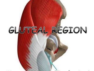
5 st under gluteus maximus obturator nerve
- 2. Thoracolumbar fascia Gluteal apponeurosis Gluteus maximus Iliotibial tract Sacrum Coccyx Hamstrings GLUTEUS MAXIMUS-ORIGIN-
- 3. Superficial fibres Deep fibres Iliotibial tract Gluteus medius Gluteal apponeurosis Gluteal tuberosity GLUTEUS MAXIMUS-INSERTION
- 4. • NERVE SUPPLY- • By the inferior gluteal nerve (L5, S1, S2). • Since finer twigs of the nerve reach the superficial part of the muscle, the gluteus maximus and medius are usually selected for intramuscular injections. • BLOOD SUPPLY- • By the superior and inferior gluteal arteries.
- 5. • ACTIONS- • It is the powerful extensor of the hip joint . • It comes into the action at the time of running, fast walking and climbing the stair-case. • But in standing and quiet walking it remains inactive and the hamstrings alone act as hip extensor. • It as the chief anti-gravity muscle of the hip joint. • It is a strong lateral rotator of hip joint. • Through the ilio-tibial tract it maintains the extended position of knee joint. • Upper fibres of act as powerful abductor of hip joint.
- 7. Iliac crest Tensor fascia latae Gluteus minimus Superior gluteal vessels and nerve Gluteus medius Pyriformis Nerve to quadratus femoris Tricipital tendon Quadartus femoris Sciatic nerve Adductor magnus Posterior cut.nerve of thigh Ischial tuberosity Hamstrings GM Pudendal n & vessels Inf gl v & n N to obt.int
- 8. • BONES AND JOINTS- • Ilium; • Ischium with ischial tuberosity; • Upper end of femur with greater trochanter; • Sacrum and coccyx • Hip joint; • Sacro-iliac joint.
- 9. • LIGAMENTS- • Sacrotuberous; • Sacrospinous • and ischiofemoral. • BURSAE- • Trochanteric bursa under gluteus maximus; • bursae over ischial tuberosity.
- 10. APPLIED ANATOMY • Intramuscular injections are given in the anterosuperior quadrant of the gluteal region. • i.e. in the glutei medius and minimus, and superficial part of maximus. • This is to avoid injury to the large vessels and nerves mainly sciatic nerve.
- 11. • When Gluteus maximus is paralysed as in muscular dystrophy- • The patient cannot stand up from sitting position without support. • Such patients, while trying to stand up rise gradually, supporting their hands first on the legs and then on the thighs; they climb on themselves.
- 12. • Our normal gait depends on – • Gluteus medius and minimus along. • The fulcrum formed by the relation of head of femur with acetabulum . • The weight transmitted through the head and neck of femur. • Normally when the body weight is supported on one limb, the glutei of the supported side raise the opposite and unsupported side of the pelvis. • Thus it prevents the drooping of pelvis on unsupported side.
- 13. • When gluteus medius and minimus are paralysed, the patient cannot walk normally. • He sways or waddles on the paralysed side to clear the opposite foot off the ground. • This is known as lurching gait; when it is bilateral it is called as waddling gait. • However, if the abductor mechanism is defective, the unsupported side of the pelvis drops and this is known as a positive Trendelenberg’s sign. • The test is positive in defects of power- • i.e. paralysis of gluteus medius and minimus; • defects of fulcrum, • abnormal weight transmission.
- 14. • All the three gluteal muscles are acting like ropes running from the pelvis, going to the anterior, posterior and lateral aspect of hip joint. • This arrangement act as guy ropes (Guy means a rope fixed to the ground to secure a tent).
- 17. Iliac crest Gluteus maximus Gluteus medius Gluteus minimus Pyriformis Greater trochanter Tricipital tendon Quadratus femoris Adductor magnus Sacrotuberous ligament Ischial tuberosity Sciatic nerve Tibial nerve Common peroneal nerve Semimembranosus Semitendinosus Long head of biceps femoris COURSE AND RELATIONS OF SCIATIC NERVE
- 18. Sacral plexus Pelvis Gluteal region Thigh Tibial component To hip joint Common peroneal component To semitendinosus To long head of biceps To semimebranosus To adductor magnus To knee joint L4 L5 S1 S2 S3 DISTRIBUTION OF TIBIAL COMPONENT OF SCIATIC NERVE
- 19. Sacral plexus For knee joint For short head of biceps femoris Common peroneal component Sciatic nerve Tibial component L4 L5 S1 S2 Pelvis Gluteal region Back of thigh DISTRIBUTION OF COMMON PERONEAL COMPONENT OF SCIATIC NERVE
- 20. APPLIED ANATOMY Compression of sciatic nerve
- 21. Foot drop
- 22. Sciatica
- 23. • Sleeping foot- • It is the concussion of the sciatic nerve when the nerve is pressed against femur by the hard surface of the chair, the nerve is damaged and leads to the paralysis of the muscles below the knee. • MOTOR LOSS- • The hamstrings are paralysed but weak flexion of knee is possible due to the action of sartorius and gracilis. • All the muscles below the knee are paralysed and the weight of foot causes it to assume the plantar flexed position or foot-drop.
- 24. • SENSORY LOSS- • There is loss of sensation below the knee, except a narrow area along the medial side of the lower part of the leg and along the medial border of the foot upto big toe which is supplied by the saphaneous nerve. • TREATMENT- • The result of operative repair of sciatic nerve injury is poor. • It is rare for active movements to return. • Sensory recovery is rarely complete. • Loss of sensation in the sole of the foot makes the development of trophic ulcers.
- 25. • SCIATIC NERVE BLOCK- • Sciatic block is an advanced nerve block technique. • The block is well suited for surgery on the leg below the knee, particularly on the ankle and foot. • It provides complete anesthesia of the leg below the knee with the exception of the medial strip of skin, which is innervated by the saphaneous nerve.
- 27. • DEFINITION- • The group of muscles in the flexor compartment of the thigh is called as hamstrings. • WHY THEY ARE NAMED AS HAMSTRINGS- • The word ‘Ham’ means the slender muscles of hip and knee along with their tendons. • These tendons of the animals are like pig, beef and goat were used for stringing in bishop’s shops hence knows hamstrings. • Furthermore, in ancient times, it was common for soldiers to slash their opponent’s horses posterior to the knees in order to cut the tendons of their posterior thigh muscles. This would bring the horse and its rider’s down. • Also they use to cut these tendons of the soldiers so they could not run; this was called as ‘hamstringing’ the enemy.
- 30. CLASSIFICATION OF HAMSTRINGS- Modified Hamstrings Long head of biceps femoris Ischial part of adductor magnus Sacrotuber ous ligament Tibial collateral ligament
- 31. • ACTIONS-
- 32. APPLIED ANATOMY
- 33. • As the two heads of biceps femoris have different nerve supply a wound in the thigh may severe a nerve paralyzing only one head and other being normal; this will not affect the length of the muscle. • In some people they are not long enough to allow them to touch their toes when they flex the vertebral column and keep their knees straight. • In other people the hamstrings are long and they can easily touch the floor with their palms. • Tendon of semitendinosus is used in the rupture of the anterior cruciate ligament for repair and replacement.
Editor's Notes
- Lateral side of the nerve is safe and medial side is of danger as the muscular branches arise on that side. The nerve may be injured by- penetrating wounds in posterior dislocation of hip, fracture dislocation of the hip, fractures of the pelvis and badly placed intramuscular injections into the gluteal region.
- When injury is complete all muscles below the knees are paralysed. It is associated with foot drop. All cutaneous sensations below the knee are lost except the area supplied by the saphaneous nerve
- It is a clinical terminology to describe the condition in which the patients have shooting and radiating pain along the sensory distribution of the sciatic nerve. The pain is experienced in the posterior aspect of the thigh, the posterior and lateral sides of the leg and lateral part of the foot. Sciatica can be caused by- prolapse of an intervertebral disc with pressure on one or more roots of the lumbar and sacral spinal nerves. pressure on the sacral plexus or sciatic nerve by an intrapelvic tumor in inflammation of sciatic nerve or its terminal branches.
- They take origin from the ischial tuberosity. They are inserted beyond the knee joint to the tibia, fibula or both the bones.They are supplied by tibial component of the sciatic nerve.They act as flexors of knee joint and the extensors of hip joint.
- They act as prime mover of flexion of the knee joint. Help in extension of hip joint especially in standing and walking. In semi-flexed knee, the semimembranosus and semitendinosus act as medial rotator. Biceps femoris act as lateral rotator of tibia on femur.
- Hamstring injuries often called as ‘pulled hamstrings’. In persons who run very fast or kick e.g. in running, jumping, baseball, football and soccer. This tears tendineous proximal attachments of the hamstrings from the ischial tuberosity. There is rupture of some blood vessels, supplying the muscles. The resultant hematoma lies in dense fascia lata this causes pain when the athlete moves or stretches the leg.