
Recommended
More Related Content
What's hot
What's hot (16)
Similar to Biol102 chp17-pp-spr10-100508132228-phpapp02
Similar to Biol102 chp17-pp-spr10-100508132228-phpapp02 (20)
More from Cleophas Rwemera
More from Cleophas Rwemera (20)
Recently uploaded
Recently uploaded (20)
Biol102 chp17-pp-spr10-100508132228-phpapp02
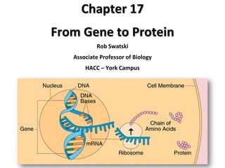
- 1. Chapter 17 From Gene to Protein Rob Swatski Associate Professor of Biology HACC – York Campus
- 2. Overview: The Flow of Genetic Information DNA information = specific sequences of nucleotides DNA protein synthesis Proteins: link genotype & phenotype Gene expression: DNA directs protein synthesis - 2 stages: transcription & translation
- 3. How was the fundamental relationship between genes & proteins discovered? - examine evidence from studies of metabolic defects
- 4. 1909: British physician Archibald Garrod 1st suggested that genes dictate phenotypes with enzymes - symptoms of inherited disease reflects inability to synthesize a certain enzyme - required understanding that cells synthesize & degrade molecules using metabolic pathways
- 6. Nutritional Mutants in Bread Mold George Beadle & Edward Tatum exposed Neurospora to x-rays - created mutants that could not survive on minimal medium (cannot synthesize certain molecules) Identified 3 classes of arginine-deficient mutants - each lacked a different enzyme needed to make arginine Developed the “one gene – one enzyme hypothesis” - each gene directs the synthesis of a specific enzyme
- 7. Minimal medium No growth: Mutant cells cannot grow and divide Growth: Wild-type cells growing and dividing EXPERIMENT
- 8. RESULTS Classes of Neurospora crassa Wild type Class I mutants Class II mutants Class III mutants Minimal medium (MM) (control) MM ornithine MM citrulline Condition MM arginine (control) Summary of results Can grow with or without any supplements Can grow on ornithine, citrulline, or arginine Can grow only on citrulline or arginine Require arginine to grow Growth No growth
- 9. CONCLUSION Wild type Class I mutants (mutation in gene A) Class II mutants (mutation in gene B) Class III mutants (mutation in gene C) Gene (codes for enzyme) Gene A Gene B Gene C Precursor Precursor Precursor Precursor Enzyme A Enzyme A Enzyme A Enzyme A Enzyme B Enzyme B Enzyme B Enzyme B Ornithine Ornithine Ornithine Ornithine Enzyme C Enzyme C Enzyme CEnzyme C Citrulline Citrulline Citrulline Citrulline Arginine Arginine Arginine Arginine
- 10. The Products of Gene Expression: A Developing Story • Some proteins aren’t enzymes… so researchers later revised the hypothesis to “one gene – one protein” • But, many proteins consist of several polypeptides, each having its own gene Beadle & Tatum’s hypothesis is now restated as the: “one gene–one polypeptide hypothesis”
- 11. Basics of Transcription & Translation RNA: the bridge between genes & the proteins they code • Transcription: synthesis of RNA under the direction of DNA - produces messenger RNA (mRNA) • Translation: synthesis of a polypeptide under the direction of mRNA - ribosomes: the sites of translation
- 12. Eukaryotes: the nuclear envelope separates transcription from translation - eukaryotic RNA transcripts are modified via RNA processing to yield finished mRNA Prokaryotes: mRNA transcripts are immediately translated without further processing
- 13. (a) Prokaryotic Cell (Bacteria) TRANSCRIPTION DNA mRNA TRANSLATION Ribosome Polypeptide
- 14. Primary transcript: initial RNA transcript from a gene before processing Central dogma: cells are governed by a cellular chain of command: DNA RNA Protein
- 15. (b) Eukaryotic cell TRANSCRIPTION Nuclear envelope DNA Pre-mRNA RNA PROCESSING mRNA
- 16. (b) Eukaryotic cell TRANSCRIPTION Nuclear envelope DNA Pre-mRNA RNA PROCESSING mRNA TRANSLATION Ribosome Polypeptide
- 17. The Genetic Code How are the instructions for assembling amino acids into proteins encoded into DNA? There are 20 amino acids, … but there are only 4 nucleotide bases in DNA How many bases correspond to an amino acid?
- 18. Codons: Triplets of Nucleotides The flow of information from gene protein is based on a triplet code - series of non-overlapping 3-nucleotide “words” Triplet: smallest unit that can code for amino acids - “AGT” = placement of serine at its correct position in the polypeptide
- 19. Transcription: one of the two DNA strands (template strand) provides a pattern for ordering the nucleotide sequence in the mRNA transcript Translation: mRNA base triplets (codons) are read in the 5 to 3 direction - each codon specifies the amino acid (1 of 20) and it’s correct position in a polypeptide
- 20. DNA template strand TRANSCRIPTION mRNA TRANSLATION Protein Amino acid Codon Trp Phe Gly 5 5 Ser U U U U U 3 3 53 G G G G C C T C A A AAAAA T T T T T G G G G C C C G G DNA molecule Gene 1 Gene 2 Gene 3 C C
- 21. Cracking the Code • All 64 codons were deciphered by the mid-1960s • Of the 64 triplets, 61 code for amino acids - 3 triplets are stop codons that end translation: UAA, UAG, UGA • The genetic code is redundant but not ambiguous • Codons must be read in the correct reading frame (correct groupings) in order to synthesize the specified polypeptide
- 22. Second mRNA base FirstmRNAbase(5endofcodon) ThirdmRNAbase(3endofcodon) UUU UUC UUA CUU CUC CUA CUG Phe Leu Leu Ile UCU UCC UCA UCG Ser CCU CCC CCA CCG UAU UAC Tyr Pro Thr UAA Stop UAG Stop UGA Stop UGU UGC Cys UGG Trp GC U U C A U U C C C A U A A A G G His Gln Asn Lys Asp CAU CGU CAC CAA CAG CGC CGA CGG G AUU AUC AUA ACU ACC ACA AAU AAC AAA AGU AGC AGA Arg Ser Arg Gly ACGAUG AAG AGG GUU GUC GUA GUG GCU GCC GCA GCG GAU GAC GAA GAG Val Ala GGU GGC GGA GGG Glu Gly G U C A Met or start UUG G
- 23. Evolution of the Genetic Code The genetic code is shared by all living things - Genes can be transcribed & translated after being transplanted from one species to another
- 24. (a) Tobacco plant expressing a firefly gene
- 25. (b) Pig expressing a jellyfish gene
- 26. Synthesis of an RNA Transcript The 3 Stages of Transcription: 1. Initiation 2. Elongation 3. Termination
- 27. Transcription: DNA RNA RNA synthesis is catalyzed by RNA polymerase - pries DNA strands apart - hooks RNA nucleotides together The RNA is complementary to the DNA template strand Follows same base-pairing rules as DNA - except uracil substitutes for thymine
- 28. Promoter: DNA sequence that RNA polymerase attaches to Transcription unit: section of DNA that is transcribed
- 29. Promoter Transcription unit DNA Start point RNA polymerase 5 5 3 3 Initiation 3 3 1 RNA transcript 5 5 Unwound DNA Template strand of DNA 2 Elongation Rewound DNA 5 5 53 3 3 RNA transcript 3 Termination 5 5 53 3 3 Completed RNA transcript
- 30. RNA Polymerase Binding & Initiation of Transcription Promoters: signal initiation of RNA synthesis - TATA box promoter is crucial in forming the initiation complex in eukaryotes Transcription factors: needed to help bind RNA polymerase & initiate transcription Transcription initiation complex: completed assembly of transcription factors & RNA polymerase bound to a promoter
- 31. Transcription initiation complex forms3 DNA Promoter Nontemplate strand 5 3 5 3 5 3 Transcription factors RNA polymerase II Transcription factors 5 3 5 3 5 3 RNA transcript Transcription initiation complex 5 3 TATA box T T T T T T A A A A A A A T Several transcription factors bind to DNA2 A eukaryotic promoter1 Start point Template strand
- 32. Elongation of the RNA Strand As RNA polymerase moves along DNA, it untwists the double helix, 10-20 bases at a time - transcription rate = 40 nucleotides/sec A gene can be transcribed simultaneously by several RNA polymerases Nucleotides are added to the 3’ end of the growing RNA molecule
- 33. Elongation RNA polymerase Non-template strand of DNA RNA nucleotides 3 end Direction of transcription (“downstream”) Template strand of DNA Newly made RNA 3 5 5
- 34. Termination of Transcription In bacteria: RNA polymerase stops transcription at end of the terminator & the mRNA can be translated without further modification In eukaryotes: RNA polymerase continues transcription after the pre-mRNA is cleaved from the growing RNA chain - polymerase eventually falls off the DNA
- 35. Eukaryotic Cells Modify RNA After Transcription Enzymes in the eukaryotic nucleus modify pre-mRNA (RNA processing) before mRNA “gene” enters cytoplasm Both ends of the primary transcript are usually altered - and some interior parts of RNA are usually cut-out & other parts spliced together
- 36. Alteration of mRNA Ends Each end of pre-mRNA is modified in a particular way: - the 5 end gets a modified nucleotide 5 cap - the 3 end gets a poly-A tail Why Modify? - Facilitates export of mRNA - Protects mRNA from hydrolytic enzymes - Helps ribosomes attach to the 5 end
- 37. Protein-coding segment Polyadenylation signal 5 3 35 5Cap UTR Start codon G P P P Stop codon UTR AAUAAA Poly-A tail AAA AAA…
- 38. Split Genes & RNA Splicing Most genes & their RNA transcripts have long noncoding regions (introns) that lie between coding regions - intron = intervening sequences (“in the way”) Exons: coding regions - expressed & translated into amino acid sequences RNA splicing: removes introns & joins exons - creates mRNA molecule with a continuous coding sequence
- 39. 5 Exon Intron Exon 5 CapPre-mRNA Codon numbers 130 31104 mRNA 5 Cap 5 Intron Exon 3 UTR Introns cut out and exons spliced together 3 105 146 Poly-A tail Coding segment Poly-A tail UTR 1146
- 40. Some RNA splicing is carried out by spliceosomes - consist of a variety of proteins & small nuclear ribonucleoproteins (snRNPs = “snurps”) - snRNPs can recognize the splice sites
- 41. RNA transcript (pre-mRNA) Exon 1 Exon 2Intron Protein snRNA snRNPs Other proteins 5 5 Spliceosome Spliceosome components Cut-out intron mRNA Exon 1 Exon 2 5
- 42. Ribozymes Catalytic RNA molecules that act as enzymes & splice RNA - not all biological catalysts are proteins!
- 43. How can RNA function as an enzyme? - can form a 3-D structure because it can base-pair with itself - some RNA bases contain functional groups - can hydrogen-bond with other nucleic acids
- 44. Some genes can encode more than 1 kind of polypeptide, depending on which segments are treated as exons during RNA splicing - the actual # of different proteins an organism can produce is much greater than its number of genes Alternative RNA Splicing
- 45. Proteins often have a modular architecture consisting of discrete regions called domains - different exons can code for different domains in a protein Exon shuffling can result in the evolution of new proteins
- 46. Gene DNA Exon 1 Exon 2 Exon 3Intron Intron Transcription RNA processing Translation Domain 2 Domain 3 Domain 1 Polypeptide
- 47. Molecular Components of Translation A cell translates mRNA message into protein with the help of transfer RNA (tRNA) tRNA molecules are not identical: - each carries a specific amino acid on one end - each has an anticodon on the other end that base- pairs with a complementary codon on mRNA
- 49. The Structure & Function of tRNA tRNA: one RNA strand, 80 nucleotides long - when flattened, it resembles a cloverleaf C Amino acid attachment site Hydrogen bonds Anticodon 3 5
- 50. Can twist & fold into an “L”-shaped 3-D molecule through hydrogen-bonding Amino acid attachment site 3 3 5 5 Hydrogen bonds Anticodon Anticodon (b) 3-D structure (c) Symbol used in this book
- 51. Translation requires 2 steps: “The Match Game” 1. tRNA and its amino acid are matched by the enzyme aminoacyl-tRNA synthetase - forms “charged tRNA” 2. tRNA anticodon and an mRNA codon are matched Flexible pairing at the 3rd base of a codon is called wobble - allows some tRNAs to bind to more than 1 codon
- 52. Aminoacyl-tRNA synthetase (enzyme) Amino acid P P P Adenosine ATP P P P P Pi i i Adenosine tRNA AdenosineP tRNA AMP Computer model Amino acid Aminoacyl-tRNA synthetase Aminoacyl tRNA (“charged tRNA”)
- 53. Ribosomes Ribosomes facilitate specific coupling of tRNA anticodons with mRNA codons in protein synthesis - 2 ribosomal subunits (large & small) are made of proteins & ribosomal RNA (rRNA)
- 54. Growing polypeptide Exit tunnel tRNA molecules Large subunit Small subunit (a) Computer model of functioning ribosome mRNA E P A 5 3
- 56. Exit tunnel A site (Aminoacyl- tRNA binding site) Small subunit Large subunit P A P site (Peptidyl-tRNA binding site) mRNA binding site (b) Schematic model showing binding sites E site (Exit site) E
- 57. A ribosome has 3 binding sites for tRNA: - A site: holds the tRNA carrying the next amino acid to be added to the chain - P site: holds the tRNA carrying the growing polypeptide chain - E site (Exit): where discharged tRNAs leave the ribosome
- 58. Amino end mRNA E (c) Schematic model with mRNA and tRNA 5 Codons 3 tRNA Growing polypeptide Next amino acid to be added to polypeptide chain
- 59. Building a Polypeptide The 3 stages of translation: 1. Initiation 2. Elongation 3. Termination All 3 stages require protein factors
- 60. Initiation of Translation Initiation stage: brings together mRNA, a tRNA with the 1st amino acid, & the 2 ribosomal subunits 1. First, the small ribosomal subunit binds with mRNA and a special initiator tRNA 2. Then the small subunit moves along mRNA until it reaches the start codon (AUG) - Initiation factors bring in the large subunit to complete the translation initiation complex
- 61. 3 35 5U U A A C G GTP GDP Initiator tRNA mRNA 5 3 Start codon mRNA binding site Small ribosomal subunit 5 P site Translation initiation complex 3 E A Large ribosomal subunit
- 62. Elongation of the Polypeptide Chain Elongation stage: amino acids are added one by one Each addition involves elongation factors and occurs in 3 steps: a. Codon recognition b. Peptide bond formation c. Translocation
- 65. Amino end of polypeptide mRNA 5 3E P site A site GTP GDP E P A E P A GDP GTP Ribosome ready for next aminoacyl tRNA E P A
- 66. Termination of Translation Termination: - occurs when a stop codon in mRNA reaches the A site The A site accepts a release factor protein - adds a water molecule instead of an amino acid - this releases the polypeptide - the translation complex comes apart
- 67. Release factor 3 5 Stop codon (UAG, UAA, or UGA) 5 3 2 Free polypeptide 2 GDP GTP 5 3
- 68. Polyribosomes Groups of ribosomes that simultaneously translate one mRNA, forming a polyribosome (polysome) - allows a cell to quickly make many copies of a polypeptide
- 69. Growing polypeptides Completed polypeptide Incoming ribosomal subunits Start of mRNA (5 end) End of mRNA (3 end) (a) Ribosomes mRNA (b) 0.1 µm
- 70. Post-Translation A protein is usually not functional immediately after translation - requires further post-translational modification It spontaneously coils and folds into its correct 3-D shape - some activated by enzymes that cleave them - others assemble into protein subunits
- 71. Targeting Polypeptides to Specific Locations Two populations of ribosomes are found in cells: - Free ribosomes: synthesize proteins that function in the cytosol - Bound ribosomes: synthesize proteins on ER and those that will be secreted from the cell Ribosomes are identical and can switch from free to bound
- 72. Polypeptide synthesis always begins and ends in the cytosol - unless the polypeptide signals the ribosome to attach to the ER Polypeptides destined for the ER or for secretion are marked by a signal peptide - a signal-recognition particle (SRP) binds to the signal peptide - the SRP brings the signal peptide & its ribosome to the ER
- 74. Point Mutations - chemical changes in just 1 base pair of a gene - a change in one DNA nucleotide can lead to the production of an abnormal protein Wild-type hemoglobin DNA mRNA Mutant hemoglobin DNA mRNA 3 3 3 3 3 3 5 5 5 5 5 5 C CT T T TG GA A A A A A AGG U Normal hemoglobin Sickle-cell hemoglobin Glu Val
- 75. Types of Point Mutations - Base-pair substitutions - Base-pair insertions or deletions
- 76. Wild type 3DNA template strand 3 35 5 5mRNA Protein Amino end Stop Carboxyl end A instead of G 3 3 3 U instead of C 5 5 5 Stop Silent mutations: have no effect on the amino acid because of redundancy in the genetic code
- 77. Wild type DNA template strand 3 5 mRNA Protein 5 Amino end Stop Carboxyl end 5 3 3 T instead of C A instead of G 3 3 3 5 5 5 Stop Missense: still codes for an amino acid, but not necessarily the right amino acid
- 78. Wild type DNA template strand 3 5 mRNA Protein 5 Amino end Stop Carboxyl end 5 3 3 A instead of T U instead of A 3 3 3 5 5 5 Stop Nonsense: changes an amino acid codon into a stop codon, nearly always leading to a nonfunctional protein
- 79. Insertions and Deletions - additions or losses of nucleotide pairs in a gene Can have a disastrous effect on the protein more often than substitutions - may produce a frameshift mutation, which alters the reading frame
- 80. Wild type DNA template strand 3 5 mRNA Protein 5 Amino end Stop Carboxyl end 5 3 3 Extra A Extra U 3 3 3 5 5 5 Stop Frameshift Mutation causing immediate nonsense (1 base-pair insertion)
- 81. Wild type DNA template strand 3 5 mRNA Protein 5 Amino end Stop Carboxyl end 5 3 3 missing missing 3 3 3 5 5 5 Frameshift Mutation causing extensive missense (1 base-pair deletion)
- 82. Wild type DNA template strand 3 5 mRNA Protein 5 Amino end Stop Carboxyl end 5 3 3 missing missing 3 3 3 5 5 5 No Frameshift Mutation, but one amino acid missing (3 base-pair deletion) Stop
- 83. What Is a Gene? We have considered a gene as: - A discrete unit of inheritance - A region of specific nucleotide sequence in a chromosome - A DNA sequence that codes for a specific polypeptide chain In summary, a gene can be defined as: - a region of DNA that can be expressed to produce a final functional product, either a polypeptide or an RNA molecule
- 84. TRANSCRIPTION RNA PROCESSING DNA RNA transcript 3 5 RNA polymerase RNA transcript (pre-mRNA) Intron Exon NUCLEUS Aminoacyl-tRNA synthetase AMINO ACID ACTIVATION Amino acid tRNACYTOPLASM Growing polypeptide 3 Activated amino acid mRNA TRANSLATION Ribosomal subunits 5 E P A A Anticodon Ribosome Codon E