2. 1
Contents
Description Page no.
1. INTRODUCTION
1.1 Introduction to electronmicroscopy
1.2 Purpose of this file
2. METHOD
2.1 TEM specimenpreparation
2.2 SEM specimenpreparation
2.3 Cryo-SEM
2.4 Staining
2.5 Use of the SEM
2.6 Use of the TEM
3. RESULTS & DISCUSSIONS
3.1 SEM micrographs
3.2 TEM micrographs
4. CONCLUSIONS
4.1 Summary of experience
5. REFERENCES
2
2
2
2
2
3
3
4
4
4
4
4
7
8
8
9
3. 2
1. INTRODUCTION
1.1 | Introduction to electronmicroscopy
Electron microscopy is extensively utilised to explore images of specimens at the
nanometre (nm) scale (John Innes Centre, 2015). The electron microscope (EM) has
the capability to observe a specimen up to 2 million times magnification. It’s
resolution and magnification greatly exceeds that of the light microscope. It does so
by emitting and controlling the path of an electron beam with the use of electrostatic
or electromagnetic lenses and detecting the interacting with the specimen (John
Innes Centre, 2015). The transmission electron microscope (TEM) is used to observe
images of ultra structures such as organelles, macromolecules and microorganisms,
including viruses and bacteria (CMRF, 2015). This is achieved through the
transmission of an electron beam through a very thin specimen, producing a two-
dimensional and grayscale image, recorded on a plane, such as a fluorescent screen
or a sensor (CMRF, 2015)(John Innes Centre, 2015). The scanning electron
microscope (SEM) produces an image by detecting secondary, scattered electrons
that have been scanned across the surface of a sample, whilst detectors build up the
image (John Innes Centre, 2015). The SEM can reveal surface characteristics of a
specimen, including texture, crystalline structure and chemical composition (Swapp,
2015).
1.2 | Purpose of this file
This investigation was carried out to gain experience and understanding in the
applications of and preparations for SEM and TEM, used to observe microorganisms
at the nm scale. This lab file was assembled to document this experience and the
images produced. Skill and practice in EM will be paramount to many investigations
in the future, as it can be used to observe an extensive variety of very small
structures. The objectives contributing to this experience were to prepare, view and
image the following specimens. Stilton and yoghurt were to be viewed using SEM, in
the hope of imaging bacteria, such as Lactobacillus. The SEM was also used to view
a biofilm comprised of an unknown bacteria and a species of ladybird, Coccinella
septempunctata. Escherichia coli and T4 bacteriophage were to be viewed using
TEM.
2. METHOD
2.1 | TEM specimenpreparation
The bacteria and viruses were prepared using the same method at room
temperature. The microorganisms were provided in separate suspensions.
4. 3
Additionally, a mixed suspension with both viruses and bacteria was prepared, in the
hope of viewing their interaction under the TEM. Fixation occurred using a EM film
100nm thick, composing of a copper grid, a colloidon and carbon. The grid was
gently placed, film side down, onto a droplet of the desired suspension for 15
minutes. A duplicate of each microorganism suspension was also fixed for 30
minutes to compare. To rinse, the grid was moved from the droplet of suspension
onto a droplet of water for 10 seconds, following which it was stained accordingly.
2.2 | SEM specimenpreparation
The C.septempunctata and the biofilm were sprayed with gold (Au) before being
mounted onto the SEM. Using this heavy metal acts like a stain as it increases the
contrast and resolution of the image and, as most specimens are electrically
insulated, the gold acts as a conductor. The outcome is referred to as a sputter
coating and although we used gold, platinum and chromium can both be used (John
Innes Centre, 2015). The Stilton was firstly treated with Carbon black, which is
primarily composed of elemental carbon and appears black on the SEM image. Its
function is also analogous to the Au used for TEM, as it allows the specimen to be
electrically conducted.
2.3 | Cryo-SEM
The yoghurt and Stilton were dehydrated before being mounted. Dehydration was
important to transfer the specimens into a solid state, preserving the structures of the
living state. This was achieved through freezing using liquid nitrogen. Liquid nitrogen
is used as it creates temperature low enough so that ice crystals will not form, as
these can absorb electrons and interfere with the image, as well as damage
biological structures. The preparation is called cryofixation and is advantageous
because it captures the specimen in a freeze frame of its solution state and produces
minimal artefacts (John Innes Centre, 2015). The Stilton underwent just plunge
freezing and freeze-drying, yoghurt was additionally freeze-fractured. Freeze-
fracturing involves cryofixing the fresh specimen.
2.4 | Staining
The virus, bacteria and mix suspensions for the SEM were stained using uranyl
acetate. Those that were fixed for 30 minutes were also stained for 30 and those that
were fixed for 15 were also stained for 15. Aqueous uranyl acetate was used
because it absorbs electrons around the structures, rather than reacting with the
specimen. This gives a negative stain. It causes stained areas to appear darker on
the image. Following staining, each individual grid was placed onto a water droplet
for 10 seconds to rinse. A paper towel was then used to absorb excess water from
the edge of each film. Pearce (et al 2015) conducted a study that used negative
staining with 2% phosphotungstic acid to view the recombinant nucleocapsid proteins
(HeV N) of the Hendra virus. The images indicated the virus’s ability to self-assemble
into helical chains and also gave information regarding the dimensions of the genetic
structures. This was an important property in the investigations’ aim to reproduce
functional Hev N for immunodiagnostic tools. Venable & Coggeshall (1964) also
suggested following the aqueous uranyl acetate with a lead citrate, as this results in
a much faster staining process, lasting only a few seconds.
5. 4
2.5 | Use of the SEM
Once the specimens were mounted on the SEM individually, each was magnified to
a point where the intricate, three-dimensional structures could be observed with great
resolution. When observing the biofilm, bacteria were located and identified. When
observing the Stilton, the bacteria had to be identified against a coarser background
of the Stilton and bacteria were located in the cracks of the cheese, as this was
assumed to be more ideal conditions for growth. This occurred very similarly with the
yoghurt specimen, which was investigated using the SEM, with particular attention to
where the freeze fracture had occurred. The ladybird was magnified to an extent
where the intricate structures not visible to the naked eye could be observed. The
feet, antenna and eyes were closely observed and surfaces were investigated for
traces of bacteria. The SEM operates by firing an electron beam from a cathode, or
‘filament’, through a vacuum column (Ding, 2015). The beam is fired through various
magnetic lenses and condensers before impacting the specimen, giving the user lots
of control over the degree of magnification. Due to this element of control, each
desired image could be located and then rescanned more frequently with the
electron beam to reveal the image in more detail. During SEM, the electrons are
scanned repeatedly across the specimen to provide topographical information (Ding,
2015). Backscattered electrons and secondary electrons are both detected to build
up an image through signal mapping (John Innes Centre, 2015).
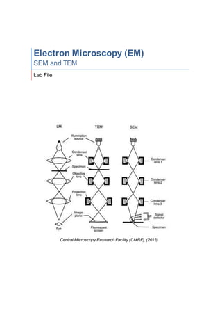
2.6 | Use of the TEM
The copper grids on which the E.coli and bacteriophage had been fixed and stained
were mounted onto the TEM specimen stage and magnified to view the two-
dimensional organisms in detail. When viewing the viruses, the magnification was,
for some images, increased to X250k, so that the details of the individual viruses
could be observed. The mixed suspension was observed to see the interaction
between the bacteria and the viruses. The TEM, like the SEM, also utilises an
electron beam inside a vacuum, which is maintained using rotary pumps and the
filament, the source of the electrons, is usually tungsten wire (CMRF, 2015). The
TEM operates on a very similar principle to a light microscope, in that the electron
beam is fired through a condenser lens, the specimen, an objective lens and a
protection lens before being detected by a fluorescent screen (the “eye” in terms of
light microscopy) (CMRF, 2015). The TEM also possesses additional tools that can
be adjusted to aid in the refining of images, including objective and intermediate
apertures (CMRF, 2015). In this investigation, the image wobbler was used to
ensure the image were as resolved as possible.
3. RESULTS & DISCUSSIONS
3.1 | SEM micrographs
6. 5
It was interesting to see the hair like structures of the antenna in Figure 1.1 were
analogous to those seen of the foot in Figure 1.2. It is broadly known that the
antenna is a key sensory organ, but seeing these structures repeated in the foot
indicates a sensory ability as well, which was confirmed in research (LAM, 2014)
This is an example of how, through using SEM, simply viewing detailed structures
more closely can indicate a lot about their function, affirming the importance of this
tool. Traces of bacteria that looked like bacilli could also be seen on the eye (Figure
1.3) The contrast of the images due to the Au preparation was also effective at
providing sufficient detail.
Figure 1.1: C.septempunctata; antenna X1,500 Figure 1.2: C.septempunctata; foot X550
Figure 1.3: C.septempunctata; eye X2,700
7. 6
The SEM was effective observing the bacteria on the biofilm to an extent where it
could be identified as a species of vibrio (Figure 2.1 & Figure 2.2). Images such as
this could be repeated in future investigations, such as to deduce the composition of
bacterial colonies and products in biofilms.
The magnification capability of the SEM enabled visibility of cocci-shaped bacteria in
the Stilton (Figure 3.1). Bacteria were difficult to locate in the yoghurt using the SEM
as they were a challenge to distinguish apart from the structural features formed by
Figure 2.1: vibrio sp. X9,000 Figure 2.2: vibrio sp. X700
Figure 3.1: Stilton cheese X2,000 Figure 3.2: Live yoghurt X19,000
8. 7
the yoghurt itself, but some cocci-shaped microorganisms were eventually observed
and presumed to be bacteria (Figure 3.2).
3.2 | TEM micrographs
Figure 4.1: E. coli Figure 4.2: T4 bacteriophage
Figure 4.3: E. coli and T4 bacteriophage Figure 4.4: T4 bacteriophage
9. 8
The negative stain, which accumulated around the biological structures, allowed both
viruses and bacteria to be observed in high definition. The stain can also be seen
accumulating on top of the bacteria in Figure 4.1, providing a little information about
it’s topography and depth. The 30-minute treatment for both microorganisms proved
to be more effective at producing images, so only those have been presented here.
Although very fine structures of the viruses (Figure 4.5)(Todar, 1012), such as the tail
fibres, could not be seen in the micrographs, the basic structure, including the head
and sheath, were clearly visible. This could have been due to many factors, including
time taken to fix and stain the microorganisms. Figure 4.3 indicated the interaction
between the viruses and the bacteria, which appears to show the T4 bacteriophage
attaching to the membrane of the E. coli, which is its host.
4. CONCLUSIONS
4.1 | Summary of experience
It was useful and informative to gain more understanding regarding the necessary
preparation for specimens being observed using the EM. The ability to utilise the
SEM and TEM to observe ultrastructures and microorganisms have proved to be an
extremely useful skill, as it could be very applicable to future investigations. It has
also been enlightening to understand how much information can be drawn from a
micrograph, which emphasises its importance.
Figure 4.5: Diagram of T4
bacteriophage (Todar, 2012)
10. 9
5. REFERENCES
Central Microscopy Research Facility (CMRF). (2015). Transmission Electron
Microscopy. Available: http://cmrf.research.uiowa.edu/transmission-electron-
microscopy. Last accessed 21st Nov 2015.
Ding, Y.. (2015). Fundamental Theory of Transmission Electronic Microscopy.
Available: http://www.nanoscience.gatech.edu/zlwang/research/tem.html. Last
accessed 23rd Nov 2015.
Fourie, J.T.. (1982). Gold in Electron Microscopy. Gold Bull.. 15 (1), pp2-6.
John Innes Centre. (2015). Electron Microscopy. Available:
https://www.jic.ac.uk/microscopy/intro_EM.html. Last accessed 20th Nov 2015.
Kaech, A.. (2015). Introduction to Electron Microscopy: Preparation. Available:
http://www.zmb.uzh.ch/teaching/Bio321/EM_Preparation.pdf. Last accessed 23rd
Nov 2015.
Learn About Nature (LAN). (2014). Ladybug Anatomy. Available: http://www.ladybug-
life-cycle.com/ladybug-anatomy.html. Last accessed 23rd Nov 2015.
Pearce, L.A., Yu, M., Waddington, L.J., Barr, J.A., Scoble, J.A., Crameri, G.S.,
McKinstry, W.J.. (2015). Structural characterization by transmission electron
microscopy and immunoreactivity of recombinant Hendra virus nucleocapsid protein
expressed and purified from Escherichia coli. Protein Expression and Purification.
116 (1), pp19–29.
Swapp, S. (2015). Scanning Electron Microscopy (SEM). Available:
http://serc.carleton.edu/research_education/geochemsheets/techniques/SEM.html.
Last accessed 21st Nov 2015.
Todar, K.. (2012). Bacteriophage. Available:
http://textbookofbacteriology.net/phage.html. Last accessed 24th Nov 2015.
Venable, J.H. & Coggeshall, R.. (1964). A simplified lead citrate stain for use in
electron microscopy. J Cell Biol.. 25 (2), pp407–408.