1. Over
the
past
years,
stem
cell
research
has
advanced
to
the
point
where
the
current
literature
is
seeking
how
best
to
implement
stem
cells
back
into
the
body.
This
is
termed
autologous
stem
cell
transportation,
and
may
be
the
key
to
treating
many
diseases
and
conditions
looking
forward.
However,
due
to
the
sensitivity
of
stem
cells
and
how
they
respond
to
their
environment,
implementing
these
back
into
the
dynamic
human
body
is
no
easy
task.
Therefore,
to
efficiently
transfer
stem
cells
back
into
the
body
for
treatment,
we
have
made
in
vitro
studies
examining
the
response
of
human
mesenchymal
stem
cells
(hMSC)
to
different
factors,
such
as
surface
patterns
and
dynamic,
mechanical
strains.
Using
a
high
throughput
device
and
nano-‐surfaces,
we
were
able
to
study
the
biomechanics
effects
of
static
and
dynamic
stresses
on
stem
cells.
Biomechanical
Effects
on
Cell
Culture:
A
Study
of
Patterns
and
Dynamics
Pablo
Maceda1,
Jason
Lee1,
Eun
Yoon1,
and
Aaron
B.
Baker1
1
Laboratory
for
Cardiovascular
Bioengineering
and
Therapeutics,
Department
of
Biomedical
Engineering,
University
of
Texas
at
Austin,
TX.
0
0.0033
0.0065
0.0098
0.013
0%
FBS 15%
FBS Stretch
RelaXve
Angle
of
Aligment
(Deg)
0.0
9.5
19.0
28.5
38.0
PaZern
So[ PaZern
SXff
WT S1KO
EllipXcal
Form
Factor
0.0
1.5
3.0
4.5
6.0
PaZern_So[ Flat_So[ PaZern_SXff Flat_SXff
WT S1KO
• Human
mesenchymal
stem
cells
(hMSC)
were
stretched
at
0.5
Hz
and
maximal
strain
of
5%
for
30
minutes
under
sine
waveform.
• Phospho-‐ERK
activation
of
stretched
hMSCs
was
measured
through
ELISA
and
compared
to
hMSCs
grown
on
both
serum-‐starved
and
15%
FBS
media.
• Murine
vascular
smooth
muscle
cells
(vSMC)
were
cultured,
with
one
strain
as
wild–
type,
and
the
other
strain
lacking
the
Syndecan1
gene.
• Both
strains
of
vSMCs
were
transferred
to
four
different
nano-‐surfaces,
each
surface
either
patterned
or
unpatterned,
and
either
soft
or
stiff.
Image
analysis
of
cells
was
conducted
using
MetaMorph.
METHODS
INTRODUCTION WT,
Pattern
+
Soft WT,
Flat
+
Soft
WT,
Pattern
+
Stiff WT,
Flat
+
Stiff
S1KO,
Pattern
+
Soft S1KO,
Flat
+
Soft
S1KO,
Pattern
+
Stiff S1KO,
Flat
+
Stiff
RESULTS
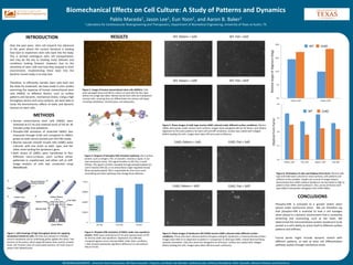
Figure
1.
CAD
drawings
of
high
throughput
device
for
applying
mechanical
stretch
to
cells.
(A)
Cells
are
cultured
on
a
flexible
silicone
membrane
and
an
underlying
piston
applies
the
stress.
Two
versions
of
the
piston,
which
apply
(B)
biaxial
strain
and
(C)
uniaxial
strain.
(D)
Trimetric
view
of
constructed
machine.
(E)
Front
view
of
system
with
labeled
parts.
A
B
C
D
Figure
3.
Diagram
of
phospho-‐ERK
activation
pathway.
(A)
A
surface
protein,
such
as
integrin,
FGF,
or
Caveolin,
receives
a
signal,
in
our
case
mechanical
stress.
This
signal
transfers
to
(B),
Ras,
a
small
GTPase.
This
signal
is
further
cascaded
through
phosphorylation
(C)
until
it
reaches
ERK
(D),
or
an
extracellular
single-‐regulated
kinase.
When
phosphorylated,
ERK
is
responsible
for
short-‐term
actin
remodeling
and
other
pathways
that
change
focal
adhesion.
Figure
4.
Phospho-‐ERK
activation
of
hMSC
under
sine
waveform
stretch.
hMSC
were
stretched
at
0.5
Hz
and
maximal
strain
of
5%
for
30
min
under
sine
waveform.
Expression
of
p-‐ERK
was
compared
against
serum-‐starved
hMSC
under
static
conditions.
t–Test
showed
statistically
significant
difference
to
cells
without
FBS
at
P<0.05.
Figure
5.
Phase
images
of
wild–type
murine
vSMC
cultured
under
different
surface
conditions.
Murine
vSMCs
were
grown
under
various
nano-‐surfaces.
Images
show
elongated
cells
for
all
factors,
and
relative
alignment
to
the
nano–patterns
for
both
soft
and
stiff
conditions.
Surface
was
coated
with
collagen
before
seeding
the
cells.
Images
were
taken
48
hours
post-‐confluency.
Figure
6.
Phase
images
of
Syndecan1-‐KO
(S1KO)
murine
vSMC
cultured
under
different
surface
conditions.
These
cells
were
cultured
without
the
gene
coding
for
Syndecan1,
a
transmembrane
protein.
Images
show
little
to
no
alignment
to
pattern
in
comparison
to
wild-‐type
vSMC,
instead
demonstrating
sporadic
orientation.
Cells
also
show
less
elongation
on
all
factors.
Surface
was
coated
with
collagen
before
seeding
the
cells.
Images
were
taken
48
hours
post-‐confluency.
Figure
8.
Orientation
of
cells
and
elliptical
form
factor.
Murine
wild-‐
type
and
S1KO
were
cultured
on
nano-‐surfaces,
with
patterns
and
stiffness
as
the
variables.
Graphs
are
a
result
of
image
analysis,
demonstrate
that
vSMCs
without
Syndecan1
are
less
likely
to
align
to
patterns
than
vSMCs
with
Syndecan1.
Also,
across
all
factors
wild-‐
type
vSMCs
have
greater
elongation
than
S1KO
vSMCs.
ACKNOWLEDGEMENTS
:
American
Heart
Association,
NIH
New
Innovator
Program,
and
Baker
Lab
Member:
Subhamoy
Das,
Anthony
Monteforte,
Peter
Voyvodic,
Adrianne
Shearer,
and
Victoria
Le
CONCLUSIONS
Phospho–ERK
is
activated
to
a
greater
extent
when
placed
under
mechanical
stress.
We
can
therefore
say
that
phospho–ERK
is
essential
to
how
a
cell
manages
when
placed
in
a
dynamic
environment
that
is
constantly
stretching
and
contracting,
such
as
the
heart.
We
determined
the
transmembrane
protein
Syndecan1
to
be
pivotal
in
a
cell’s
ability
to
orient
itself
to
different
surface
patterns
and
stiffness.
Future
works
might
include
dynamic
stretch
with
different
patterns,
as
well
as
stem
cell
differentiation
pathway
studies
through
mechanical
strain.
Figure
2.
Image
of
human
mesenchymal
stem
cells
(hMSCs).
Cells
were
passaged
twice
and
left
to
culture
on
same
dish
for
four
days
before
this
image
was
taken.
Mesenchymal
stem
cells
are
multipotent
stromal
cells,
meaning
they
can
differentiate
into
various
cell
types,
including
osteoblasts,
chondrocytes,
and
adipocytes.