This document describes an experiment to develop a method for selecting aptamers against protein targets generated through in vitro transcription and translation of genes. Specifically, they attempt to select aptamers against the human U1A protein, a component of the nuclear spliceosome, where the U1A protein was produced through in vitro transcription and translation of its gene, and was also biotinylated to allow for immobilization during aptamer selection. The results showed that the selected aptamer sequences closely mimicked the natural RNA binding sequences and structures of U1A, demonstrating the potential of this method for high-throughput aptamer generation against proteomes.


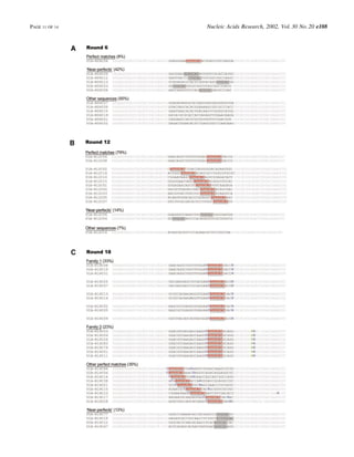
![Eighteen rounds of selection were performed against
biotinylated U1A. In the ®rst 12 rounds of selection 20 cycles
of PCR were carried out, while in the last six rounds this
number was decreased to 16 cycles to prevent overampli®ca-
tion of the selected pool; these parameters had been empiric-
ally determined during previous automated selection
experiments (6). In the ®rst 12 rounds of selection the wash
buffer was 1Q SBB. In the last six rounds of selection the
stringency of the selection was increased by progressively
increasing the monovalent salt concentration, as detailed in
Table 2.
The progress of selection was monitored every six rounds
(6, 12 and 18) by placing [a-32P]-radiolabeled ribo- and
deoxyribonucleotides in the ampli®cation reactions and
resolving ampli®cation products by gel electrophoresis. The
automated protocol included a provision to archive aliquots of
the reverse transcription (10 ml, 10% of the total reaction) and
in vitro transcription (10 ml, 10% of the total reaction)
reactions. After the automated protocol had run its course, the
aliquots (in standard stop dye) were run on 8% acrylamide±
7 M urea (19:1 acrylamide:bisacrylamide) denaturing gels.
Decade RNA Markers (Ambion) were used as size standards.
The gels were visualized using a PhosphorImager SI
(Amersham Pharmacia Biotech). In addition, single point
binding assays were carried out with equimolar concentrations
of protein and RNA samples (50 nM), as described below (see
also Fig. 5).
High throughput sequencing
Aliquots (1 ml of a 50 ml archive) of RT±PCR reactions from a
given round of selection were further ampli®ed and then
ligated into a thymidine-overhang vector (TA Cloning Kit;
Invitrogen). Templates for sequencing reactions were gener-
ated from individual colonies via 50 ml colony PCR reactions
(22) with standard M13 sequencing primers ¯anking the
insertion site of the thymidine-overhang vector (primers M13
forward and M13 reverse). The PCR products were puri®ed
away from primers, salt and enzyme using a MultiScreen96-
PCR clean-up plate (Millipore). Aliquots of the colony PCR
reactions (5 ml) were developed on a 4% agarose gel to verify
the insertion of aptamers into vectors.
Cycle sequencing reactions were performed using a CEQ
DTCS Quick Start Kit (Beckman-Coulter) and the vendor's
modi®ed M13 sequencing primer (primer ±47 seq). Reaction
assembly and cycling conditions were performed largely as
described in the vendor's instructions. Approximately
100 fmol of puri®ed colony PCR products were used as
templates and reactions were performed with half of the
master mix concentration recommended in the instructions in
order to minimize reagent use (4 ml of master mix in a 20 ml
sequencing reaction, rather than 8 ml). Unincorporated dye
was removed by size exclusion chromatography, as described
in Beckman-Coulter Technical Application Information
Bulletin T-1874A (http://www.beckman.com/Literature/
BioResearch/T-1874A.pdf). Brie¯y, 45 ml of dry Sephadex
G50 (Amersham-Pharmacia Biotech) was placed into each
well of a MultiScreen HV plate using a MultiScreen 45 ml
Column Leader (Millipore). The Sephadex chromatography
resin was allowed to hydrate in 300 ml of water for 3 h at room
temperature. After incubation, the resin was packed by
centrifugation for 5 min at 1100 g. The columns were rinsed
once with 150 ml of water and packed again at the same speed
for the same time. The 20 ml sequencing reactions were loaded
onto the tops of the columns using a multichannel micro-
pipettor and spun again for 5 min at 1100 g. Puri®ed samples
were recovered from a CEQ Sample microplate (Beckman-
Coulter) that had been placed below the chromatography
plates before centrifugation. Recovered samples were dried
under vacuum at room temperature. Pellets were resuspended
in 40 ml of deionized formamide and developed on a CEQ
2000XL eight channel capillary DNA sequencer (Beckman-
Coulter). Aptamer secondary structures were predicted using
RNAstructure 3.6 by Mathews et al. (23).
Cellular expression of U1A
The plasmid pJH-hisU1A was transformed into BL21 cells
(Stratagene). A 1 ml starter culture grown from a single
plasmid was used to inoculate 50 ml of fresh LB. The culture
was grown to an OD600 of ~0.6 at 37°C and protein expression
was induced by the addition of IPTG to a ®nal concentration of
840 mM. The induced culture was grown for an additional 3 h
at 37°C. Cells were pelleted at 5000 g and lysed in B-PER
(Pierce), 10 U DNase (Invitrogen) and 10 mM MgCl2 in a total
volume of 5 ml. After incubation at room temperature for
15 min, cellular debris was pelleted at 13 000 g. The
supernatant was further puri®ed by nickel-chelation chroma-
tography. IMAC resin (2 ml; Amersham-Pharmacia Biotech)
was equilibrated with 4 ml of water, followed by 4 ml of
charging buffer (50 mM NiCl2). The column was equilibrated
with 10 ml of protein binding buffer (PBB; 1Q PBB = 50 mM
Tris pH 7.5, 100 mM NaCl, 5 mM imidazole). Clari®ed
supernatant (~15 ml) was loaded and the column was washed
with 4 ml of 1Q PBB and 15 ml of wash buffer (50 mM Tris
pH 7.5, 100 mM NaCl, 50 mM imidazole). The U1A protein
was eluted by the addition of 3 ml of elution buffer (50 mM
Tris pH 7.5, 100 mM NaCl, 500 mM imidazole); 500 ml
fractions were collected for gel analysis. Fractions containing
signi®cant amounts of U1A were pooled and dialyzed in
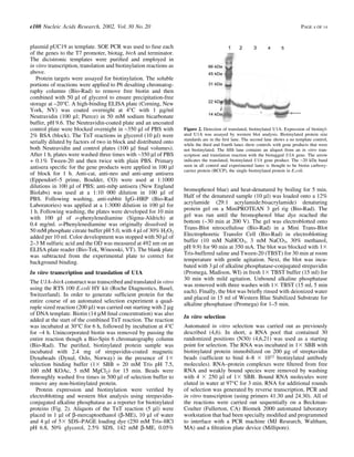
Table 2. Conditions for the
automated selection of anti-U1A
aptamers
See Materials and Methods for details.
The binding ability of the pool was
assayed at 0, 6, 12 and 18 rounds.
PAGE 5 OF 14 Nucleic Acids Research, 2002, Vol. 30 No. 20 e108](https://image.slidesharecdn.com/a788c7db-8662-4c28-8ca7-6d3688384d1c-160130181227/85/Cox2002-Automated_selection_of_aptamers_against_protein_targets_translated_in_vitro_from_gene_to_aptamer-5-320.jpg)
![50 mM Tris pH 7.5, 100 mM NaCl with four buffer exchanges
of 500 ml every 3 h at 4°C in order to remove imidazole from
the preparation. Finally, the absorbance at 280 nm was used to
determine protein concentration (extinction coef®cient =
5442 M±1 cm±1).
Binding constants
Plasmids containing individual aptamers were used to gener-
ate transcription templates via PCR. Transcription reactions
were carried out with an AmpliScribe T7 RNA transcription
kit (Epicentre, Madison, WI) according to the manufacturer's
instructions, except that incubation was at 42°C rather than
37°C. Aptamers were puri®ed on denaturing polyacrylamide
gels (24), dephosphorylated and radiolabeled with [g-32P]ATP
(25). Radiolabeled RNA was extracted with phenol:chloro-
form (1:1) and unincorporated nucleotides were removed
using size exclusion spin columns (Princeton Separations,
Adelphia, NJ).
Nitrocellulose ®lter binding assays were employed to
determine the dissociation constants of aptamer±protein
complexes (25). A standard protocol was automated using
the Biomek 2000 automated laboratory workstation and a
modi®ed Minifold I ®ltration manifold (Schleicher & Schuell,
Keene, NH). RNA samples in 1Q SSB were thermally
equilibrated at 25°C for 30 min. The RNA concentration for
binding reactions was kept constant at a ®nal concentration of
200 pM while the concentration of U1A ranged from 1 mM
down to 17 pM (11 different concentrations). Equal volumes
(60 ml) of RNA and U1A were incubated together for 30 min at
room temperature in 1Q SBB. The binding reactions (100 ml)
were ®ltered through a sandwich of Protran pure nitrocellulose
(Schleicher & Schuell) and Hybond N+ nylon transfer
membrane (Amersham Pharmacia Biotech) that had been
assembled in the modi®ed Minifold I vacuum manifold. The
®lters were washed three times with 125 ml of 1Q SBB. The
amount of radiolabeled RNA captured from a reaction onto the
nitrocellulose membrane was quanititated using a Phosphor-
Imager SI (Amersham Pharmacia Biotech). The log of U1A
concentration was plotted against the amount of RNA bound.
Data were ®tted to the equation y = (a´b)/(b + x) + C, where C
is the fraction of RNA bound to the nitrocellulose at zero
protein concentration, b is the maximum fraction bound, x is
the fraction of RNA bound to U1A and a is the dissociation
constant for the RNA±protein complex. Assays were per-
formed in triplicate and standard deviations were calculated.
The 21 nt synthetic RNA (AAUCCAUUGCACUCCGGA-
UUU) previously employed in structural studies of the U1A
protein was used as a positive control (12).
RESULTS AND DISCUSSION
From gene to biotinylated protein
While we have previously automated the in vitro selection of
aptamers that target proteins (4±6), our methods are currently
limited to puri®ed proteins. Ultimately, the need to purify
protein targets individually would drastically reduce the speed
with which aptamers could be generated against proteomes
and would therefore reduce the utility of aptamers as reagents
for proteome analysis. Therefore, we have sought to increase
selection throughput by generating protein targets via in vitro
transcription and translation. Moreover, in order to manipulate
protein targets during automated selection, we have attempted
to introduce a biotin tag during the in vitro synthesis
procedure.
A number of kits were available for the in vitro transcription
and translation of genes. Ultimately, we found that the Roche
RTS 100 E.coli HY kit worked well with the automated
selection procedures we had previously established. In order to
biotinylate translated proteins, templates were modi®ed
(Fig. 1) so that translated proteins would contain an N-ter-
minal peptide tag (MAGGLNDIFEAQKIEWHEDTGGSS)
that was an ef®cient substrate for BPL, the product of the birA
gene (26±28). The e-amino group of the single lysine residue
in the BPL recognition peptide becomes covalently linked to
biotin (29). Biotin ligase can either be co-translated or added
as a separate reagent.
Existing BPL present in E.coli S30 extracts proved
insuf®cient to ef®ciently biotinylate target proteins.
Therefore, the MBP gene (malE) was expressed in tandem
with a downstream birA cistron. Following gel separation, an
anti-MBP antibody was used to con®rm that roughly equal
amounts of MBP were synthesized. However, when
avidin±HRP was used as a probe it was apparent that only
the dicistronic mbp±birA template directed ef®cient biotinyl-
ation. The band at ~20 kDa that stains intensely with
avidin±HRP is E.coli biotin carboxyl-carrier protein
(BCCP), which is present in the S30 extracts used for in vitro
translation (Fig. 2). MBP and three other biotinylated protein
targets were immobilized in Neutravidin-coated ELISA wells
and detected with cognate sera (Fig. 3). These results indicated
that in vitro translated and biotinylated proteins could fold into
native structures that present epitopes similar to those found
in vivo. The amounts of in vitro translation and biotinylation
reactions necessary to saturate microwells for selection
experiments proved to be relatively small (1% of reaction
volume), indicating that this procedure should be useful for
multiplex formats, such as the acquisition of aptamers against
multiple proteins in an organismal proteome.
We have previously carried out selections against chemical-
ly biotinylated target proteins (4,6). Unfortunately, chemical
biotinylation generally generates a population of molecules
with differing amounts of biotinylation at different conjuga-
tion sites, typically a-amino groups on a protein. Thus,
multiple different epitopes are presented during selection. A
biotinylation tag obviates this problem and a relatively
homogeneous set of epitopes should be present during the
selection. Additionally, chemical biotinylation may block an
active or allosteric site of the protein, while speci®c
biotinylation at the N-terminus is less likely to interfere with
function.
In vitro selection of aptamers that bind to translated,
biotinylated U1A
As in our previous automated selection experiments, biotinyl-
ated protein targets are loaded onto streptavidin beads and
incubated with RNA libraries. Bound RNA molecules are
sieved from unbound by ®ltration; the use of beads facilitates
robotic manipulation. The beads are directly transferred to a
thermal cycler and RNA is prepared for the next round of
selection by a combination of reverse transcription, PCR and
in vitro transcription. The entire selection procedure can be
e108 Nucleic Acids Research, 2002, Vol. 30 No. 20 PAGE 6 OF 14](https://image.slidesharecdn.com/a788c7db-8662-4c28-8ca7-6d3688384d1c-160130181227/85/Cox2002-Automated_selection_of_aptamers_against_protein_targets_translated_in_vitro_from_gene_to_aptamer-6-320.jpg)


![Pells et al [2015] PLoS ONE 10[7] e0131102](https://cdn.slidesharecdn.com/ss_thumbnails/f79bb09e-8eb1-41e7-8042-28c0aa4a48c6-150720142907-lva1-app6891-thumbnail.jpg?width=640&height=640&fit=bounds)


