
Recommended
More Related Content
What's hot
What's hot (20)
Similar to Tongue
Similar to Tongue (20)
More from Chitransha03
More from Chitransha03 (16)
Recently uploaded
Recently uploaded (20)
Tongue
- 1. TONGUE
- 2. The tongue is a mobile muscular organ situated in the floor of the mouth.
- 6. Root Root of Tongue Attached to – Above - Styloid Process & Soft Palate Below – Mandible & Hyoid bone
- 7. TIP Anterior free end, lies behind upper incisors.
- 8. Body Dorsum Oral Part Pharyngeal Part Posterior most Part Inferior Surface
- 10. BODY The bulk of tongue between the root and tip is called body. It has dorsal and ventral surfaces and right and left lateral margins.
- 11. Dorsal surface It is divided by a V-shaped sulcus, the sulcus terminalis into two parts, viz. 1. Anterior two-third or oral part. 2. Posterior one-third or pharyngeal part.
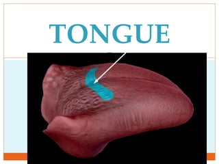
- 13. The apex of the sulcus terminalis is marked by a blind foramen, FORAMEN CAECUM, which indicates the point of origin of the thyroglossal duct in IUL.
- 14. The oral part presents the following features: 1. A Median Furrow, representing the bilateral origin of the tongue. 2. Large number of Papillae.
- 15. The pharyngeal part presents the following features: 1. A large number of lymphoid follicles, which together constitute the lingual tonsil. 2. A large number of mucus and serous glands.
- 16. Papillae Of The Tongue These are projections of mucuos membrane which gives the anterior 2/3rds of the tongue its characteristic roughness.
- 18. Vallate Papillae Largest papillae Size- 1-2 mm in diameter. 8 to 12 in number. Arrange in V-shaped row in front of sulcus terminalis
- 19. Filiform papillae Smallest & Devoid of taste buds. Conical projections with sharply pointed tips. Filiform papillae are located abundantly on the dorsum of tongue and responsible for its velvety appearance.
- 20. Fungiform papillae These are mushroom shaped, numerous near tip & margins of tongue. They are visible as discrete red pinheads.
- 21. Foliate Papilla Vertical grooves & ridges on lateral border in front of sulcus terminalis. Rudimentry in humans.
- 22. Pharyngeal Part Posterior 1/3rd Lies behind the palatoglossal arches.
- 23. Devoid of papillae but appears rough due to the presence of numerous lymphatic follicles in the underlying submucosa, that collectively termed lingual tonsil.
- 24. Inferior surface of the tongue 1. Frenulum linguae: a median-fold of mucus membrane connecting the tongue to the floor of the mouth.
- 26. 2. Deep lingual veins: on either side of frenulum linguae (lingual nerve & lingual artery are medial to the vein but not visible). 3. Plica fimbriata: a fimbriated fold of mucous membrane lateral to the lingual vein.
- 27. Muscles of the Tongue ExtrinsicIntrinsic
- 28. INTRINSIC MUSCLES 4 paired intrinsic muscles originate and insert within the tongue. No attachment outside the tongue. These muscles alter the shape of the tongue.
- 30. INTRINSIC MUSCLES Superior longitudinal Inferior longitudinal Transverse Vertical
- 31. Origin: submucous fibrous layer & median fibrous septum Insertion: mucous membrane.
- 32. superior longitudinal beneath the mucous membrane • shortens the tongue • makes the dorsum concave inferior longitudinal close to inferior surface between genioglossus and hyoglossus • shortens the tongue • makes the dorsum convex transverse extends from median septum to the margin makes the tongue narrow and elongated vertical at the border of the anterior part of the tongue makes the tongue broad and flattened
- 33. Movements Intrinsic Muscles Superior Longitudinal : shortens the tongue, makes the dorsum concave Inferior longitudinal : shortens the tongue, makes the dorsum convex Transverse : makes the tongue narrow and elongated Vertical : makes the tongue broad and flattened
- 34. Movements Extrinsic Muscles Genioglossus : Protrusion Styloglossus: Retraction Hyoglossus: Depression Palatoglossus: Elevation
- 35. EXTRINSIC MUSCLES Genioglossus: superior genile tubercle Hyoglossus: greater cornu of hyoid bone Styloglossus: styloid process Palatoglossus: palatine aponeurosis
- 36. Genioglossus superior genile tubercle whole of the tongue & hyoid bone. •Protrudes the tongue when acting together with its counterpart of opposite side Hyoglossus greater cornu of hyoid bone & adjecent part of hyoid bone Side of tongue •Depresses the sides of the tongue •Makes the dorsal surface convex Styloglossus Tip of styloid process Side of tongue •Draws the side of the tongue upwards and backwards Palatoglossus Oral surface of palatine aponeurosis Side of tongue •Pulls up the root of the tongue •Approximates palatoglossal arches
- 38. VASCULAR SUPPLY
- 39. ARTERIAL SUPPLY Lingual artery: divides into : Dorsal lingual artery Deep lingual artery Sublingual artery Tonsillar branch of the facial artery. Ascending pharyngeal artery.
- 41. VENOUS DRAINAGE Deep lingual vein is the principal vein Venae comitantes accompanying the lingual artery. Venae comitantes accompanying the hypoglossal nerve. All these veins terminate into IJV.
- 42. LYMPHATIC DRAINAGE Tip - drain to submental nodes. The right and left halves of the anterior 2/3rds drain in submandibular nodes. Posterior part & posterior most part- drains to deep cervical nodes. Whole lymph finally drain into jugulo-omohyoid LN.
- 44. Development of tongue Development begins at the 4th week of IUL. The tongue develops in relation to the1,2,3,4 pharyngeal arches in the floor of the developing mouth.
- 46. At the end of the 4th week of IUL, a small median triangular swelling called Tuberculum Impar develops in the floor of primitive pharynx, just cranial to Foramen Cecum. Soon after this, two lateral oval swellings called Lingual Swellings develop on each side of tuberculum impar.
- 49. Caudal to tubercular impar, another large median swelling called hypobranchial eminence (copula of His) develops in the floor of primitive pharynx in relation to 3,4 pharyngeal arches.
- 50. The hypobranchial eminence soon subdivides into large cranial part and small caudal part.
- 51. Posterior 1/3 of the tongue: The posterior one-third of the tongue including circumvallate papillae develops from cranial part of hypobranchial eminence.
- 52. The posterior most part of the tongue and epiglottis develop from the caudal part of the hypobranchial eminence.
- 53. Development of Musculature of tongue: Derived from occipital myotomes Nerve supply is by hypoglossal nerve.
- 54. NERVE SUPPLY Motor supply: All the muscles of tongue are supplied by the Hypoglossal Nerve except palatoglossus which is supplied by cranial root of accessory via pharyngeal plexus.
- 55. Sensory supply Anterior 2/3rd except circumvallate papillae Posterior 1/3rd & circumvallate papillae Posteri- or most part General sensations Lingual nerve Glossopha -ryngeal nerve Vagus nerve through internal laryngeal branch Special sensations Chorda tympani Glossoph- ryngeal Vagus nerve
- 56. APPLIED ANATOMY Aglossia: absence of tongue. Hemiglossia: half tongue:- It occurs if one of the lingual swelling fails to develop. Microglossia: Tongue is too small. Macroglossia: Tongue is too large.
- 57. Tongue tie (ankyloglossia): when frenulum of tongue extends to the tip of the tongue, thus preventing its protrusion and causing difficulty in speech. Bifid tongue: In this condition, the anterior portion of the tongue splits into two parts.