
Recommended
More Related Content
What's hot
What's hot (20)
Similar to Changes in the cardiovascular system during pregnancy
Similar to Changes in the cardiovascular system during pregnancy (20)
More from ChinjuJoseSajith
More from ChinjuJoseSajith (20)
Recently uploaded
Recently uploaded (20)
Changes in the cardiovascular system during pregnancy
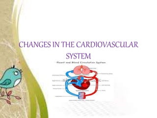
- 1. CHANGES IN THE CARDIOVASCULAR SYSTEM
- 2. CHANGES IN THE CARDIOVASCULAR SYSTEM For the care of women with normal pregnancies as well as for the management of women with pre-existing cardiovascular diseases, an understanding of these changes is important.
- 3. THE HEART • The heart muscle, particularly the left ventricle, hypertrophies leading to enlargement of the heart. • The growing uterus pushes the heart upwards and to the left During pregnancy the heart rate and stroke volume (the amount of blood pumped by the heart with each beat) increases. • This is due to the increased blood volume and increased oxygen requirements of the maternal tissues and the growing fetus.
- 4. • The cardiac output increases markedly by the end of the first trimester. • In the third trimester, a rise, fall or no change at all has been shown to occur, depending on individual variations. • Although the cardiac output is increased in pregnancy, the blood pressure does not rise because of the reduction in peripheral resistance.
- 5. • The capacity of veins and venules increases The increased production of vasodilator prostaglandin also contributes to this. • During the mid-trimester, changes in blood pressure may occur causing fainting. In later pregnancy, hypotension may occur in 10 per- cent of women in unsupported supine position. This is termed as the supine hypotensive syndrome.
- 6. • The pressure of the gravid uterus compresses the vena cava, reducing the venous return. Cardiac output is reduced by 25-30 percent and the blood pressure may fall by 10-15 percent, which produces the feelings of dizziness, nausea and even fainting.
- 7. • Poor venous return in late pregnancy along with increased distensibility and pressure in the veins of the legs, vulva, rectum and pelvis can lead to edema in the lower leg, varicose veins and hemorrhoids. • There is increased blood flow to the breasts throughout pregnancy and dilated veins may be seen on the surface of the breasts along with enlargement and tingling from early pregnancy
- 8. • Blood flow increases to the uterus, kidneys, breasts and skin during pregnancy. Much of the cardiac output goes to the utero- placental circulation, which is about 750 ml per minute .
- 9. THE BLOOD VOLUME • The increase in blood volume in pregnancy varies according to the size of the woman, the number of pregnancies she has had, her parity and whether the pregnancy is singleton or multiple • The increase begins at about tenth week gestation and progresses up to 30th-34th week of gestation • The increase may be as much as 100 percent in some women.
- 10. A higher circulating volume is required for the following functions: To provide extra blood flow for placental circulation. To supply the extra metabolic needs of the fetus To provide extra perfusion of kidneys and other organs To counterbalance the effects of increased arterial and venous capacity To compensate for blood loss at delivery
- 11. Plasma Protein During the first 20 weeks of pregnancy, the plasma protein concentration reduces as a result of the increased plasma volume. This leads to lowered osmotic pressure, contributing to edema of the lower limbs seen in late pregnancy . In the absence of disease, moderate edema is seen as physiological
- 12. IRON METABOLISM • Iron requirement increases significantly in the last trimester, during which time iron absorp- tion from the gut is enhanced. • The purpose of iron supplementation in pregnancy is to prevent iron deficiency in the mother, not to raise the hemoglobin.
- 13. CLOTTING FACTORS • The clotting and fibrinolytic systems undergo major changes during pregnancy. Plasma fibrinogen (factor 1) increases from the third month of pregnancy progressively until term. Prothrombin (factor 2) increases only slightly. Factors 7, 8, 9 and 10 increase leading to change in coagulation time from 12-8 minutes
- 14. • The capacity for clotting is thus increased, in preparation for the prevention of hemorrhage at placental separation. However, there is a higher risk of thrombosis, embolism.
- 15. White Blood Cells • The neutrophils increase in pregnancy, which enhances the blood's phagocytic and bactericidal properties. • In the second and third trimester the action of the polymorphonuclear leukocytes may be depressed, perhaps accounting for the increased susceptibility of pregnant women to infection. • The lymphocyte and monocyte numbers remain the same through- out pregnancy.
- 16. IMMUNITY • Immune response is reduced in pregnancy. • Levels of immunoglobulins IgA, IgG and IgM decrease steadily from the 10th week to the 30th week and then remain at these levels until term resulting in reduced immune response. • Antibody titers against viruses, such as measles, influenza A and herpes simplex, are reduced in proportion to the hemodilution effect, therefore viral resistance is unchanged