1. Essential idea: Sexual reproduction involves the
development and fusion of haploid gametes.
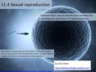
11.4 Sexual reproduction
By Chris Paine
https://bioknowledgy.weebly.com/
Both sperm and egg cells are the product of reduction division
and it is the process of fertilisation that creates a unique diploid
(zygote) cell which can develop into a new organism.
http://blogs.discovermagazine.com/science-sushi/files/2015/06/sperm_egg.jpg
2. Understandings
Statement Guidance
11.4.U1 Spermatogenesis and oogenesis both involve mitosis,
cell growth, two divisions of meiosis and differentiation.
11.4.U2 Processes in spermatogenesis and oogenesis result in
different numbers of gametes with different amounts of
cytoplasm.
11.4.U3 Fertilization in animals can be internal or external.
11.4.U4 Fertilization involves mechanisms that prevent
polyspermy.
Fertilization involves the acrosome
reaction, fusion of the plasma
membrane of the egg and sperm and
the cortical reaction.
11.4.U5 Implantation of the blastocyst in the endometrium is
essential for the continuation of pregnancy.
11.4.U6 HCG stimulates the ovary to secrete progesterone
during early pregnancy.
11.4.U7 The placenta facilitates the exchange of materials
between the mother and fetus.
11.4.U8 Estrogen and progesterone are secreted by the
placenta once it has formed.
11.4.U9 Birth is mediated by positive feedback involving
estrogen and oxytocin.
3. Applications and Skills
Statement Guidance
11.4.A1 The average 38-week pregnancy in humans
can be positioned on a graph showing the
correlation between animal size and the
development of the young at birth for other
mammals.
11.4.S1 Annotation of diagrams of seminiferous tubule
and ovary to show the stages of
gametogenesis.
11.4.S2 Annotation of diagrams of mature sperm and
egg to indicate functions.
5. 11.4.S2 Annotation of diagrams of mature sperm and egg to indicate functions.
Structure of the mature egg
Haploid (n) contains 23 chromosomes to
be passed from mother to child
Consists of a glycoprotein that protects
the egg and prevents the entry of sperm.
Not required – will break down
Contains nutrients to support the early
development of fertilised egg
Makes the zona pellucida impenetrable to
sperm (after fertilisation) to prevent
polyspermy*
Provides nutrients to support the early
development of fertilised egg
Diagram from: http://www.slideshare.net/gurustip/reproduction-ahl-1062218
Cortical
granules
Can you match the annotations to the labels?
6. 11.4.S2 Annotation of diagrams of mature sperm and egg to indicate functions.
Structure of the mature egg
Haploid (n) contains 23 chromosomes to
be passed from mother to child
Consists of a glycoprotein that protects
the egg and prevents the entry of sperm.
Not required – will break down
Contains nutrients to support the early
development of fertilised egg
Makes the zona pellucida impenetrable to
sperm (after fertilisation) to prevent
polyspermy*
Provides nutrients to support the early
development of fertilised egg
Diagram from: http://www.slideshare.net/gurustip/reproduction-ahl-1062218
Cortical
granules
7. 11.4.S2 Annotation of diagrams of mature sperm and egg to indicate functions.
Structure of the mature sperm
Haploid (n), contains 23 chromosomes to be passed
from father to child
Contains enzymes which can digest the zona pellucida
Possesses helical mitochondria which provide the ATP
(energy) for swimming (and other processes)
Contains protein fibres and microtubules to strengthen
and allow the tail to move respectively.
Can you match the annotations to the labels?
8. 11.4.S2 Annotation of diagrams of mature sperm and egg to indicate functions.
Structure of the mature sperm
Haploid (n), contains 23 chromosomes to be passed
from father to child
Contains enzymes which can digest the zona pellucida
Possesses helical mitochondria which provide the ATP
(energy) for swimming (and other processes)
Contains protein fibres and microtubules to strengthen
and allow the tail to move respectively.
9. Nature of science: Assessing risks and benefits associated with scientific research—the risks to human male fertility were
not adequately assessed before steroids related to progesterone and estrogen were released into the environment as a
result of the use of the female contraceptive pill. (4.8)
http://www.theguardian.com/envi
ronment/2012/jun/02/water-
system-toxic-contraceptive-pill
https://www.arhp.org/publications-and-
resources/contraception-journal/august-2011
https://www.sciencedaily.com/releases/2010/12/101208125813.htm
Assessing risks and benefits associated with scientific research: pollution
from the female contraceptive pill
Use these and other links. How serious is the pollution
threat from the female contraceptive pill?
Out for the count: Why levels of sperm
in men are falling http://www.independent.co.uk/news/science/out-for-the-
count-why-levels-of-sperm-in-men-are-falling-1954149.html
Don't blame the pill
for estrogen in drinking water
£30bn bill to purify
water system after toxic impact of
contraceptive pill
Birth Control Hormones In Water:
Separating Myth From Fact
10. 11.4.U1 Spermatogenesis and oogenesis both involve mitosis, cell growth, two divisions of meiosis and differentiation. AND 11.4.U2
Processes in spermatogenesis and oogenesis result in different numbers of gametes with different amounts of cytoplasm.
11. 11.4.S1 Annotation of diagrams of seminiferous tubule and ovary to show the stages of gametogenesis.
divide to produce spermatocytes
spermatogonia
12. 11.4.S1 Annotation of diagrams of seminiferous tubule and ovary to show the stages of gametogenesis.
divide to produce spermatocytes
spermatogonia
13. 11.4.U1 Spermatogenesis and oogenesis both involve mitosis, cell growth, two divisions of meiosis and differentiation. AND 11.4.U2
Processes in spermatogenesis and oogenesis result in different numbers of gametes with different amounts of cytoplasm.
http://highered.mheducation.com/sites/007249
5855/student_view0/chapter28/animation__spe
rmatogenesis__quiz_1_.html
http://www.cengage.com/biology/discipline_content/ani
mations/spermatogenesis.html
14. 11.4.U1 Spermatogenesis and oogenesis both involve mitosis, cell growth, two divisions of meiosis and differentiation. AND 11.4.U2
Processes in spermatogenesis and oogenesis result in different numbers of gametes with different amounts of cytoplasm.
Roles of hormones are not required, but it is interesting to note that FSH and LH have roles
in males as well as females
15. 11.4.U1 Spermatogenesis and oogenesis both involve mitosis, cell growth, two divisions of meiosis and differentiation. AND 11.4.U2
Processes in spermatogenesis and oogenesis result in different numbers of gametes with different amounts of cytoplasm.
contains the primary oocyte surrounded by a single layer of supporting
follicle cells
contains the secondary oocyte, ready for ovulation
outer layer of cells in the ovary
16. 11.4.S1 Annotation of diagrams of seminiferous tubule and ovary to show the stages of gametogenesis.
17. 11.4.S1 Annotation of diagrams of seminiferous tubule and ovary to show the stages of gametogenesis.
contains the primary oocyte surrounded by a single layer of supporting
follicle cells
contains the secondary oocyte, ready for ovulation
outer layer of cells in the ovary
18. 11.4.U1 Spermatogenesis and oogenesis both involve mitosis, cell growth, two divisions of meiosis and differentiation. AND 11.4.U2
Processes in spermatogenesis and oogenesis result in different numbers of gametes with different amounts of cytoplasm.
https://youtu.be/2-VKgdhfNpY
Human ovulation captured on film:
19. 11.4.U1 Spermatogenesis and oogenesis both involve mitosis, cell growth, two divisions of meiosis and differentiation. AND 11.4.U2
Processes in spermatogenesis and oogenesis result in different numbers of gametes with different amounts of cytoplasm.
Image edited from: http://www.ib.bioninja.com.au/_Media/oogenesis_med.jpeg
Oogenesis production of ova (female gametes)
during fetal development large
numbers of oogonia are formed by
mitosis.
oogonia enlarge (growth)
and undergo meiosis, but
stop in prophase I (until
puberty). They are now
termed primary oocytes
and are held in primary
follicles.
(at puberty) some follicles develop each month in response to
FSH:
• the oocyte completes the first meiotic division
• Division of the cytoplasm is unequal creating a polar body
• the secondary oocyte continues into meiosis II and halts
at prophase II
polar bodies eventually degenerate
Secondary oocytes develop along with the follicle. When the follicle is mature it
rupture to release the secondary oocyte with a small number of cells (the mature
egg) into the fallopian tube. The remaining follicle cells remain in the ovary to
form the corpus luteum (which secretes progesterone).
The oocyte completes meiosis II
(forming the ovum) if the cell is
fertilized and another polar body
1
2
4
3b
5
3a
20. 11.4.U1 Spermatogenesis and oogenesis both involve mitosis, cell growth, two divisions of meiosis and differentiation. AND 11.4.U2
Processes in spermatogenesis and oogenesis result in different numbers of gametes with different amounts of cytoplasm.
Oogenesis resources:
http://highered.mheducation.com/sites/0072495
855/student_view0/chapter28/animation__matur
ation_of_the_follicle_and_oocyte.html
http://highered.mheducation.com/olcweb/cgi/pluginpop.cgi?it=swf::6
40::480::/sites/dl/free/0072495855/63089/28_02_1.swf::Structure%2
0of%20the%20Ovary%20and%20the%20Developmental%20Sequence
%20of%20the%20Ovarian%20Follicles
http://www.wiley.com/college/jenkins/0470227583/ani
mations/index_25_03_01.html
21. 11.4.U1 Spermatogenesis and oogenesis both involve mitosis, cell growth, two divisions of meiosis and differentiation. AND 11.4.U2
Processes in spermatogenesis and oogenesis result in different numbers of gametes with different amounts of cytoplasm.
Compare and contrast the processes of spermatogenesis and oogenesis (8 marks)
22. 11.4.U1 Spermatogenesis and oogenesis both involve mitosis, cell growth, two divisions of meiosis and differentiation. AND 11.4.U2
Processes in spermatogenesis and oogenesis result in different numbers of gametes with different amounts of cytoplasm.
Compare and contrast the processes of spermatogenesis and oogenesis
Oogenesis Spermatogenesis
Cell division Begin with mitosis and later on involve meiosis
Growth Involve cell enlargement before meiosis
Product Haploid cells (gametes)
Differentiation Produce specialised gametes
Location Eggs/ova produced in the ovaries Sperm produced in the testes
Initiated During development of fetus During puberty
Pauses During prophase I and between
prophase II and metaphase II
None
cytokinesis Unequal, producing polar bodies Equal
Number of
gametes
One ova, polar bodies degenerate Four sperm
Release 14th day, midpoint of the menstrual
cycle
Continuous production, released during
sexual intercourse
Ceases At the menopause Continuous until death
(8 marks)
23. 11.4.U3 Fertilization in animals can be internal or external.
http://www.bio1100.nicerweb.com/Locked/media/SAVE/ch31/31_08.jpg
https://i.ytimg.com/vi/q50Yphp1gzI/maxresdefault.jpg
In some aquatic species fertilisation is
external (e.g. fish and amphibians) ;
eggs are released followed shortly by
sperm. This method of fertilisation is
susceptible to environmental variation
and therefore animals that use it often
produce large quantities of eggs and
sperm to compensate for losses.
Terrestrial animals (e.g. reptiles, birds and
mammals) are mostly internal fertilisers to
prevent dehydration of gametes or the
developing embryo. Sperm is deposited into
the female, in easy reach of the ova, during
intercourse.
Internal and external Fertilization
24. 11.4.U4 Fertilization involves mechanisms that prevent polyspermy.
There is more to fertilisation than the fusion of the gametes
http://www.abpischools.org.uk/res/coResourceImport/
modules/genome/en-flash/fertilisation.swf
Why can only a single sperm fertilise an ova,
what prevents polyspermy?
How does the sperm penetrate the zona
pellucida?
What causes the pause in the ova’s meiosis
process to continue?
25. 11.4.U4 Fertilization involves mechanisms that prevent polyspermy.
1. The sperm pushes through the
follicular cells and binds to
receptors in the zona pellucida
Fertilisation
https://commons.wikimedia.org/wiki/File:Acrosome_reaction_diagram_en.svg
2. Enzymes are released from the
acrosome and digest the
glycoprotein based zona pellucida
http://www.vivo.colostate.edu/hbooks/pat
hphys/reprod/fert/fert.html
26. 11.4.U4 Fertilization involves mechanisms that prevent polyspermy.
1. The sperm pushes through the
follicular cells and binds to
receptors in the zona pellucida
Fertilisation
https://commons.wikimedia.org/wiki/File:Acrosome_reaction_diagram_en.svg
2. Enzymes are released from the
acrosome and digest the
glycoprotein based zona pellucida
2. The membranes of the sperm and
the ova fuse this stimulates:
a. By exocytosis cortical granules
(vesicles) release proteases (enzymes)
into the zona pellucida causing the
zona pellucida to ‘harden’ and
become inpenetrable to (subsequent)
sperm, preventing polyspermy.
a. An influx of Ca2+ into the ova which
prompts the completion of meiosis II
http://www.vivo.colostate.edu/hb
ooks/pathphys/reprod/fert/fert.ht
ml
27. 11.4.U4 Fertilization involves mechanisms that prevent polyspermy.
1. The sperm pushes through the
follicular cells and binds to
receptors in the zona pellucida
Fertilisation
https://commons.wikimedia.org/wiki/File:Acrosome_reaction_diagram_en.svg
2. Enzymes are released from the
acrosome and digest the
glycoprotein based zona pellucida
2. The membranes of the sperm and
the ova fuse this stimulates:
a. By exocytosis cortical granules
(vesicles) release proteases (enzymes)
into the zona pellucida causing the
zona pellucida to ‘harden’ and
become inpenetrable to (subsequent)
sperm, preventing polyspermy.
a. An influx of Ca2+ into the ova which
prompts the completion of meiosis II
4. The nucleus of the sperm cell is deposited into
the ova’s cytoplasm and subsequently fuses
with the ova’s nucleus forming a diploid
zygote (cell).
28. 11.4.U5 Implantation of the blastocyst in the endometrium is essential for the continuation of pregnancy.
Blastocyst formation occurs in the fallopian
tubes and uterus prior to implantation.
When the blastocyst reaches the uterus, it
will embed itself in the endometrium.
Once implanted the developing embryo will
gain nutrients and oxygen from the
endometrium tissue fluid which is supplied,
in turn, by a the endometrium’s capillary
network.
http://www.ib.bioninja.com.au/_Media/blastocyst_med.jpeg
http://www.as.wvu.edu/~sraylman/physiology/cleavage_impl
ant.swf
Implantation of the blastocyst
A ball of cells called a morula form after a series
of mitotic divisions
The ball of cells continues to divide, but
unequally forming a fluid-filled cavity in the
middle - this is now termed a blastocyst which
consists of:
• Inner mass of cells (develops into the
embryo)
• Outer layer (develops into the placenta)
• A fluid filled cavity
29. 11.4.U6 HCG stimulates the ovary to secrete progesterone during early pregnancy.
So can you explain why during
pregnancy …
… menstruation ceases?
… no further mature eggs are
released?
(think about what you know from 6.6)
31. 11.4.U8 Estrogen and progesterone are secreted by the placenta once it has formed.
*The placenta takes over the hormonal role of the corpus luteum at about
week ten of the pregnancy:
• HCG initially maintains the corpus luteum
• Estrogen maintains the lining of the uterus
• Progesterone maintains the endometrium and prevents contractions
*
32. 11.4.U7 The placenta facilitates the exchange of materials between the mother and fetus.
Chorionic villi increase surface area for
exchange of substances
Placental (chorionic) cells secrete
hormones, e.g. HCG, oestrogen and
progesterone.
33. 11.4.U9 Birth is mediated by positive feedback involving estrogen and oxytocin.
The process of birth is stimulated by the rise in
estrogen levels.
As estrogen increases it is no longer inhibited by
progesterone and therefore it initiates
contracting in the (smooth) muscular wall of the
uterus.
The contractions stimulate stretch receptors
signal the brain to release oxytocin from the
pituitary gland.
Oxytocin also stimulates the muscle of the
uterine wall and contractions to grow stronger.
The contractions again stimulate stretch
receptors causing more oxytocin
Contractions continue for short time after birth
to eject the placenta. As the stretch receptors are
no longer stimulated oxytocin levels fall and
contractions cease.
Positive
feedback
Hormonal control of birth
34. 11.4.A1 The average 38-week pregnancy in humans can be positioned on a graph showing the correlation
between animal size and the development of the young at birth for other mammals.
http://jeb.biologists.org/content/208/9/1731
The graph shows the relationship
between (adult) body mass and
gestation period (pregnancy) in a
range of mammals.
(g)
(days)
Adult size and development of newborn young in mammals
Altricial mammals give birth to
relatively helpless, incompletely
developed offspring. Precocial
mammals give birth to offspring
that are mobile and able to defend
themselves. These are in reality
extremes on a scale.
Although there is a definite positive
correlation between body mass and
gestation period there are mammals
with the same gestation period but
widely varying body masses (by an
order greater than 103).
The general rule is that animals with a long gestation
periods give birth to offspring who are more
developed at the time of birth.
humans