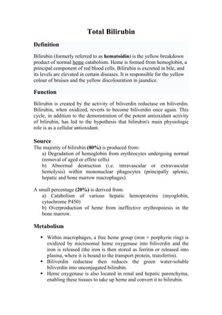
1. Total Bilirubin
Definition
Bilirubin (formerly referred to as hematoidin) is the yellow breakdown
product of normal heme catabolism. Heme is formed from hemoglobin, a
principal component of red blood cells. Bilirubin is excreted in bile, and
its levels are elevated in certain diseases. It is responsible for the yellow
colour of bruises and the yellow discolouration in jaundice.
Function
Bilirubin is created by the activity of biliverdin reductase on biliverdin.
Bilirubin, when oxidized, reverts to become biliverdin once again. This
cycle, in addition to the demonstration of the potent antioxidant activity
of bilirubin, has led to the hypothesis that bilirubin's main physiologic
role is as a cellular antioxidant.
Source
The majority of bilirubin (80%) is produced from:
a) Degradation of hemoglobin from erythrocytes undergoing normal
(removal of aged or effete cells)
b) Abnormal destruction (i.e. intravascular or extravascular
hemolysis) within mononuclear phagocytes (principally splenic,
hepatic and bone marrow macrophages).
A small percentage (20%) is derived from:
a) Catabolism of various hepatic hemoproteins (myoglobin,
cytochrome P450)
b) Overproduction of heme from ineffective erythropoiesis in the
bone marrow.
Metabolism
Within macrophages, a free heme group (iron + porphyrin ring) is
oxidized by microsomal heme oxygenase into biliverdin and the
iron is released (the iron is then stored as ferritin or released into
plasma, where it is bound to the transport protein, transferrin).
Biliverdin reductase then reduces the green water-soluble
biliverdin into unconjugated bilirubin.
Heme oxygenase is also located in renal and hepatic parenchyma,
enabling these tissues to take up heme and convert it to bilirubin.
2. Birds lack biliverdin reductase, thus they excrete heme breakdown
products as biliverdin rather than bilirubin.
Unconjugated or free bilirubin is then released into plasma where it
binds to albumin. Uptake of unconjugated bilirubin occurs in the
liver and is carrier-mediated. The carrier-mediated uptake is shared
with unconjugated bile acids and dyes such as BSP.
Once within the hepatocyte, unconjugated bilirubin is transported
with ligand (Y protein) or other proteins (e.g. Z protein) and the
majority is conjugated to glucuronic acid by glucuronyl transferase.
The remainder is conjugated to a variety of neutral glycosides (,
xylose).
In the horse, the majority of bilirubin is conjugated to glucose.
Bilirubin must be conjugated before it can be excreted into bile
(conjugation makes bilirubin water soluble).
Bilirubin is secreted into the intestine.
In the intestine, bacterial degrade it to urobilinogen.
Urobilinogen is reabsorbed (about 10%) or broken down (90%)
into urobilin and stercobilin (both of which are excreted in the
feces).
Of the resorbed urobilinogen, most is taken up by the liver
(enterohepatic circulation, i.e. the urobilinogen is absorbed into the
portal vein, taken up by the liver and re-excreted into bile, whilst
the rest bypasses the liver and is excreted into the urine.
3. Fig.1
Conjugated bilirubin is not normally found in the urine of domestic
animals, although small to (1+) amounts of conjugated bilirubin may be
seen in concentrated urine from dogs (particularly males), due to the low
canine renal threshold for bilirubin.
In all species (but dogs, in particular), bilirubinuria may precede an
increase in serum bilirubin in cholestatic disorders. Remember, only
conjugated bilirubin can be excreted in urine as it is water soluble.
Circulating bilirubin exists in two main forms as determined by the Van
den Bergh reaction, which differentiates bilirubin into conjugated (direct)
and unconjugated (indirect) forms.
4. Bilirubin (in blood) is in one of two forms:
(I) Direct-reacting (conjugated) Bilirubin.
(II) Indirect-reacting (unconjugated) Bilirubin.
(I) Direct-reacting (conjugated) Bilirubin
Conjugated bilirubin (direct-reacting). This form reacts in the diazo
reaction without the addition of alcohol.
Increases in conjugated bilirubin occur with:
Hemolysis.
Liver disease.
Cholestasis.
N.B. increased conjugated bilirubin in blood will produce bilirubinuria,
which in all species, excluding the dog, is diagnostic for cholestasis.
In horses, if conjugated bilirubin comprises > 25% of total bilirubin
values, cholestasis likely exists (a concurrent bilirubinuria will be
present). Extrahepatic bile duct obstruction produces the most marked
increases in total bilirubin (20-30 mg/dL).
(II) Indirect-reacting (unconjugated) Bilirubin
Free bilirubin (indirect-reacting or unconjugated). This is a relatively
insoluble, nonpolar form requiring the addition of alcohol in the diazo
reaction to allow color formation.
Increases in unconjugated bilirubin occur with:
Hemolysis.
Liver disease.
Cholestasis.
Fasting in horses.
5. In many instances, if unconjugated bilirubin dominates, hemolysis (or in
the case of horses, fasting) is the likely cause of the icterus.
Water
Abb. Name(s) Reaction
Soluble?
Reacts quickly when dyes are
Yes (bound to
"Conjugated" or added to the blood specimen to
"BC" glucuronic
"Direct bilirubin" produce azobilirubin "Direct
acid)
bilirubin"
Reacts more slowly. Still
produces azobilirubin. Ethanol
"Unconjugated" or No, but makes all bilirubin react
"BU"
"Indirect bilirubin" fat soluble promptly then calc: Indirect
bilirubin = Total bilirubin -
Direct bilirubin
Total bilirubin measures both BU and BC. Total and direct bilirubin
levels can be measured from the blood, but indirect bilirubin is calculated
from the total and direct bilirubin.
Measurement method
Originally the Van den Bergh reaction was used for a qualitative estimate
of bilirubin.
Causes of hyperbilirubinemia
Clinical icterus is observed when total bilirubin values exceed 1.5
mg/dL.
1. Artifact. 4. Cholestasis.
2. Hemolysis. 5. Physiologic.
3. Liver disease. 6. Inherited.
(1) Artifact:
6. Hemolysis (destruction of red cells, whether through extravascular or
intravascular hemolysis will increase the production of unconjugated
bilirubin) and lipemia (even mild) will cause artifactually high
bilirubin values.
As bilirubin is unstable in light, samples stored for several days, in the
presence of light, may have falsely reduced bilirubin values.
(3) Liver disease:
Hepatic disease may cause increases in both unconjugated and
conjugated bilirubin.
(4) Cholestasis:
This is defined as decreased bile flow due to:
a. Physical obstruction of bile flow.
b. Functional defects in the transporters that deliver bile salts or
bilirubin into the biliary system.
a) Physical obstructions to bile flow can be:
1. Intrahepatic (hepatocyte swelling due to hepatic lipidosis in cats).
2. Extrahepatic (bile duct obstruction from pancreatic neoplasia,
cholelithiasis, Fasciola hepatica in cattle).
b) Functional defects in bile salt or bilirubin transporters
1. Secondary to inflammatory cytokines (endotoxemia) and drugs.
2. Defects in these transporters also occur with physical obstructions
to bile flow.
Cholestasis will result in bilirubinemia with a higher conjugated than
unconjugated bilirubin. There is often a concurrent bilirubinuria (excess
conjugated bilirubin in blood is excreted into the urine, because it is
water soluble).
(5) Physiologic:
a. Fasting. b. Neonatal.
b.
7. a) Fasting: In horses, fasting will produce a hyperbilirubinemia due to
unconjugated bilirubin.
b) Neonatal: Young animals, especially foals, often have jaundice (due
primarily to unconjugated bilirubin). This is due to multifactorial causes,
including:
1. Hemolysis of fetal red blood cells.
2. Decreased liver uptake of bilirubin.
3. Immaturity of hepatic conjugation mechanisms.
4. Poor albumin binding.
9. Intravascular hemolysis
Fig.2
Intravascular hemolysis results from the rupture or lysis of red
blood cells within the circulation, and the release of their
hemoglobin into the plasma.
Haptoglobin binds the liberated free hemoglobin.
If intravascular hemolysis continues, the hemoglobin is present in
excess amount (>20 mg/dL) resulted in hemoglobinemia and
hemoglobinuria.
The remaining hemoglobin is oxidized to met-hemoglobin, which
disassociates into a free heme and globin chains.
10. The oxidized free heme (met-heme) binds to hemopexin and the
met-heme and hemopexin complex (met-heme/Hpx) is taken up by
hepatocytes and macrophages within the spleen, liver and bone
marrow (only hepatocyte uptake is illustrated in the image above).
Similarly, the hemoglobin/haptoglobin complex is taken up by
hepatocytes and macrophages (to a lesser extent).
Within these cells, the hemoglobin disassociates into heme and
globin chains. The globins are broken down to amino acids, which
are then used for protein synthesis.
The heme is oxidized by heme oxygenase forming biliverdin and
releasing iron.
The iron can be transferred to apotransferrin (the iron transport
protein) in plasma or can be stored within cells as ferritin (i.e. the
iron is bound to the storage protein, apoferritin).
The remaining porphyrin ring (biliverdin) is degraded to
unconjugated bilirubin by biliverdin reductase.
If the hemoglobin/haptoglobin complex is internalized by
macrophages, the unconjugated bilirubin is released into the
plasma, where it binds to albumin (to render it water-soluble) and
is taken up by hepatocytes.
Thus, with intravascular hemolysis, increases in bilirubin are
usually due to unconjugated bilirubin (indirect) and are likely of
macrophage (rather than hepatocyte) origin.
The intravascular hemolysis is usually accompanied by
extravascular hemolysis which is the source of most of the
unconjugated bilirubin observed in hemolytic anemia.
Because haptoglobin is consumed during intravascular hemolysis,
serum values of this protein usually decline with intravascular
hemolytic anemias or when hemoglobin is liberated into plasma by
artifactual lysis of red cells in vitro.
Since heme oxygenase is also present in renal tubular cells, the
renal epithelium is capable of converting hemoglobin to bilirubin.
However, this only occurs when there is intravascular hemolysis
with hemoglobinuria (i.e. the renal epithelium does not take up
unconjugated bilirubin or hemoglobin from blood!).
The renal epithelium absorbs the filtered hemoglobin from the
urine, converting it to unconjugated bilirubin and then conjugating
it for excretion into the urine (see fig.3).
This may be responsible for some of the bilirubinuria seen in
animals with intravascular hemolysis, however in most of these
animals, there is concurrent cholestasis that is responsible for the
bilirubinuria (which is conjugated).
11. Fig.3
Note that red cells can also lyse or rupture in vitro (either in the blood
collection tube or during collection). When this occurs, the hemolysis is
considered an artifact and does not indicate the animal has a hemolytic
anemia.
12. Extravascular hemolysis
Fig.4
Extravascular hemolysis occurs when RBCs are phagocytized by
macrophages in the spleen, liver and bone marrow.
Extravascular hemolysis is the most common form of hemolytic
anemia in animals.
It usually occurs alone (without intravascular hemolysis), but will
always (to some extent) accompany intravascular hemolysis.
Note that during the normal aging of red cells in the circulation,
effete red cells are destroyed by macrophages, i.e. extravascular
13. hemolysis is always occurring to some degree. However, this is a
physiologic process and does not result in anemia or excessive
unconjugated bilirubin production.
With extravascular hemolysis, the erythrocytes are degraded
within macrophages, so hemoglobin is not released free into the
cytoplasm.Thus, we do not see hemoglobinemia or hemoglobinuria
with extravascular hemolysis alone, unless it is accompanying
intravascular hemolysis.
Within macrophages, the hemoglobin is broken down into its
constituents, i.e. the heme ring and globin chains.
The globins are broken down to amino acids, which are then used
for protein synthesis.
The porphyrin ring of heme is oxidized by microsomal heme
oxygenase, producing biliverdin and releasing the iron.
The iron can then be exported into plasma through iron channels,
where it binds to apotransferrin forming transferrin or can be stored
within cells as ferritin, with time, ferritin becomes oxidized and
degrades to form hemosiderin.
Hemosiderin can be visualized within macrophages as a dusky
blue-gray pigment and can be definitively stained with Prussian
blue (which turns hemosiderin blue).
Biliverdin is reduced by biliverdin reductase to unconjugated
bilirubin (water insoluble).
The unconjugated bilirubin is released into the plasma, where it
binds to albumin (to render it water-soluble) and is taken up by
hepatocytes.
14. Jaundice
Definition
Jaundice known as icterus, is a yellowish discoloration of the skin, the
conjunctival membranes over the sclerae, and other mucous membranes.
Jaundice is most frequently caused by an increase of bilirubin in the
circulation, although it can be caused by other substances such as
carotene or certain drugs. Conjugated bilirubin causes more jaundice than
unconjugated bilirubin because of its higher water solubility and easier
absorption into tissues.
General circulation
.Fig. 5. Normal enterohepatic circulation of bile pigments
15. Classification of Jaundice
Jaundice is classified into three categories, depending on which part of
the physiological mechanism the pathology affects. The three categories
are:
• (I) Pre-hepatic: The pathology is occurring prior the liver.
• (II) Hepatic: The pathology is located within the liver.
• (III) Post-hepatic: The pathology is located after the conjugation
of bilirubin in the liver.
In both pre-hepatic and post-hepatic jaundice types, the function of the
liver itself is not impaired. In many of these situations, the liver is, in fact,
functioning at its maximum capacity in a compensatory effort to alleviate
the problems caused by other factors. This is not the case with hepatic
jaundice where the abnormalities are caused by an intrinsic liver defect or
disease.
16. (I) Pre-hepatic (Hemolytic Jaundice)
Pre-hepatic jaundice is caused by an increased production and release of
bilirubin most commonly due to:
1- Hemolytic process.
2- Ineffective erythropoiesis.
Increased hemolysis may be due to:
a. Variety of hemolytic anemias.
b. Exposure to chemicals.
c. Hemolytic antigen antibody reactions.
d. Disease such as some cancers.
e. Drugs coating red blood cells.
Ineffective erythropoiesis
Is a pathologic process where a very low proportion of red cells
formed in the bone marrow enter the circulation and those remaining in
the bone marrow are prematurely destroyed. An increase in the amount of
bilirubin released from the bone marrow results and is called early
labeled bilirubin since it has not been circulating within the red blood
cells for 120 days.
The rate of hemolysis and the ability of the liver to transport,
conjugate, and excrete bilirubin will determine the degree of jaundice in a
patient. In most cases of pre-hepatic jaundice, the production of bilirubin
is well below the capacity of the liver to conjugate and excrete it. Serum
bilirubin levels may still be essentially normal when there is a 50%
reduction in red cell survival as long as liver function is normal. Liver
function tests are helpful in the diagnosis of pre-hepatic jaundice. The
increase in bilirubin is the most obvious abnormality, being primarily of
the unconjugated type. Depending on the degree of hemolysis, varying
amounts of bilirubin enter the liver and corresponding amounts of
conjugated bilirubin are found in the intestine. This causes an increased
formation of urobilinogen in the gut (that is excreted in the feces or
absorbed into the enterohepatic circulation and ultimately excreted in the
urine.
17. There should be no bilirubin found in the urine because the increase is
of the unconjugated type, which is not filtered by the glomerulus of the
kidney. Liver enzyme assays should be normal in this condition except in
conditions where there is hemolysis. In these situations, lactic
dehydrogenase (LD) will be increased due to the high concentration of
LD found within red cells that is now released into the plasma. It is
occurred as a result of excessive destruction of RBCs.
General circulation
Fig. 6, Hemolytic crisis. Note the increase in the quantities of
unconjugated bilirubin (indirect reacting) in the serum (unable to pass the
renal filter), stercobilin in the stool (imparting a darker color to the stool),
and urinary urobilinogen. Increased urinary urobilinogen may be partly
due to secondary liver damage (less re-excreted into the bile and hence
lost to the serum and urine) in addition to the increased quantity of bile
pigments metabolized owing to erythrocyte hemolysis. If secondary liver
damage is extensive from hemosiderosis or bile pigment overload, some
bilirubin glucuronide may be regurgitated and lost to the urine (not in
diagram). RE, Reticuloendothelium
18. (II) Hepatic (Toxic Jaundice)
Jaundice of the hepatic type can be subdivided into two types:
(1) Retention jaundice.
(2) Regurgitation jaundice.
(1) Retention jaundice
It results from a defect in the transport of bilirubin into the hepatocyte.
In this type of jaundice:
Conjugated bilirubin is less than 0.2 mg/dl.
Urine bilirubin negative.
Urine urobilinogen is decreased or normal.
(2) Regurgitation jaundice
It occurs when the hepatic cell is damaged or defective or the excretion of
products from the hepatocyte is impaired.
If there is a regurgitation type of jaundice present, uptake, conjugation
and excretion impairment are present because of damaged liver cells.
In this type of jaundice:
Increased total bilirubin, conjugated bilirubin, and urine bilirubin
levels.
Urine urobilinogen level is increased because uptake is blocked.
Fecal urobilinogen may be decreased.
Stool color is lighter than usual.
Conjugation enzyme deficiencies.
Gilberts disease, and Crigler-Najjar syndrome are examples of causes of
retention jaundice, and Dubin-Johnson syndrome, Rotor syndrome, viral
hepatitis, and neoplastic conditions are examples of regurgitation
jaundice.
Laboratory values will vary within the category of hepatic jaundice.
Although the total bilirubin concentration will invariably be increased,
the relative amounts of unconjugated and conjugated bilirubin vary
according to the defect in the disease process. In general, a decreased
19. amount of bilirubin reaches the intestines because of the malfunctioning
liver and results in a decreased amount of urobilinogen being formed and
excreted into the feces. This is reflected in less urobilinogen being
absorbed into the enterohepatic circulation and a decreased amount of
urobilinogen being excreted into the urine. A very small amount of
urobilinogen is normally excreted in the urine so a lower than normal
value is difficult to determine. If the conjugated bilirubin concentration is
increased, an increased urine bilirubin can also be expected.
General circulation
Fig. 7, Hepatocellular pathology. Increased levels of bilirubin conjugates
(direct reacting) can be present in the serum; lesser amounts of
unconjugated bilirubin may also be elevated in the serum owing to a
decreased uptake of the pigment. During recovery from cholestasis,
increased serum levels of direct-reacting covalently bound bilirubin
conjugates (biliprotein) may persist without bilirubinuria. Observe the
presence of bilirubin glucuronide and increased amounts of urobilinogen
in the urine. Increased urinary urobilinogen is due to the inability of the
.altered hepatic cells to re-excrete this pigment into the bile
20. (III) Post-hepatic (Obstructive Jaundice)
Post-hepatic jaundice is caused by a blockage of the flow of bile from
the liver. Although the liver itself is not the cause of the problem, bile
produced by the liver cannot be released into the intestines and overflows
back into the blood. Although a complete blockage of the flow of bile is
uncommon, partial and intermittent obstructions are likely, and the
jaundice found in conjunction with this condition varies.
The most common obstructions are:
Stones within the common bile duct.
Obstructing neoplasm of the pancreas or other organs in close
proximity to the ducts.
Strictures severe enough to cause a blockage.
Stones are usually formed in the gallbladder and rarely cause symptoms
until they travel through the small ducts and lodge there.
In Post-hepatic jaundice
The increase in bilirubin is almost entirely of the conjugated type.
Because of the requisite obstruction, the quantity of bilirubin
reaching the intestines is decreased, resulting in clay-colored feces.
This color is due to the decreased formation of urobilinogen from
bilirubin in the intestines and its decreased excretion.
There should be little or no urobilinogen but large quantities of
bilirubin in the urine.
The kidney provides the only route of excretion for the increased
levels of conjugated bilirubin in the plasma, and the yellow-orange
urine color reflects this excretion of bilirubin.
Often there is no correlation between the plasma concentration of
conjugated bilirubin and the concentration of bilirubin excreted in
the urine.
Much of the conjugated bilirubin in obstructive conditions circulates
covalently bound to albumin and is called delta bilirubin. Since delta
bilirubin is protein-bound, it cannot pass the glomerulus of the kidney,
and therefore urinary bilirubin concentrations are less than expected when
the serum concentrations of conjugated bilirubin are significantly
elevated.
21. Fig.8, Extrahepatic obstruction. Note regurgitation to the serum and
subsequently the urine of all bilirubin diglucuronides conjugated in the
liver. Biliprotein may also be present in the serum during cholestasis.
Urinary urobilinogen and fecal stercobilin are absent.