
Recommended
More Related Content
What's hot
What's hot (20)
Similar to PJP Pneumonia Case: 74yo Female Develops Pneumocystis Jirovecii Pneumonia on Immunosuppression
Similar to PJP Pneumonia Case: 74yo Female Develops Pneumocystis Jirovecii Pneumonia on Immunosuppression (20)
More from Brian Locke
More from Brian Locke (17)
Recently uploaded
Recently uploaded (20)
PJP Pneumonia Case: 74yo Female Develops Pneumocystis Jirovecii Pneumonia on Immunosuppression
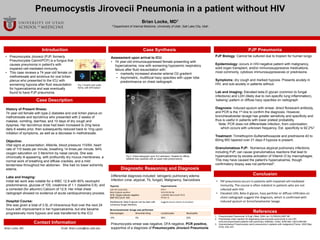
- 1. Case Description PJP Pneumonia Pneumocystis Jirovecii Pneumonia in a patient without HIV Brian Locke, MD1 1 Department of Internal Medicine, University of Utah, Salt Lake City, Utah References Brian Locke, MD Email Brian.Locke@hsc.utah.edu Contact Information Introduction • Pneumocystis Jirovecii (PJP, formerly Pneumocystis Carinii/PCP) is a fungus that causes pneumonia in patient’s with impaired cell-mediated immunity. • This case reviews a 74 year-old female on methotrexate and sirolimus for oral lichen planus who presented to the ICU with worsening hypoxia after fluid resuscitation for hypercalcemia and was eventually found to have PJP pneumonia. History of Present Illness: 74 year-old female with type-2 diabetes and oral lichen planus on methotrexate and tacrolimus who presented with 2 weeks of malaise, vomiting, diarrhea, and 10 days of dry cough and dyspnea. Her tacrolimus dose had been increased to 2mg twice daily 6 weeks prior, then subsequently reduced back to 1mg upon initiation of symptoms, as well as a decrease in methotrexate. Objective: Vital signs at presentation: Afebrile, blood pressure 110/64, heart rate of 110 beats per minute, breathing 14 times per minute, 94% oxygen saturation on 3 liters/min by nasal canula. She was chronically ill-appearing, with profoundly dry mucus membranes, a normal work of breathing and diffuse crackles, and a mild tenderness throughout her abdomen. She had no lower extremity edema. Labs and Imaging: Initial lab work was notable for a WBC 12.9 with 80% neutrophil predominance, glucose of 105, creatinine of 1.1 (baseline 0.8), and a corrected (for albumin) Calcium of 12.9. Her initial chest radiograph showed no evidence of acute cardiopulmonary process. Hospital Course: She was given a total of 3.5L of intravenous fluid over the next 24 hours with improvement in her hypercalcemia, but she became progressively more hypoxic and was transferred to the ICU Conclusion • PJP pneumonia occurs in patients with impaired cell-mediated immunity. The course is often indolent in patients who are not infected with HIV. • Elevated LDH, Beta-D glycan, hazy perihilar or diffuse infiltrates on chest radiograph suggest the diagnosis, which is confirmed with induced sputum or broncheoalveolar lavage. Diagnostic Reasoning and Diagnosis Assessment upon arrival to ICU: • 74 year-old immunosuppressed female presenting with hypercalcemia, now with worsening hypoxemic respiratory failure after fluid resuscitation with: • markedly increased alveolar-arterial O2 gradient • Asymmetric, multifocal hazy opacities with upper lobe predominance on chest radiograph. 1. Pneumocystis Pneumonia. N Engl J Med. 2004 Jun 10;350(24):2487-98. 2. Polymerase chain reaction for diagnosing pneumocystis pneumonia in non-HIV immunocompromised patients with pulmonary infiltrates. Chest. 2009 Mar;135(3):655-661. 3. Granulomatous Pneumocystis carinii pneumonia in patients with malignancyThorax. 2002 May; 57(5): 435–437. Case Synthesis Differential diagnosis included: Iatrogenic pulmonary edema Infection (viral, atypical, Tb, fungal), Malignancy, Sarcoidosis Pneumocystis smear was negative, DFA negative, PCR positive, supportive of a diagnosis of Pneumocystis Jirovecii Pneumonia PJP Biology: Cannot be cultured due to tropism for human lungs Epidemiology: occurs in HIV-negative patient with malignancy, solid organ transplant, and/or immunosuppressive medications, most commonly, cytotoxic immunosuppressives or prednisone. Symptoms: dry cough and marked hypoxia. Presents acutely in HIV, and sub-acutely in patients without. Lab and imaging: Elevated beta-D glycan (common to fungal infections) and LDH (likely due to non-specific lung inflammation), ‘batwing’-pattern or diffuse hazy opacities on radiograph Diagnosis: Induced sputum with smear, direct florescent antibody, and PCR is the 1st-line to confirm the diagnosis. However, broncheoalveolar lavage has greater sensitivity and specificity and thus is useful in patients with lower pretest probability. Note: PCR does not differentiate infection, from colonization, which occurs with unknown frequency. Est. specificity is 92.2%2 Treatment: Trimethoprim-Sulfamethoxazole and prednisone 40 to 60mg BID tapered over 21 days if hypoxia is present. Granulomatous PJP: Numerous atypical pulmonary infections, including PJP, can cause granulomatous reactions that lead to hypercalcemia by excess activation of Vitamin D by macrophages. This may have caused the patient’s hypercalcemia, though confirmatory biopsy was not performed3. Hypoxia Hypercalcemia LDH 327 (ULN 253) Beta-D-glycan positive AFB, fungal cultures negative BNP 633 (ULN 100) PTH 7 Vit D 25 OH 56 1,25OHD 184.0 (ULN 79.3) PTHrP <2 Multifactorial. Beta-D-glycan can be seen with numerous fungal infections Suggests excess vitamin D activation Broncheoalveolar lavage was performed: Macrophages Bronchial lining Lymphocytes Neutrophils 10% 6% 59% 25% Fig 1:Trophic and cystic forms, with DFA below1 Fig 2: Chest radiograph upon ICU admission. Notable for diffuse bilateral hazy opacities with an upper lobe predominance.
Editor's Notes
- Figure 1: add MHC data? Png for graphs