
Recommended
Recommended
More Related Content
What's hot
What's hot (20)
Similar to In-Vitro Transcription and Transfection of HCV Genomic Replicons
Similar to In-Vitro Transcription and Transfection of HCV Genomic Replicons (20)
Recently uploaded
Recently uploaded (20)
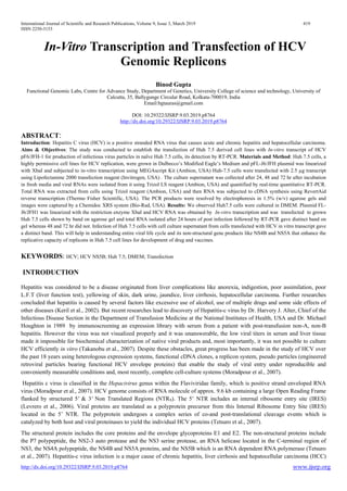
In-Vitro Transcription and Transfection of HCV Genomic Replicons
- 1. International Journal of Scientific and Research Publications, Volume 9, Issue 3, March 2019 419 ISSN 2250-3153 http://dx.doi.org/10.29322/IJSRP.9.03.2019.p8764 www.ijsrp.org In-Vitro Transcription and Transfection of HCV Genomic Replicons Binod Gupta Functional Genomic Labs, Centre for Advance Study, Department of Genetics, University College of science and technology, University of Calcutta, 35, Ballygunge Circular Road, Kolkata-700019, India Email:bgtauras@gmail.com DOI: 10.29322/IJSRP.9.03.2019.p8764 http://dx.doi.org/10.29322/IJSRP.9.03.2019.p8764 ABSTRACT: Introduction: Hepatitis C virus (HCV) is a positive stranded RNA virus that causes acute and chronic hepatitis and hepatocellular carcinoma. Aims & Objectives: The study was conducted to establish the transfection of Huh 7.5 derived cell lines with In-vitro transcript of HCV pF6/JFH-1 for production of infectious virus particles in naïve Huh 7.5 cells, its detection by RT-PCR. Materials and Method: Huh 7.5 cells, a highly permissive cell lines for HCV replication, were grown in Dulbecco’s Modified Eagle’s Medium and pFL-J6/JFH plasmid was linearized with XbaI and subjected to in-vitro transcription using MEGAscript Kit (Ambion, USA) Huh-7.5 cells were transfected with 2.5 µg transcript using Lipofectamine 2000 transfection reagent (Invitrogen, USA) . The culture supernatant was collected after 24, 48 and 72 hr after incubation in fresh media and viral RNAs were isolated from it using Trizol LS reagent (Ambion, USA) and quantified by real-time quantitative RT-PCR. Total RNA was extracted from cells using Trizol reagent (Ambion, USA) and then RNA was subjected to cDNA synthesis using RevertAid reverse transcription (Thermo Fisher Scientific, USA). The PCR products were resolved by electrophoresis in 1.5% (w/v) agarose gels and images were captured by a Chemidoc XRS system (Bio-Rad, USA). Results: We observed Huh7.5 cells were cultured in DMEM. Plasmid FL- J6/JFH1 was linearized with the restriction enzyme XbaI and HCV RNA was obtained by In-vitro transcription and was transfected to grown Huh 7.5 cells shown by band on agarose gel and total RNA isolated after 24 hours of post infection followed by RT-PCR gave distinct band on gel whereas 48 and 72 hr did not. Infection of Huh 7.5 cells with cell culture supernatant from cells transfected with HCV in vitro transcript gave a distinct band. This will help in understanding entire viral life cycle and its non-structural gene products like NS4B and NS5A that enhance the replicative capacity of replicons in Huh 7.5 cell lines for development of drug and vaccines. KEYWORDS: HCV; HCV NS5B; Huh 7.5; DMEM; Transfection INTRODUCTION Hepatitis was considered to be a disease originated from liver complications like anorexia, indigestion, poor assimilation, poor L.F.T (liver function test), yellowing of skin, dark urine, jaundice, liver cirrhosis, hepatocellular carcinoma. Further researches concluded that hepatitis is caused by several factors like excessive use of alcohol, use of multiple drugs and some side effects of other diseases (Keril et al., 2002). But recent researches lead to discovery of Hepatitis-c virus by Dr. Harvery J. Alter, Chief of the Infectious Disease Section in the Department of Transfusion Medicine at the National Institutes of Health, USA and Dr. Michael Houghton in 1989 by immunoscreening an expression library with serum from a patient with post-transfusion non-A, non-B hepatitis. However the virus was not visualized properly and it was unanswerable, the low viral titers in serum and liver tissue made it impossible for biochemical characterization of native viral products and, most importantly, it was not possible to culture HCV efficiently in vitro (Takanobu et al., 2007). Despite these obstacles, great progress has been made in the study of HCV over the past 18 years using heterologous expression systems, functional cDNA clones, a replicon system, pseudo particles (engineered retroviral particles bearing functional HCV envelope proteins) that enable the study of viral entry under reproducible and conveniently measurable conditions and, most recently, complete cell-culture systems (Moradpour et al., 2007). Hepatitis c virus is classified in the Hepacivirus genus within the Flaviviridae family, which is positive strand enveloped RNA virus (Moradpour et al., 2007). HCV genome consists of RNA molecule of approx. 9.6 kb containing a large Open Reading Frame flanked by structured 5' & 3' Non Translated Regions (NTRS). The 5’ NTR includes an internal ribosome entry site (IRES) (Levrero et al., 2006). Viral proteins are translated as a polyprotein precursor from this Internal Ribosome Entry Site (IRES) located in the 5’ NTR. The polyprotein undergoes a complex series of co-and post-translational cleavage events which is catalyzed by both host and viral proteinases to yield the individual HCV proteins (Tetsuro et al., 2007). The structural protein includes the core proteins and the envelope glycoproteins E1 and E2. The non-structural proteins include the P7 polypeptide, the NS2-3 auto protease and the NS3 serine protease, an RNA helicase located in the C-terminal region of NS3, the NS4A polypeptide, the NS4B and NS5A proteins, and the NS5B which is an RNA dependent RNA polymerase (Tetsuro et al., 2007). Hepatitis-c virus infection is a major cause of chronic hepatitis, liver cirrhosis and hepatocellular carcinoma (HCC)
- 2. International Journal of Scientific and Research Publications, Volume 9, Issue 3, March 2019 420 ISSN 2250-3153 http://dx.doi.org/10.29322/IJSRP.9.03.2019.p8764 www.ijsrp.org worldwide. A protective vaccine is not yet available and therapeutic options are still unlimited. Current therapies include pegylated interferon-α combined with ribavirin but this treatment is not so effective and only responsive to some patients infected with particular genotype (Moradpour et al,. 2007). All the genotypes do not respond to this drug.. The hepatitis C virus is usually detectable by PCR in the blood within one to three weeks after infection, and antibodies to the virus are generally detectable within 3 to 15 weeks or, chronic refers to infection with the hepatitis C virus persisting for more than six months. Clinically, it is often asymptomatic (without symptoms) and it is mostly discovered accidentally (e.g. usual checkup). The natural course of chronic hepatitis C varies considerably from one person to another. It is transmitted through the blood, items contaminated with infected blood and Sexual transmission is rare (Shimotohno et al., 2000). It is very difficult to study its proper life cycle due to absence of robust cell culture and small animal model. But now recent development of HCV replicon systems has made progress for the study of HCV translation and RNA replication in human hepatoma-derived Huh-7 cells derived for HCV genotype 2a replicon (JFH-1) In- vitro by Wakita and colleagues (Lohmann et al., 1999) . Moreover, these replicons do not replicate efficiently without adaptive mutation so Huh-7 cells are derived to Huh-7.5 cells and Huh 7.5.1 cells for better replication study. This system provides a powerful tool for producing infectious HCV viral particles in Huh 7.5 cell lines by In-Vitro transcription of genomic pFL-JG/JFH by transfection detecting the presence of RNA by RT-PCR and maintaining the replicon in culture cell line, Time course replication of Hepatitis-c virus for analysis of host-virus interactions that should facilitate the discovery of antiviral drugs and vaccines for this important human pathogen (Moradpour et al., 2007). MATERIALS AND METHODS The hepatic Huh 7.5 cells (a kind of gift from Prof. Charles M. Rice, The Rockefeller University, New York, NY, USA (Blight et al., 2002) were maintained in complete DMEM (Dulbecco's Modified Eagle's medium, Invitrogen, USA) supplemented with 10% FBS (Fetal Bovine Serum, Invitrogen), 100µg/ml Penicillin, 100µg/ml streptomycin (HiMedia, India), Glutamax 1% (Invitrogen), Hygromycin (50µl/ml), Puromycin (1µg/ml) at 37o C in 5% CO2 and grown for 5-6 days depending upon how much seeding is needed with frequent changing and checking the media and cells. 10µg of pFL-J6/JFH1 plasmid was linearized with XbaI and purified by purification as per manufacturer’s protocol. 1µg of Linearized and Purified pFL-J6/JFH genotype 2a construct(a kind of gift from Prof.Charles M. Rice) was subjected to in vitro transcription for the synthesis of HCV genomic replicon using T-7 Mega script kit (Ambion, USA) as per manufacturer’s protocol. In-vitro transcribed genomic HCV RNA was delivered to overnight grown naive Huh 7.5 cells in a 60 mm tissue culture dish using Lipofectamin 2000 transfecting reagent (Invitrogen), as per manufacturer’s protocol (Mandal et al., 2016) Total cellular RNA was isolated by TRIZOL (Invitrogen) using manufacturer’s protocol. Then after Reverse Transcriptase Polymerase Chain Reaction (RT-PCR) for β-actin, NS5B was done by using previously designed and purified oligonucleotides suitable for priming for β-actin (300nM), NS5B (1µM) of HCV genome. For β- actin, the forward primer sequence were HCV 5’ UTR: forward, 5’- GCGGGAAATCGTGCGTGACATT-3’; reverse, 5’- GATGGAGTTGAAGGTAGTTTCGTG-3’. For NS5B , forward , 5’-ACATCAAGTCCGTGTGGAAGG-3’; reverse, 5’-AGCCCGGGCGAGTGGAGTGG-3’ , Sybr green real- time PCR master mix reagent (Invitrogen) was used for the Assays in a Mastercycler ep RealPlex2 (Eppendorf , Germany) as described earlier (Mandal et al., 2016) Synthesis of first strand cDNA for β-actin, NS5B: RT (Reverse Transcription) reaction was done for First strand cDNA synthesis for β-actin, NS5B by taking total RNA sample (after isolation from transfected Huh 7.5 cells) along with 2 µl of specific primer. Initially RNA and primer were heated at 65o C for 5 minutes along with 25mM dNTP (200µM) and then quickly chilled in ice for 10 minutes, and then spinned down. β-actin (300nM), NS5B (1µM) was taken. cDNA synthesis was done on Realplex 2 Thermal cycler (Eppendorf). Double stranded cDNA was amplified by PCR using 1 µl of first strand cDNA (1.2µg). PCR for β-actin, 0.6µl of Mgcl2 and for NS5B, 0.75µl of Mgcl2 was added. PCR was done by following thermal profile: Initial denaturation at 94o C for 5 minutes followed by 30 cycles of denaturation, annealing and extension at 94o C for 30 seconds, 58o C for 30 seconds and 72o C for 30 seconds respectively, followed by a final extension at 72o C for 10 minutes. The reaction volume for each reaction was 20µl. Each reaction mix contained ------ Reagents for 1X 4.5X Stock final conc. Buffer 2 µl 9 µl 10X 1X Forward primer 1 µl 4.5 µl 12.5µM 1µM Reverse primer 1 µl 4.5 µl 12.5µM 1µM 25mM dNTP 1 µl 4.5 µl 5mM 200µM Mgcl2 0.6 µl 2.7 µl 50mM 1.5mM Enzyme (Taq polymerase) 0.5 µl 2.25 µl 5U/ µl 2.5 units/µl
- 3. International Journal of Scientific and Research Publications, Volume 9, Issue 3, March 2019 421 ISSN 2250-3153 http://dx.doi.org/10.29322/IJSRP.9.03.2019.p8764 www.ijsrp.org H2O (Double distilled water) 12.9 µl 58.05 µl cDNA 1µl DNA was electrophoresed on 1.5% Agarose gel, run for 60 minutes at 70-80 volts on 50X TAE buffer system, stained with ethidium bromide and visualized under UV-light in a UV-trans illuminator analysed on a Gel Doc (Bio-Rad) using standard protocol. No destaining of the gel was required. Infection of Naïve Huh 7.5 cells with viral supernatant: And also to study the HCV infection in Huh 7.5 cell line, Viral supernatant from the previous experiment were used to infect naïve Huh 7.5 cell line and left for 24 hours. Then the media was changed and fresh media (supplemented with G-418) was added and incubated. After 48 hours RNA was isolated by TRIZOL method using standard protocol and then RT-PCR was done. Then the sample was loaded on the 2% Agarose gel for electrophoresis for the confirmation of the infection. Time course study of hepatitis-c virus: Time course study means for how much time the virus remains infective. For this, Viral supernatant was taken and infected the naïve Huh 7.5 cells and left for 24 hours for infection. Then the media was changed and the Replicon maintaining media was added and left for 48 hours for post infection. After 48 hours RNA was isolated by TRIZOL method using standard protocol and then RT-PCR was done and the products were analyzed by Agarose gel electrophoresis using standard methods. RESULTS: I. Culture of Huh 7.5 cells: Cells appeared as spindle shaped. First day had less confluency but third day got more confluent culture. II. In-vitro transcription of pFL-J6/JFH Linearization of plasmid pFL-J6/JFH1 with the restriction enzyme XbaI I were optimized at 37o C for 4 hours and was In- Vitro transcribed by using Linearized plasmid. The full length HCV genomic replicon was obtained which was confirmed by presence of distinct band on agarose gel. . For control : pTRI-Xef transcript (1.96 kb) was used which gave successful band on agarose gel. Fig1: Linearization of pFL-J6/JFH1 Vector with Xba I. . Here, plasmid was found to be fully linearized at above optimum condition. Lane 1 loaded with uncut pFL-J6/JFH1 gave three forms of plasmid and lane 2 loaded with XbaI I treated plasmid gave single form of plasmid. This Linearized plasmid was used for In vitro transcription. Lane 1: Uncut pFL-J6/JFH1, Lane 2: pFL-J6/JFH1 linearized with XbaI
- 4. International Journal of Scientific and Research Publications, Volume 9, Issue 3, March 2019 422 ISSN 2250-3153 http://dx.doi.org/10.29322/IJSRP.9.03.2019.p8764 www.ijsrp.org Fig2: In vitro transcription using linearized pFL-J6/JFH1 to generate the full length HCV genome. Lane 2 was loaded with pTRI-Xef transcript (1.96kb) as a control and lane 3 with the pFL-J6/JFH1 transcript (9.6kb) full length which was in-vitro transcribed using linearized one. . Lane2: pTRI-Xef transcript (1.96 kb), Lane 3: pFL-J6/JFH1 transcript (9.6 kb) The presence of distinct band shows the successful in vitro transcription. III. HCV replicon Transfection: Total RNA was isolated after 48 hours post transfection followed by RT-PCR for NS5B region gave a distinct band on agarose gel confirmed successful transfection. Fig8: Transfection of 24hrs grown Huh7.5 cells with 2.5µg HCV in vitro transcript using Lipofactamin 2000 reagent (Invitrogen). Different lanes in the gel were loaded with different samples as given below:- Lane1: NS5B (PCR control) Lane2:NS5B (Transfected Huh7.5 cells), Lane3:NS5B (Naive Huh7.5 cells) Thus from the picture above it is clear that PCR for NS5B occurred properly showing the presence of HCV RNA. Thus, experiment giving successful transfection. IV. Infection of HCV in Huh 7.5 cells: The cell culture supernatant was obtained from tissue culture dish containing cells transfected with HCV In- Vitro transcript gave a successful band on agarose gel. 1 2 3 1 2 3 4 5 6 7 8 1 2 3 4 5 6 7 8
- 5. International Journal of Scientific and Research Publications, Volume 9, Issue 3, March 2019 423 ISSN 2250-3153 http://dx.doi.org/10.29322/IJSRP.9.03.2019.p8764 www.ijsrp.org Fig9: Infection of Huh7.5 cells with cell culture supernatant from cells transfected with HCV in vitro transcript Lane 1: PCR control β actin was loaded and distinct band was obtained. Lane2: RT control β actin, was loaded and distinct band was obtained. Lane3: β- actin (naïve Huh 7.5), it also showed the distinct band confirming its presence in naïve cells. Lane4: was loaded with β actin obtained from 24 hour grown infected Huh 7.5 cells. A distinct band was obtained confirming PCR has occurred correctly. Lane5: was loaded with NS5B (PCR control) Lane 6: was loaded with NS5B (24 hour Infected Huh 7.5 cells). This lane shows the clear distinct band confirming the successful infection. Lane 7- was loaded with the NS5B obtained by 48 hour of post infection. This lane does not show any band pattern suggesting no infection established after 48 hours. Lane 8-was loaded with the NS5B obtained by 72 hour of post infection. Here it also does not have any band suggesting no establishment of infection. DISCUSSION: FL-J6/JFH1 is a full-length, chimeric, genotype 2a HCV genome containing the core-NS2 coding region from the J6 HCV isolate and the NS3-NS5B coding region of HCV strain JFH-1. This genome includes nucleotides (nt) 1-300 of the JFH-1 strain, nt 301- 3430 of the J6 strain, and nt 3431-9678 of the JFH-1 strain. These genomes were created by using standard molecular biology techniques and were maintained as cloned cDNAs within the plasmids pFL-J6/JFH (Hugle et al., 2001) The pFL-J6/JFH plasmids were now linearized at the 3’end of the HCV cDNA by XbaI digestion. The linearized DNA was then purified and used as a template for in-vitro transcription. Infectious RNAs were synthesized with T7 RNA Polymerase via standard In- vitro transcription reactions. Then the RNA was purified using standard protocol and finally quantified. From the data provided above, distinct DNA band of NS5B shows that pFL-J6/JFH plasmid construct is able to replicate efficiently and produce infectious virus particles in Huh 7.5 cultured cells whereas β-actin is used as PCR control because it is housekeeping gene of mammalian cells that is always expressed and when loaded on gel after PCR always showed a distinct band confirming that PCR has occurred correctly. This cell lines are thus capable of contributing to efficient viral production and infection in this system. Also to see that the viral supernatant is infective or not, the naïve Huh 7.5 cells were infected with the viral supernatant and by following the protocol, RT- PCR and gel electrophoresis it was found that it can generate the infection and produce infectious viral particles in naïve Huh 7.5 cell lines. CONCLUSION: Thus it was possible to produce the infectious HCV viral particles in Huh 7.5 cell lines by In-vitro transcription of genomic pFL- JG/JFH by transfection and it could be also serially passaged to naïve cells. The ability of this cell culture technique created an opportunity to address the aspects of virus life cycle, its infective nature, the time course of infection that somehow created the knowledge of new investigative opportunities for the discovery of antiviral drugs and vaccines. But the main challenging question is that ‘For how much time these cells remain infective?’ For this, the time course replication study was done by first taking the viral supernatant and infecting the naïve Huh7.5 and following incubation detecting the presence of RNA by RT-PCR and again taking the viral supernatant for the next experiment and again infecting the naïve Huh 7.5 and so on. By this it was possible to get the infectivity for just 24 hour as shown by the data above. But in general it is difficult to get the infectivity for so long time that could be preserved, it automatically disappears. Due to this it has not been possible to maintain the replicon in culture cell line and future genetic studies on HCV proteins and RNA element; it still remains in the dark. Hence due to this, the dangerous HCV viral lifecycle still remains unclear and development of drug and vaccines also remains to be discovered. REFERENCES:
- 6. International Journal of Scientific and Research Publications, Volume 9, Issue 3, March 2019 424 ISSN 2250-3153 http://dx.doi.org/10.29322/IJSRP.9.03.2019.p8764 www.ijsrp.org .Alter, H. J., and L. B. Seeff. (2000). Recovery, persistence and sequelae in hepatitis C virus infection: a perspective on the long- term outcome. Semin. Liver Dis. 20:17–25. Bachmair, A., D. Finley, and A. Varshavasky.( 1986). In vivo half-life of a protein is a function of its amino-terminal residue. Science 234:179–186. Blight, K.J., J.A. Mckeating, and C.M.Rice. (2002). Highly permissive cell lines for subgenomic and genomic hepatitis c virus RNA replication. J. Virol.76:13001-13014. Friebe, P., V. Lohmann, N. Krieger, and R. Bartenschlager.(2001). Sequences in the 5 nontranslated region of hepatitis C virus required for RNA repli-cation. J. Virol. 75:12047–12057. Hugle, T., F. Fehrmann, E. Bieck, M. Kohara, H.-G. Krausslich, C. M. Rice, H. E Blum, and D. Moradpour. (2001). The hepatitis C virus nonstructural protein 4B is an integral endoplasmic reticulum membrane protein. Virology 284:70–81. Kato, Takanobu , Tomoko Date, Asako Murayama, Kenichi Morikawa, Daisuke Akazawa & Takaji Wakita(2006). Cell culture and infection system for hepatitis C virus. Natures protocols.1:2334-2339. Kolykhalov, A. A., S. M. Feinstone, and C. M. Rice. (1996). Identification of a highly conserved sequence element at the 3 terminus of hepatitis C virus genome RNA. J. Virol. 70:3363–3371. Levrero.M. (2006), Viral hepatitis and liver cancer: the case of hepatitis C. Nature Publishing Group. 25:3834-3847. Lohmann, V., F. Korner, J. O. Koch, U. Herian, L. Theilmann, and R. Bartenschlager. (1999). Replication of subgenomic hepatitis C viru RNAs in a hepatoma cell line. Science 285:110-113. Lohmann, V., F. Korner, A. Dobierzewska, and R. Bartenschlager.(2001).Mutations in hepatitis C virus RNAs conferring cell culture adaptation.J. Virol. 75:1437–1449. Mandal A, Ganta KK, Chaubey B: Combinations of siRNAs against La autoantigen with NS5B or hVAP-A have additive effect on inhibition of HCV replication. Hepat Res Treat 2016; 2016: 1–11. Moradpour, Darius, Francois Penin and Charles M. Rice.(2007). Replication of hepatitis C virus.Nature Publishing Group.5:453- 462. Pietschmann, T., V. Lohmann, A. Kaul, N. Krieger, G. Rinck, G. Rutter, D. Strand, and R. Bartenschlager. (2002). Persistent and transient replication of full-length hepatitis C virus genomes in cell culture. J. Virol. 76:4008–4021. Reed, K. E., J. Xu, and C. M. Rice. (1997). Phosphorylation of the hepatitis C virus NS5A protein in vitro and in vivo: properties of the NS5A-associated kinase. J. Virol. 71:7187–7197. Reed, K. E., A. E. Gorbalenya, and C. M. Rice.(1998). The NS5A/NS5 proteins of viruses from three genera of the family Flaviviridae are phosphorylated by associated serine/threonine kinases. J. Virol. 72:6199–6206. Rijnbrand, R., P. J. Bredenbeek, P. C. Haasnoot, J. S. Kieft, W. J. M. Spaan, and S. M. Lemon. (2001). The influence of downstream protein-coding se-quence on internal ribosome entry on hepatitis C virus and other flavivirus RNAs. RNA 7:585–597. Shimotohno, K. (2000). Hepatitis C virus and its pathogenesis. Semin. Cancer Biol. 10:233-240. Suzuki, Tetsuro, Koji Ishii, Hideki Aizaki, Takaji Wakita (2007). Hepatitis C viral life cycle. Advanced drug delivery Reviews, Elsevier. Science Direct. ADR-11596: 1-8. Takanobu Kato, Takuya Matsumura, Theo Heller, Satoru Saito, Ronda K. Sapp, Krishna Murthy, Takaji Wakita, and T. Jake Liang (2007). Production of infectious Hepatitis C virus of various Genotypes in Cell Cultures. American society of micro,USA 81 :4405-4411. Tanji, Y., T. Kaneko, S. Satoh, and K. Shimotohno.(1995). Phosphorylation of hepatitis C virus-encoded nonstructural protein NS5A. J. Virol. 69:3980– 3986.
- 7. International Journal of Scientific and Research Publications, Volume 9, Issue 3, March 2019 425 ISSN 2250-3153 http://dx.doi.org/10.29322/IJSRP.9.03.2019.p8764 www.ijsrp.org Walewski, J. L., T. R. Keller, D. D. Stump, and A. D. Branch. (2001). Evidence for a new hepatitis C virus antigen encoded in an overlapping reading frame. RNA 7:710–721. World Health Organization (1997). Hepatitis C: global prevalence. Wkly. Epidemiol. Rec. 72:341-344. Zhong. P, Pablo Gastaminza, Guofeng Cheng, Sharookh Kapadia, Takanobu Kato, Dennis R. Burton, Stefan F. Wieland, Susan L. Uprichard, Takaji Wakita, and Francis V. Chisari (2005). Robust hepatitis C virus infection in vitro. Proc. Natl. Acad. USA 102:9294-9299.