2. Learning objectives
At the end of this chapter, the student will be able to:
State the different laboratory wares.
Describe the use of laboratory wares.
Explain the general cleaning and care of
laboratory wares.
2
3. Outline
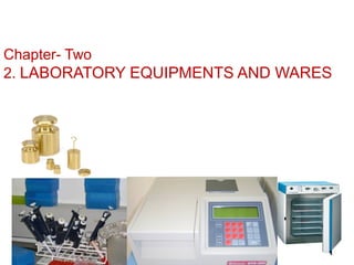
2. LABORATORY EQUIPMENTS AND WARES
2.1: General laboratory wares
2.1.1 Classification of Laboratory glass wares
2.1.2 Pipettes
2.1.3 Burettes
2.1.4 Flasks
2.1.5 Beakers
2.1.6 Cylinders 3
4. 2.1: General laboratory wares
LABORATORY GLASSWARES AND
PLASTICWARES
Definition: laboratory glassware and plastic wares are
materials used in clinical laboratory for:
measuring
pipetting
transferring
Preparation of reagents
Storage etc.
4
5. General laboratory …
Most of the routine laboratory wares used to be
of glass, but recent advantage made in the use
of plastic resin to manufacture a wide range of
plastic ware having led to a gradual replacement
of glass wares with durable plastic ware.
5
6. 2.1.1 Classification of Laboratory glass wares
A. can be divided in to five main types according to
their composition
1. Glass with high thermal resistance – borosilicate glass
can resist about 500o
c and low alkaline contact.
2. High silica glass- contains 96% silicon, It is thermal
endurable, chemically stable and electric resistant.
3. Glass with high resistance to alkali- Boron free, used in
strong alkali low thermal resistance. 6
7. Classification of Laboratory glass……
4. Low actinic glass – amber color to protect light
5. Standard flint glass- soda lime glass, poor resistance to
increased temp. Contains free soda in its walls
B . Based on their use
a) volumetric wares
b) Semi-volumetric Glass wares
c) Non- volumetric glass wares.
7
8. Classification of Laboratory glass……
a)Volumetric wares
Apparatus used for measurement of liquids
Can be made either from glass or plastic . it includes :
Volumetric flasks
Graduated centrifuge tubes
Graduated serological pipette
Medicine dropper
Burettes
Micropipettes
Diluting or thoma pipettes etc
8
9. Classification of Laboratory glass……
b). Non- volumetric glass wares: are not calibrated to hold a
particular or exact volume, but rather are available for various
volumes, depending on the use desired .
Erlenmeyer flask
Round bottom flask
Flat bottom flask
Beaker
Centrifuge tube
Test tube
Pasture pipette
9
10. Classification of Laboratory glass……
C).Semi-volumetric Glass wares: are used for approximate
measurement. it includes;
Graduated cylinder
Graduated specimen glass
Beakers
Conical flask
Medicine droppers with or with out calibration mark
Graduated beaker with double beaks
Graduated glass
10
11. 2.1.2 Pipettes
There are several types each having their own advantages and
limitations.
They are designated as class “A” or “B” according to their
accuracy.
Class “A” pipettes are the most accurate and the tolerance
limits are well defined that is, +0.01, + 0.02 and 0.04 ml for
2, 25, and 50 ml pipettes respectively.
Class “B” pipettes: are less accurate but quite satisfactory
for most general laboratory purposes. 11
12. Pipettes …
Significant errors will result if the temperature
of the liquid pipetted is widely different from
the temperature of calibration.
The usual temperature of calibration is 20o
C
and this is marked on the pipette.
12
13. 2.1.2.1 Volumetric pipettes
Volumetric pipettes are calibrated to deliver a constant
volume of liquid.
The most commonly used sizes are 1, 5, and 10ml
capacities.
Less frequently used sizes are those which deliver 6, 8, 12,
and so on ml.
They have a bulb mid – way between the mouthpiece and
the tip.
13
14. Volumetric …
The main purpose of the bulb is to decrease the
surface area per unit volume and to diminish the
possible error resulting from water film.
The Volume (capacity) and calibration
temperature of the pipettes are clearly written on
the bulb.
They should be used when a high degree of
accuracy is desired.
14
15. Volumetric pipettes……
The pipette is first rinsed several times with a little
of the solution to be used, and then filled to just
above the mark.
Then the liquid is allowed to fall to the mark and
the tip is carefully wiped with filter paper.
The contents are allowed to drain in to the
appropriate vessel. A certain amount of liquid will
remain at the tip and this must not be blown out.
15
16. Volumetric …
N.B: The reliability of the calibration of the volumetric
pipette decreases with an increase in size and therefore,
special micropipettes have been developing for chemical
microanalysis.
16
17. 2.1.2.2 Graduated or measuring pipettes
Graduated pipettes consist of a glass tube of uniform
bore with marks evenly spaced along the length.
The interval between the calibration marks depends
up on the size of the pipette.
Two types calibration for delivery are available:
A. One is calibrated between two marks on the stem
(Mohr).
B. The other has graduation marks down to the tip
(serological pipette)
17
18. Graduated or measuring…….
These pipettes are intended for the delivery of
predetermined volumes.
The serological pipette must be blown out to deliver
the entire Volume of the liquid and it has an etched
ring (pair of rings) near the mouth end of the pipette
signifying that it is a blow out pipette.
Measuring pipettes are common only in 0.1, 0.2, 0.5,
1.0 5.0, and 10.0 ml sizes. 18
19. Graduated measuring…
The liquid is delivered by allowing it to fall from one
calibration mark to another.
N.B. The classification of pipettes may not always be based
on the presence or absence of a bulb and etched ring.
19
20. A. B C D.
A. Volumetric (transfer) B. Ostwald folin (transfer). C. Measuring (Mohr) D. Serological (Graduated)
20
22. 2.1.2.3 Micropipettes
Micropipettes are frequently used in
Medical chemistry
Virology
Immunology and serology laboratories.
This is because in these laboratories often only
small quantities of materials are available for
measurement. 22
23. Micropipettes …
They are found in different capacities such as
5, 10, 25, 50, 100 and 1000 micro liter.
There are also other kinds of pipettes that are
used in medical laboratories.
Example: Toma pipette, Pasteur pipette,
automatic pipettes and others.
23
25. 2.1.3 Burettes
• Burettes are used for measuring variable quantities of
liquid that are used in volumetric titrations.
• They are made in capacities from 1 to100 milliliters.
• They are long graduated tubes of uniform bore and are
closed at the lower end by means of a glass stopper, which
should be lightly greased for smooth rotation.
Fig. Burette
25
26. 2.1.4 Flasks
There are four types of flaks having 25 to 6,000
milliliter (ml) capacities.
2.1.4.1 Conical (Erlenmeyer) flasks
Conical (Erlenmeyer) flasks are useful for
titrations and also for boiling solutions when it
is necessary to keep evaporation to a minimum.
Some have a side arm suitable for attachment to
a vacuum pump.
26
27. Flask …
2.1.4.2 Flat bottomed round flasks
Flat-bottomed round flasks are convenient
containers to heat liquids.
These flasks are widely used in the preparation of
bacteriological culture media.
27
28. Flasks …
2.1.4.3 Round bottomed flasks
Round bottomed flasks can with stand higher
temperatures than the flat- bottomed type.
they may be heated in a necked flame or in an electro-
thermal mantle. As a result used for boiling.
28
29. Flasks …
2.1.4.4 Volumetric flasks
Volumetric flasks are
flat - bottomed
pear-shaped vessels with long narrow necks
fitted with ground glass stoppers.
29
30. Flasks …
Volume metric ….
Most flasks are graduated to contain a certain volume, and
these are marked with the liters.
A horizontal line etched round the neck denotes the stated
volume of water at given temperature.
They are used to prepare various kinds of solutions.
The neck is narrow so that slight errors in reading the
meniscus results in relatively small volumetric differences
(minimizes volumetric differences or errors). 30
31. A. Conical B. Flat bottomed C. Flat bottomed D.Volumetric
31
33. 2.1.5 Beakers
Beakers have capacities from 5 to 5,000 ml.
They are usually made up of heat resistant glass and are
available in different shapes.
The most commonly used is the squat form, which is
cylindrical and has a spout.
There is also a tall form, usually with out a spout
33
36. Cylinders…
36
Measurement of liquids can be
made quickly with these vessels,
but a high degree of accuracy is
impossible because of the wide
bore of the cylinders.
37. 2.1.7 Test tube
Test tubes are made of hardened glass or plastic
materials that can withstand actions of chemicals,
thermal shock and centrifugal strains.
They are used to hold samples and solutions during
medical laboratory procedures.
These include simple round hollow tubes conical
centrifuge tubes, vaccutainer tubes. Test tubes can be
with or with out rims (lips).
Test tubes with out rim are satisfactory.
37
39. 2.1.8 Reagent bottles
Reagent bottles are used to store different types of
laboratory reagents.
They are made from glass or plastics. Depending on their
use, they are available in various sizes and type.
39Dropping bottle
40. 2.1.9 Petri dishes
Petri dishes are flat glass or plastic containers, which
have a number of uses in the medical laboratory.
They are used predominantly for the cultivation of
organisms on solid media.
They are made with diameters of 5 to 14
centimeter.
40
41. 2.1.10 Funnels
There are two types of funnels that are widely used in a
medical laboratory. These are filter funnel and separating
funnel.
2.1.10.1 Filter Funnels
Filter funnels are used for pouring liquids into narrow
mouthed containers, and for supporting filter papers during
filtration.
They can be made from glass or plastic materials.
41
42. Funnel …
2.1.10.2 .Separating funnels
Separating funnels are used for separating
immiscible liquids of different densities. Example,
ether and water.
42
43. 2.1.11. Pestle and mortar
Pestle and mortar are used for grinding solids, for
example, calculi and large crystals of chemicals.
After each use always clean the pestle and mortar
thoroughly.
This is because chemicals may be driven into the
unglazed surfaces during grinding, resulting in
contamination when the apparatus is next used..
43
44. 2.1.12 Laboratory Cuvettes (Photometry)
used for photometric readings in instruments or
used for measurements of absorbance.
Glass Cuvettes resist many laboratory reagents like
organic solvents, whereas plastic Cuvettes are
affected by many reagents and become cloudy,
hence affecting the absorbance’ of the reacting
mixture and so lack accuracy & precision.
44
45. Laboratory Cuvettes …
Can be glass, quartz, or plastic
Require uniform thickness, density, composition
Should be uniformly calibrated
45
46. 2.1.13. Pasture pipette
They are non-volumetric glassware used in
transferring liquid.
It has a long –drown-out tip with a rubber bulb or teat
to suction.
Eye droppers or medicine droppers can use instead of
pasture pipettes.
46
47. Precautions when using glassware
1. All glassware must be handled carefully.
2. Breakage can some times be dangerous and may result in
the loss of valuable and irreplaceable materials.
3. Flasks and beakers should be placed on a gauze mat when
they are heated over a Bunsen flame.
4. Test tubes exposed to a naked flame should be made of
heat resistant glasses.
5. If liquids are to be heated in a bath or boiling water, the
glass contents should be heat resistant.
47
48. 2.2 Medical laboratory Equipment
Learning objectives ;
Identify the types and uses of laboratory balances.
Explain the advantages of laboratory
refrigerators.
Describe the importance of ovens, water baths
and incubators.
State the use of photometers and desiccators.
48
49. Learning objectives…
Identify the types and uses of microscopes.
State the basic components centrifuge.
Discuss pH in terms of ion activity and units.
Describe the main components of a pH meter
including their role in analysis.
49
50. Out line
2.2 lab equipment
2.2.1: Microscope
2.2. 2: Equipment for purifying water
2.2.3: Equipment for weighing
2.2.4: Equipment for pipetting and dispensing
2.2.5: Laboratory centrifuges
2.2.6: laboratory autoclaves, ovens
50
51. Out line…
2.2.7: Incubator, water bath, heat block
2.2.8: Colorimeter
2.2. 9: Mixers
2.2.10: Refrigerators
2.2.11: Desiccators
2.2.12: PH
meter
2.2.13: Safety cabinets
2.3: Care and cleaning of laboratory equipments and wares
51
52. 2. 2 Medical laboratory Equipment
2.2.1: THE MICROSCOPE
Used to visualize minute objects (animate and
inanimate), that cannot be seen by our naked eye.
It is a magnifying lens.
It was invented by Anton van Leeuwenhoek –
founder of microscope.
52
53. Microscope …
2.2.1.1 Types of microscope
1. Light field microscope ;- are the group of microscope that uses
light.
This includes:
a. Compound light(bright) field:
Compound microscope is a light microscope, which is
routinely used in medical laboratories of hospitals and/or
health centers.
b. Dark field microscope or dark ground illumination
Makes some living micro-organisms which can not be seen by
ordinary transmitted lighting.
53
54. Microscope …
Principle of light microscope
The light enters a special condenser which has a
central blacked-out area so that the light cannot
pass directly to enter the objective.
The only light entering the eye comes from the
micro-organisms themselves, no light entering the
eye directly from light source.
54
55. Microscope …
In the way small micro-organisms are seen brightly
illuminated against a black background, like stars in a
night sky.
Importance of Dark field microscope
Used for examining-
Treponema palladium
Borreliae in blood
Microfilariae in blood
55
56. Microscope …
c) Phase contrast microscope
Makes use of this ability of waves to help or hinder
each other to produce variations increase the contrast
achieved by placing annulus in condenser and phase
plate in the objective.
56
57. Microscope …
Used for examination of
Unstained bacteria
Urine sediments
Haemoparasites
Amoebae in faecal preparations
Trypanosomes in blood, cerebrospinal fluid,
lymph gland fluid.
57
58. Microscope …
d) Fluorescence microscope
widely used in the immunodiagnosis
Principle:
Ultraviolet light may be used to illuminate particles or
micro-organisms which have been previously stained
with fluorescing dyes.
These dyes transform the invisible ultraviolet light to
visible light.
58
59. Microscope …
Value of fluorescence microscope
Examination of sputum and c.s.f for acid fast bacilli
(AFB) using an auramine staining technique.
Examination of acridine orange stained
Trichomonas vaginalis flagellates.
59
60. Microscope …
2. Electron Microscope: - as the name suggests, employ
a beam of electrons produced by an electron gun to
produce the magnified image.
Mainly used in
Negative staining
Sample stained with potassium phosphotungestate
Examination of viruses
NB. The beam can not pass through the metallic back
ground of the microscope.
60
61. Microscope …
2.2.1.2 Major parts of microscope
A. Frame work of the microscope
This includes:
An arm (stand): - The basic frame of the microscope
to which the base, body and stage are attached.
A stage: - the table of the microscope where the slide
or specimen is placed.
A foot or base: - is the rectangular part up on which
the whole instruments rest.
61
62. Microscope …
B. Focusing system
This encompasses:
Coarse and fine focusing adjustments
Course adjustment: - The course focusing
adjustment is controlled by a pair of large knobs
positioned one on each side of the body. Give rough
image.
Fine adjustment: - it moves the stage so slowly that
and give clear image .
62
63. Microscope …
Condenser adjustments: - The condenser is focused
usually by rotating a knob to one side of it.
This moves the condenser up or down.
The condenser aperture is adjusted by the iris
diaphragm, which is found just below the
condenser.
The principal purpose of the condenser is to
condense the light required for visualization.
63
64. Microscope …
C. Magnification system
This comprises:
Objectives: - Objectives are components that
magnify the image of the specimen to form the
primary image.
For most routine laboratory work 10x, 40x and
100x (oil immersion) objectives are adequate.
Eyepiece:- Eyepiece is the upper optical component
that further magnifies the primary image and brings
the light rays to a focus at the eye point.
64
65. Microscope …
Eye piece:
It consists of two lenses mounted at the correct
distance.
It is available in a range of magnifications usually
of 10x, 15x and sometimes as high as 20x.
N.B: Based on their number of eyepiece microscopes can
be classified as monocular, binocular microscopes etc.
65
66. Microscope …
D. Illumination system
Condenser and iris
Condenser is a large lens with an iris diaphragm.
The condenser lens receives a beam from the light
source and passes it into the objective.
The iris is a mechanical device mounted underneath
the Condenser and controls the amount of light entering
the condenser.
66
67. Microscope …
Mirror
Mirror is situated below the condenser and iris.
It reflects the beam of light from the light source up
wards through the iris into the condenser.
The mirror is used to reflect ray or electrical light.
67
70. Microscope …
Filters
Light filters are used in the microscope to:
Reduce the intensity of light.
Increase contrast and resolution.
Adjust the color balance of the light to give the best
visual effect.
Provide monochromic light.
Absorb light.
Transmit light of selected wavelength.
Protect the eye from injury caused by ultra-violet
light.. 70
73. Microscope …
2.2.2.3 Working principle of the microscope
The magnified image of the object (specimen) is first
produced by a lens close to the object called the
objective.
This collects light from the specimen and forms the
primary image.
A second lens near the eye called the eyepiece (ocular)
enlarges the primary image converting it into one that
can enter the pupil of the eye.
73
74. Microscope …
The magnification of the objective multiplied by that of
the eyepiece gives the total magnification of the image
seen in the microscope
Example:
Objective Eyepiece Total
Magnification Magnification Magnification
10X 10X 100X
40X 10X 400X
100X 10X 1000X
74
75. Microscope …
Objectives
Low power (10X) Objective
Used for the initial scanning and observation in
most microscopic work.
When using 10 X
Close iris diaphragm.
Lower the condenser.
75
76. Microscope …
High -dry power (40X) Objective
Is used to study un stained specimens such as
stool and urine sediments for more detailed
examination.
When using 40 X
open the iris diaphragm half way.
raise the condenser half way.
76
77. Microscope …
Oil immersion (100X) Objective
Routinely used for morphologic examination of blood
films and microbes.
An oil immersion lens requires that special
grade of oil (immersion oil) be placed b/n
the objective and the slide.
The oil is used to increase the intensity of
light.
When using 100 X
open the iris diaphragm completely.
raise the condenser completely. 77
78. Microscope …
2.2.2.4 Resolving power of the microscope
It may be defined as the ability to level closely
adjacent structural details as being actually separate
and distinct.
The increase in magnifying power is always linked to
an increase in resolving power.
The higher the resolving power of an objective, the
closer can be the fine lines or small dots in the
specimen which the objective can separate in the
image. 78
79. Microscope …
The resolving power of an objective is dependent on
what is known as the numerical aperture (NA) of the
objective.
The numerical aperture is a designation of the amount of
light entering the objective from the microscope field, i.e.
the cone of light collected by the front lens of the
objective (an index or measurement of the resolving
power).
79
80. Microscope …
Numerical aperture is dependent on the diameter of
the lens and the focal length of the lens.
E.g. Res. power of:
Human eye- 0.25 mm
Light microscope- 0.25µm
Electron microscope- 0.5 nm
80
81. Microscope …
Numerical Aperture
Defined as the product of the refractive index of the
medium outside the lens (n) and the sine of half the
angle of the cone of light absorbed by the front lens of
the objective (u) or
Is a number that expresses the ability of a lens to
resolve fine detail in an object being observed.
81
82. Microscope …
E.g. 0.25 on X10 objective
0.65 on X40 objective
1.25 on X100 objective
The greater the N.A the greater the resolving power.
The following are the usual numerical apertures of
commonly used objectives:
10 X objective ----------- NA 0.25
40 X objective ----------- NA 0.65
100 X (immersion oil) objective ------- NA 1.25
82
83. Microscope …
Total magnification
is the product of the objective and the eye piece
magnification
Useful magnification range
is calculated as:
(500-1000)x NA of that objective
E.g. The useful magnification range when an Eyepiece with
magnification of 10x & an objective with magnification
40x & NA of 0.65 is: 325-650.
83
85. Microscope …
Large diameter
Shorter focal length
Very high NA
Very high resolution
Very high useful magnification
85
86. Microscope …
Small Diameter
Long focal length
Very low NA
Very low r.p
Very low useful magnification
86
87. Microscope …
Small diameter
Short focal length
Low NA
Low resolution
Low useful magnification
Therefore the wider the angles of the cone of light the
higher the NA of the objective and greater is the
objectives resolving power and useful magnification.
87
88. Microscope …
2.2.2.5 Working principle of an oil immersion objective
When a beam of light passes from air into glass it is
bent and when it passes back from glass to air it is
bent back again to its original direction.
This has effect on oil immersion objective and affects
the NA of the objective and consequently it’s resolving
power.
88
89. Microscope …
The bending effect on the objective can be avoided by
replacing the air between the specimen and the lens
with oil, which has the same optical properties as
glass, i.e. immersion oil.
The oil provides better resolution and a brighter image
by collecting extra oblique light.
89
91. Microscope …
2.2.2.5 Routine use of the microscope
A microscope must always be used with gentleness,
care and the following should be noted.
1. Place the microscope on a firm bench so that it does
not vibrate.
a. Make sure that it is not be exposed to direct sun
light.
b. The user must be seated at the correct height for the
convenient use of the microscope. 91
92. Microscope …
2. Select the appropriate source of light.
3. Place the specimen on the stage, making sure that
the under side of the slide is completely dry.
4. Select the objective to be used.
It is better to begin examination with 10x
objective.
92
93. Microscope …
5. Bring the objective as close as possible to the slide
preparation.
6. Adjust the light source until the illumination of image is at
its brightest.
7. Focus the condenser.
8. Adjust the aperture (opening) of the condenser iris
according to the specimen being examined.
93
94. Microscope …
The wider the condenser aperture, the brighter will be
the specimen and the smaller be the details, which
can be resolved.
The smaller the aperture, the greater will be the
contrast.
Certain specimens, example stained and mounted
specimens give little glare illuminated image with fine
detail. 94
95. Microscope …
Other specimens, like urine, unstained cerebrospinal
fluid and saline mounted fecal specimens give much
glare and require a reduced source of light to increase
contrast.
9. Examine the specimen by systematically moving the
slide with the mechanical stage.
N.B: The image of the specimen will be up side down
and will move in the opposite direction to the side.
10. For a higher magnification, swing the 40x objective into
place.
Focus the 40x objective, using the fine adjustment. 95
96. Microscope …
If for any reason the image is not visible, lower the
objective until it is nearly but not quite touching the
specimen.
Then looking through the eyepiece, focus up wards
with the fine adjustment until the image comes into
view
11.For the highest magnification, add a drop of immersion
oil to the specimen and swing the 100x oil immersion
objective into place, then open the iris fully to fill the
objective with light.
Example. Stained blood smear, acid-fast stain, etc 96
97. Microscope …
2.2.2. 6 Care, Cleaning and Repair of microscope
The microscope is one of the most expensive and
delicate instruments.
Good microscopy practice should include:
I. Daily cleaning and quality control(QC) check
a. Using a clean cloth, wipe any dust from stage
and other surfaces of microscope.
97
98. Microscope …
b. Using lens tissue clean dry objective.
Clean 100X objective with tissue dampened with xylene.
Never use alcohol to clean the oil because it will dissolve
the cement holding the lens.
c. Carry out a QC check to ensure the lenses are
completely clean.
98
99. Microscope …
II- Care when using the microscope
1. Do not use force for any mechanism.
2. Check stage and under side of the specimen, re
DRY and CLEAN.
3. Cover wet preparation with cover slip.
4. Use non-drying oil immersion.
5. Put eyepieces that are not in use in closed container.
6. Always lift and carry the microscope well supported
with hands.
7. Protect the microscope from dust, moisture and
direct sunlight.
99
100. Precautions when using microscope
Never dip the objectives in xylene or ethanol, as this
may cause the lenses to become detached.
Never use ordinary paper to clean the lenses.
Never touch the lenses with your fingers.
Never clean the support or the stage with xylene or
acetone.
100
101. Microscope …
III- At the end of the Day’s
Turn the switch off.
Clean using a soft tissue.
Do not leave the objective of eyepiece open.
Decontaminate the stage with 70% alcohol
dampened cloth.
Cover with its dust cover.
101
102. 2.2.2: Equipment for purifying water
2.2.1: DISTILLER
A process by which impure water is boiled and the
steam condensed on cold surface (condenser) to give
pure distilled water is called distillation.
Distilled water is free from dissolved salts and clear
colorless, odorless and tasteless. It is sterile too.
The apparatus is called distiller.
A considerable volume of cool running water is
required to operate or to condense the steam.
102
103. Equipment for purifying
2.2. 2: DEIONIZER
Deionizer is an apparatus used to produce ion free water.
A deionizer is an apparatus for demineralizing water by
means of cartridges filled with ion-exchange resin.
Deionization is a process in which chemically impure
water is passed through anion and cation exchange
resins to produce ion free water.
Deionized water has low electrical conductivity, near
neutral pH and is free from water-soluble salts but is not
sterile. 103
104. 2.3: Equipment for weighing/Balances
Balances are essential laboratory instruments that are
widely used for weighing of various substances (powders,
crystals and others) in the laboratory.
For instance, to prepare reagents, stains and culture media,
balances are required to weigh accurately and precisely
within the needed range.
They should be kept carefully clean and located in an area
away from heavy traffic, large pieces of electrical equipment,
and open windows.
To minimize any vibration, as interference that may happen,
a slab of marble is placed under the balance.
104
105. Balances …
Balances in medical laboratory may be:
2.3.1. Rough balances (mechanical balances)
2.3.2. Analytical balances/electrical/
2.3.1 Rough balances
Rough balances are several types. Some of them use
sliding scale, some have a single or double pan (s)
and others utilize dial - operated fractions.
They are used for weighing substances, which do not
call for extreme accuracy. 105
106. Balances …
While operating, they do not require mains electricity
or battery power and are currently less expensive
than analytical balances of the similar sensitivity.
Some rough balances weigh accurately to 0.1 gm of a
substance.
Two - pan balance is a rough balance, which has two
copper pans supported by shafts.
It is used:
To weigh large amounts (up to several kilo
grams).
When a high degree of accuracy is not required.
The sensitivity of a two pan balance is 0.5 gm. 106
107. Balances …
The sensitivity of a balance is the smallest weigh that
moves the pointer over one division of the scale.
For routine laboratory purposes the sensitivity of a
balance can be considered to be the smallest weigh
that it will measure accurately.
Usually the larger the amount of substance to go into
a reagent, the least accuracy is required.
107
109. Balances …
2.3. 2 Analytical balances
Nowadays analytical and electronic balances (single
pan balances that use an electron magnetic force
instead of weights) are the most popularly used
balances in medical laboratories to provide a
precision and accuracy for reagent and standard
preparation.
Analytical balance is a highly sensitive instrument.
It may have two pans suspended from a cross beam,
inside a glass case.
It requires mains electricity or battery (D.C) supplied
power.
109
110. Balances …
These balances are used:
1. To weigh small quantities usually in mili gram (mg) range.
2. When great accuracy is required. E.g., 2.750mg,
0.330 mg, 5.860mg, etc.
Its sensitivity is 0.5 mg to 1 mg depending on the model.
N.B: The accuracy of a balance should be checked regularly
as recommended by the manufacturer.
110
113. Balances …
Before starting to weigh, zero the balance as directed
by the manufacturer. If using a beam balance, check
the position of the beam.
Weigh the chemicals at room temperature in a
weighing scoop or small beaker. And Never put the
chemicals directly on the balance pan.
113
114. Balances …
Use and care……..
When adding or removing a chemical, remove the
container to avoid spilling any chemical on the
balance.
When using an analytical double pan balance,
bring the pans to rest before adding or removing a
chemical.
Always use forceps to add or remove weighs.
Protect the weights from dust, moisture and fungal
growth. 114
115. Balances …
Use small brush to remove any chemical, which
may have been spilt on the balance.
A container of self - indicating silica gel should be
kept inside the analytical balance case to remove
any moisture present in the atmosphere.
Keeps the balance clean, being particularly careful
not to let dirt accumulate near the pivots and
bearings.
115
116. 2.2.4: Equipment for pipetting and
dispensing
There are different types of devices used for pipetting
and dispensing specimens. Some of them are:
Simple bulb aspirator- this is simple inexpensive
device suitable for use with graduated capillaries.
Thumb wheel aspirator – it can be used with
capillaries, shell- back pipettes, example, Sahli or
WBC pipettes ad most small bore graduated pipettes,
example measuring up to 0.5ml.
116
117. Equipment for pipetting …
Automatic pipetter – it use plastic or glass tips and
models are available for measuring single volumes or
several different volumes. Automatic pipetters have a
greater precision and accuracy.
PVC bulb pipette filler – its tapered and flexible end
enables all types of pipettes up to 10 ml volume to be
inserted easily and safely in to the end and to be held
securely.
117
118. Equipment for pipetting …
Pi- pump2500, pipette filler – it is highly recommended
for the controlled filling and dispensing of fluid from
pipettes.
Bottle top dispenser - it is used to measure a fixed
volume of fluid or several different volumes of fluid.
118
119. Equipment for pipetting and ……
Bottle top hand operated dilutor – this is the most
expensive of the devices described above. It is used for
measuring accurately and precisely, specimen and
reagent.
Plastic bulb pipettes – Plastic bulb pipettes have many
uses in a medical laboratory. They can be
decontaminated in disinfectant, wash, and reused many
times.
119
120. 2.2.5: Laboratory centrifuges
Centrifuge: is equipment that is used to separate
solid matter from a liquid suspension by means of
centrifugal force.
They sediment particles (cells, bacteria, casts,
parasites, etc.) suspended in fluid by exerting a force
greater than that of gravity.
The suspended materials are deposited in the order
of their weight.
There are many types of centrifuges, but the basic
principle is the same, that is, the all use centrifugal
force.
120
121. Centrifuge….
When a body is rotated in circular movement at speed,
centrifugal force is created that drives the body away
from the center of the circular movement.
The greater the outward pull due to rotation, that is
centrifugal force, the more rapid and effective is the
sedimentation.
As a result, heavier elements are thrown to the bottom of
the tube followed by lighter particles.
121
123. Centrifuge….
Centrifugal force increases with the speed of rotation
that is the revolution of the rotor per minute and the
radius of rotation.
The actual sedimentation achieved at a given speed
depends therefore, on the radius of the centrifuge.
Most techniques requiring centrifugation will usually
specify the required relative centrifugal force (RCF)
expressed in gravity.
123
124. Centrifuge….
2.2.5.1 Basic components of centrifuges
Central Shaft: - It is a part that rotates when
spinning is effected manually.
Head: - It is a part that holds the bucket and
connected directly to the central shaft or
spindle.
Bucket or tube: - Are portions that hold test
tubes containing a given sample to be spined.124
126. Centrifuge….
2.2.5.2 Classifications of centrifuges
A. Hand centrifuges
These centrifuges are:
Operated by hand or water pressure and they
are most commonly used in small laboratory for
routine purposes.
Used for preparation of urinary sediments and
to concentrate parasites from the given
specimen and it is not advisable to use them to
separate serum from whole blood.
126
127. Centrifuge….
B.Electrical Centrifuges
Electrical centrifuges are those centrifuges that are
operated by electrical power and produce high
centrifugal force.
They are used in most medical laboratories.
127
128. Centrifuge….
Based on their tube angle rotation, there are two
types.
A. Swing out head:- This is the most frequently used
type and the head is designed to swing the tubes to the
horizontal position during centrifugation process.
B. Fixed head: - They have different angles. They are
useful for certain laboratory techniques. Example, for
agglutination tests in blood grouping using test tube
128
129. Centrifuge….
2. 2 .5. 3 Kinds of centrifuges
1. Micro-centrifuges
They are used for spinning small tubes as in
blood bank laboratories.
2. Medium size centrifuges.
Are used for centrifuging of urine specimens for
microscopic analysis of urinary sediments.
129
130. Centrifuge….
3. Large centrifuges
They are widely applied in bacteriology and medical
chemistry laboratories.
A centrifuge may have a preset speed or more often
there is a knob by which the laboratory personnel
control the speed.
The speed is given in revolution per minuets (rpm).
Small models are designed to centrifuge volumes up
to 200 ml at maximum speeds of 3,000 - 4,000 rpm.
Large models will centrifuge volumes up to 2,200 ml
with maximum speeds of 5,000 rpm.
130
131. Centrifuge….
A centrifuge may have built in timer or may have to be
timed with a watch. Some centrifuges may have a
temperature gauge in order to keep the temperature
constant as it spines.
4. Cyto-centrifuge
Specific use
Spreading of cells across slide
Body fluids
Microscopic – morphologic slides
5. Ultracentrifuges
High-speed
Up to 90,000 – 100,000 rpm; 178,000 g
More common in research
131
132. Centrifuge….
2.2.5.3 Use and care of centrifuges
Although most centrifuges are fitted with an
imbalance detector, lid interlock, and an automatic
braking system, it is important for laboratory workers
to know how to use a centrifuge correctly to prevent it
from damage and breakages.
These include:
Reading the manufacturer’s instructions.
Placing a centrifuge on a firm level bench out of
direct sunlight, towards the back of the bench.
Whenever possible using plastic tubes made
from polystyrene or autoclavable.
132
133. Centrifuge….
Always closing the centrifuge top before turning it on.
Always balancing the tubes that are being centrifuged.
Tubes of the same weight should be placed directly
opposite to each other.
Tubes should also be of the same size and should
also contain the same amount of liquid.
Increasing spinning speed gradually is important.
133
134. Centrifuge….
Give the centrifuge a chance to come up to that speed
and then turn up the dial a little further until it reaches the
desired 3,000 rpm.
Five minutes are the usual time required to centrifuge
most substances.
Never open the centrifuge while it is still spinning.
Never try to slow it down with your hand. Most
centrifuges have a brake, which will cause the
centrifuge to stop faster. 134
135. 2.2.6: laboratory autoclaves, ovens
2.6.1 AUTOCLAVE
Autoclave is an instrument that operates by creating
high temperature under steam pressure.
Autoclaving is the most common, effective, reliable
and practical method of sterilizing laboratory
materials.
Temperature of 1210
c, which will kill spores with in 15
minutes and at 15 psi /pound/.
At this particular temperature, pressure and time, all
135
137. Autoclaves …
Precautions in the use of autoclaves
The following guidelines can help to minimize risks
while working with autoclaves.
1. Proper use and care of autoclaves.
2. Regular inspection of the chamber, door seals
and gauges.
3. The steam should be saturated and free from
chemicals that could contaminate the items being
sterilized.
137
138. Autoclaves …
4. Materials to be autoclaved should be in containers
that allow ready removal of air and permit good
heat penetration.
5. The chamber of the autoclave should be loosely
packed so that steam will reach the load evenly.
6. Operator should wear protective gloves for
protection when opening the autoclave.
138
139. Autoclaves …
6. Thermocouples should be placed at the
center of each load in order to determine
proper operating cycles.
7. Ensure that the relief valves of pressure
cooker autoclaves do not blocked.
139
140. 2.6.2 OVENS
Hot - air ovens are instruments that are used for
drying of chemicals and glass wares.
They are also used for the sterilization of various
glass wares and metal instruments.
They consist of double walls that are made of copper
or steel.
They are heated by circulation of hot air from gas
burners between the metal walls or by electrical
mains.
There is a thermometer on the top of the ovens and
generally an automatic device (thermostat) is fitted to
regulate the temperature.
140
142. 2.2.7: Incubator and water bath.
2.2.7.1 INCUBATOR
Incubation at controlled temperature is required for
bacteriological cultures, blood transfusion,
Serology, Hematology and clinical Chemistry tests.
For bacteriological cultures, an incubator is
required whereas for other tests a dry heat block or
a water bath may be used.
142
143. Incubator …
For the incubator, the air inside is kept at a specific
temperature (usually at 370
c). When tubes are kept
inside the incubator, they take the temperature of
the incubator.
The appropriate temperature is obtained by means
of temperature regulator and is maintained by a
thermostat. This permits a more accurate
temperature control.
143
144. Incubator …
Use and Care of Incubator
Read carefully the manufacturer’s instruction.
Make sure the incubator is positioned on a level surface
and that none of the ventilation openings are blocked.
If the incubator does not have a temperature display,
insert a thermometer in the vent hole through the roof of
the incubator. Adjust the thermostat dial until the
thermometer shows the correct reading, i.e., 35 - 37Oc
for the routine incubation of bacteriological cultures.
144
145. Incubator …
Before incubating cultures and tests, check the
temperature of the incubator.
Clean the incubator regularly; making sure it is
disconnected from its power supply.
Every three to six months check the condition of the
incubator
At the time of purchase, it is advisable to buy a spare
thermostat and thermometer if these are of special
type and are not available locally. 145
147. 2.2.7.2. WATER BATH
The water bath, like the incubator, is required for
controlled temperature incubation of culture and
liquids, and many other laboratory tests.
The temperature of the water bath is thermostatically
controlled and can be set at any desired level ranging
usually from 20o
C to 100o
C.
The heating coil may be of immersion type or
enclosed in a case, some models have propellers to
help to circulated water so that identical temperature
is maintained throughout the water bath.
147
148. WATER BATH …
Use and care of water bath
Maintain the minimum level in the water bath with
chemically pure water. Avoid use tap water. Avoid use
of water as salts from tap water may get deposited on
coil and so affect its function
Always use a thermometer to check that the
temperature is stable at the desired level.
148
149. WATER BATH …
Make sure that the substance being incubated is
below the surface of water in the bath
It is advisable to cover the tubes, flasks or plates
during incubation to avoid contamination and dilution
as a result of condensation of water from the lid of the
water bath.
Clean the water bath regularly.
149
150. 2.2.8: Colorimeter/ (Photometer)
Colorimeter is an instrument used to measure the
concentration of a substance in a sample by
comparing the amount of light it absorbs with that
absorbed by a standard preparation containing a
known amount of the substance being tested.
In a test, a colored solution of the substance being
measured or a colored derivative of it is produced
this is measured in a colormeter colored solutions
absorb light at a given wavelength in the visible
spectrum.
150
151. Colorimeter …
Biological samples contain many substances that can
be determined quantitatively or qualitatively
A constant source of radiant energy
Some optics for focusing the light
a colored filter
a cuvette holder
Light- sensitive detector( Converts light energy to
electrical energy)
Read out device
151
152. Spectrophotometer
Is similar to a colorimeter except that instead of using a
filter to select the color of the light to pass through the
sample, the white light is separated into a
rainbow( spectrum of colors) using a prism or diffraction
grating.
Continuous adjustment of λ with the help of prisms or
diffraction gratings.
152
153. 2.2.9: Mixers
are instruments used for preparation of reagents for
mixing or dissolving purpose.
Also used for homogenization.
153
154. 2..2.10: Refrigerators
Refrigerators are physical means of preserving
various laboratory specimens.
They suppress the growth of bacteria and maintain
the specimens with little alteration.
In addition to this, they are also used in the medical
laboratory to preserve some reagents such as:
Pregnancy tests kits.
Rapid plasma reagin (RPR) test kits,
Culture media are also preserved.
Blood grouping anti sera and others which
are kept in the refrigerators to prevent their
deterioration. 154
155. 2.2.11: Desiccators
Desiccators are instruments, which are used for
drying of chemicals or to keep other chemicals from
being hydrated.
As chemicals stay for long period of time out of
dessicators, they sometimes absorb water
The chemical is dried in an oven at 110o
c for 1 hour,
and then it is placed in a desecrator over night before
weighing on the analytical balance.
The purpose of the oven is to remove the water and
that of the desicator is to store the chemical at an
ambient temperature where it cannot reabsorb water.
155
156. Desiccator…….
A desiccators contains substances called drying
agents.
These absorb the water in the air of the desiccators.
The most commonly used drying agents (desiccants)
are calcium chloride and concentrated sulfuric acid.
The chemical that is to be dried is placed in another
bottle or test tube and is put on top of the desiccants
present in a securely closed desiccators.
156
157. 2.2.12 . PH meter
Definition: is an instrument which is used to measure
Potential of ion hydrogen (i.e. acidity or alkalinity of a
substance) or Is an instrument used to measure the PH
or H+ ion concentration.
Potential of hydrogen pH scale is 0 – 14
Acid pH: 0-6.9
Neutral pH: 7.0
Alkaline pH: 7.1-14.0
157
158. PH meter …
1. Glass bulb electrode( PH- electrode)
2. Reference( Calomel) electrode
3. Potentiometer (Sensitive meter) which measures the
electric volt.
The glass bulb electrode contains a solution of a
certain fixed PH or H+ conc.
When the electrodes are placed in a solution of
unknown PH, an electrical potential is produced
between them( i.e the solution and the H+ ions in
the PH-electrode).
158
159. PH meter …
This potential which is proportional to the H+ ion
concentration of the test solution, is measured with
the aid of reference electrode which is compared
to the potential of the PH-electrode.
The mili volt(MV) potential difference is displayed
as digital or galvanometric readings(PH0-14)
OMV=7.0
159
160. 2.2.13: Safety cabinets
Safety Cabinets are designed to protect the laboratory
personnel, the laboratory environment and work
materials from exposure to infectious aerosols and
splashes that may be generated when manipulating
materials containing infectious agents, such as
primary cultures, stocks and diagnostic specimens.
These cabinets could be chemical or biological
N.B: It is extremely important to use gloves as a
personal means of protection from various
infectious agents while working in medical
laboratories.
160
161. Biological safety cabinets/BSC/
Are the principal equipment used to provide physical
containment
Are used as primary barriers to prevent the escape of
aerosols into the laboratory environment.
Certain BSC can also protect the test/specimen from
air born contamination
The selection of BSC is based on:
The hazard of the agent in the test
The potential of the the laboratory technique to
produce aerosols and
The need to protect the test from airborne
contamination
161
162. BSC …
Three types of BSC are available- Class I ,Class II, Class
III.
Class I –BSC
This is an open fronted work chamber which is exhaust
ventilated to provide personnel and environmental
protection only by means of an inward air flow away from
the operator, the exhaust air being filtered through a
HEPA filter before being discharged from the cabinet
Are used with agents with low to moderate risk
162
163. BSC …
Class II- BSC
Is a partially open fronted work chamber which
provides protection for personnel and the surrounding
environment against biological hazards by means of
barrier airflow at the work opening.
A quantity of air equal to the barrier air is exhausted
from the cabinet through a HEPA filter.
163
164. BSC …
Also provides product(test) protection against
contamination by means of HEPA filtered air flowing into
the cabinet
Class III-BSC
Is a totally enclosed and gas-tight structure
Work procedures in the cabinet are carried out through
replaceable arm length gloved sleeves.
Is supplied with air through a HEPA filter and exhausted
through two HEPA filters mounted in series
Suitable for use with all categories of biological agents
HEPA- high efficiency particulate air
164
166. 2.3: Care and cleaning of laboratory
equipments and wares
Care of glassware
All glass ware must be handled carefully.
Breakage can some times be dangerous and may
result in the loss of valuable and irreplaceable
materials.
Flasks and beakers should be placed on a gauze mat
when they are heated over a Bunsen flame. Gauze
mat is made from asbestos and its function is to
distribute the heat evenly.
166
167. Care …
Test tube exposed to a naked flame should be made
of heat resistant glass such as Pyrex.
If liquid are to be heated in a bath or boiling water the
glass contents should be heat resistant.
When diluting concentrated acids, thin walled
glassware should be heat resistant.
Because the heat evolved by the procedure often
cracks thick glassware.
167
168. Care of …
Containers and their corresponding ground glass
stopper should be numbered.
Because it is used to ensure direct matching when
stoppers are replaced.
Because of the danger of chemical and
bacteriological contamination, pipettes should never
be left lying on the bench.
168
169. Cleaning of glass wares
It is clear that volumetric glass wares and glass
apparatus must be absolutely clean, otherwise
volumes measured will be inaccurate and chemical
reactions are affected adversely.
One gross method generally used to test for cleanness
is to fill the vessel with distilled water and then empty it
and examine the walls to see whether they are
covered by a continuous thin film of water.
Imperfect wetting or the presence of discrete of
droplets water indicates that vessel is not sufficiently
clean.
169
170. Cleaning of ….
A wide variety of methods have been suggested for
the cleaning of most glassware.
Wide varieties of methods have been suggested for
the cleaning of most glassware.
In all cases, glassware for the clinical laboratory must
be:
Physically clean
Chemically clean
Bacteriologically clean or sterile
170
171. Cleaning of new glassware
New glass ware should be appropriately treated and
cleaned before use.
Newly purchased soft glass ware (soda lime) should
be treated overnight with 5% HCl.
This will neutralize free alkali found on the surface.
171
172. Cleaning of new glassware …
This treatment is not necessary for borosilicate glass
(hard glass).
Acid treated glassware must be first rinsed with tap
water followed by through rinsing with distilled
deionnized water.
Newly purchased borosilicate glass ware is cleaned
with detergent followed by washing with tap water and
then rinsing with distilled water. 172
173. General cleaning procedure
1). Preliminary rinsing
Rinse all glassware immediately after use.
Remember, dry glassware, like the dry dishes after a
meal, is difficult to clean, stains, markings, proteins
and other materials may get stubborn due to drying.
Rinse twice in cold or warm water.
2) Soaking in detergent solution
Place in detergent solution (2%).Detergents can be in
either solution or powder form. Do not use too
concentrate detergent solution which may not be
completely removed and this will affect the test result.
173
174. General……
Dissolve the detergent completely before putting in
the glassware.
Preliminary soaking in the detergent can save time,
reduce contamination problems and make the final
washing greatly simplified. Soak for one hour.
174
175. General. .….
3).Scrubbing
Scrub thoroughly with good quality brush (choose
appropriate brush for the type of glassware being
cleaned). A milled abrasive may help cleaning but the
abrasive should not scratch the glass.
Make sure that the brush reaches all parts of
the glassware, inside and the out side.
4). Washing
Wash each glassware one by one under running
water. Wash each item 5 times or more.
175
176. General ……
5). Rinsing
Rinse each glassware with distilled water or
deionnized water at least three times.
6). Drying
Place in a wire baskets and dry glassware completely
by keeping it in an oven (1400
c).If an oven is not
available, dry the glassware on the drying rack at
room temperature over night.
Dry the burette in the inverted position on the burette
stand. Glassware dried in the hot air oven should be
in an inverted position to ensure complete drainage of
water as it dries. 176
177. General……
7). Plugging
The clean dry glassware should be put away in a cup
board to protect it from dust.
It is recommended that containers should be plugged
with non – absorbent cotton wool or the mouth
covered with little cups made from wrapping paper or
preferably thin sheeting of paraffin wax.
177
178. Special cleaning of glass ware
If the glassware becomes dirty due to coagulated
organic matter ( for example dried blood) or other
substances , it must be cleaned with chromic acid
cleaning solution.
Potassium dichromate (or sodium dichromate) and
sulphuric acid are both power full corrosive solutions and
the mixture makes it even more so.
The following cleaning solutions may be used in special
cases:
Diluted hydrochloric acid - 50% concentrated HCl
in water removes iron stains.
178
179. Special cleaning …
Use nitric acid for stains due to Nesslers reagents
(iodine).
Remove grease by boiling with weak alkali solution
(sodium carbonate).Never use strong alkalis
(sodium hydroxide, potassium hydroxide).
Ordinary grease can also be removed with acetone
and ether (flammable).For removing silicone
grease use sulphuric acid.
Note: All the cleaning reagents must be washed away
and the glassware rinsed finally with distilled water or
Deionized water.
179
180. 1. Cleaning of pipettes
Pipettes should be placed in a vertical position with
the tips up in a jar of cleaning solution in order to
avoid the breakage of their tips. A pad of glass wool is
placed at the bottom of the jar to prevent breakage.
After soaking for several hours, the tips are drained
and rinsed with tap water until all traces of cleaning
solution are removed.
180
181. Cleaning of pipette…
The pipettes are then soaked in distilled water for at
least an hour.
Filing with water, allowing the pipette to empty, and
observing whether drops formed on the side within
the graduated portion make a gross test for
cleanness.
Formation of drops indicates greasy surfaces after
the final distilled water rinse the pipettes are dried
in an oven at not more than 110o
c.
181
182. Cleaning of pipette…
Most laboratories that use large numbers of pipettes
daily use a convenient automatic pipette washer.
These devices are made of metal or polyethylene and
can be connected directly to hot and cold water
supplies. Polyethylene baskets and jars may be used
for soaking and rinsing pipettes in chromic acid
cleaning solution.
182
183. 2. Cleaning of flasks, beakers, cylinders
and other glass wares
Pour warm cleaning solution into each vessel and
stopper or cover carefully.
Each vessel should be manipulated so that all
portions of the wall are repeatedly brought into
contact with the solution.
This procedure should be followed for at least five
minutes.
183
184. Cleaning of flasks …
The cleaning solution can be poured from one vessel
to another and then returned to its original container.
The vessels should then be rinsed repeatedly with tap
water four times and finally rinsed three times with
distilled water.
It is important that the necks of volumetric flasks
above the graduation mark be clean because when
solutions are diluted in the flask drops of water may
adhere to an unclean wall and may invalidate the
measurement of volume.
184
185. 3. Plastic wares
Plastic wares are usually manufactured from
polymers of polyethylene, polypropylene and
TEFLON.
These plastics are chemically inert and unaffected by
acid /alkali.
Plastic wares includes any article made of plastic
that is intended for laboratory use, including, but not
limited to beakers, bottles, Petri dishes, flasks,
funnels, jars, tubes and stoppers.
185
186. 4. Cleaning of plastic wares
After each use Laboratory plastic wares should be
immediately soaked in water or if contaminated
soaked overnight in a suitable disinfectant such as
0.5% w/v sodium hypochlorite or bleach.
Most plastic ware is best clean in a warm detergent
solution followed by at least two rinses in clean water
and ideally a final rinse in distilled water.
The articles should then be left to drain and dry
naturally or dried in a hot air oven, set at a
temperature the plastic can withstand.
186
187. Cleaning of …
A brush or harsh abrasive cleaner should not be used
on plastic ware.
Stains or precipitates best removed using dilute nitric
acid or 3% v/v acid alcohol.
187
188. Summary questions
1. Explain the general cleaning and care of laboratory
wares.
2. Explain the general cleaning and care of laboratory
wares.
3. Explain the types and uses of microscope.
188
189. References
1. Linne Jean Jergenson, Basic techniques of medical
laboratory 4th
ed. 2000.
2. WHO, Manual of basic techniques for a health
laboratory 2000.
3. Chees brough M.District Laboratory manual for
tropical courtiers, Cambridge Univerity press, 2000 (Vol
).
4. Chees brough M.District Laboratory manual for
tropical courtiers, Cambridge Univerity press, 2000 (Vol
II).
5. Seyoum B. Introduction to medical laboratory
technology students lecture note series 2002.
6. www.CDC.gov 189
190. End of slide
Next chapter will be : Collection, handling and
shipment of laboratory specimens
190
Editor's Notes
Source :www.CDC.gov
The plastic ware used in the laboratory should be of high quality.
The plastic wares are also cheaper and safer to use than glassware.
The glass wares have the minor advantage of being re-usable and autoclavable.
The glass ware is heavier, more costly and easily broken. In fact, in this age of good awareness of the dangers posed by hepatitis and human immunodeficiency viruses (HIV), most of the plastic wares are disposable, thereby cutting down on the cost of cleaning.
The plastic ware are fashioned and shaped exactly like the glass wares.
High thermal resistance glass is widely used in the laboratory because of their high mechanical, thermal and chemical resistance.
Volumetric wares are calibrated to hold specific volume of liquid, however, non-volumetric aare not calibrated to hold specific amount of liquid.
A conical flat-bottomed laboratory flask with a narrow neck.
A beaker is a simple container for stirring, mixing and heating liquids commonly used in many laboratories. Beakers are generally cylindrical in shape, with a flat bottom. Most also have a small spout (or "beak") to aid pouring as shown in the picture.
a simple glass pipette drawn into a capillary tube at one end, used with a rubber teat fitted to the other (Pasture pipette)
Source for the fig. :Seyoum B. Introduction to medical laboratory technology students lecture note series 2002.
Source for the fig. :Seyoum B. Introduction to medical laboratory technology students lecture note series 2002.
Whole blood or serum or plasma is often measured and when such viscous fluids are used these pipettes are convenient.
Source ; www. CDC.gov
Source for the fig. :Seyoum B. Introduction to medical laboratory technology students lecture note series 2002.
Source for the fig. :Seyoum B. Introduction to medical laboratory technology students lecture note series 2002.
Source ; www. Indigo.com
Source ; www.CDC.com
Source ; www. Indigo.com
Source for the fig. :Seyoum B. Introduction to medical laboratory technology students lecture note series 2002.
Source ; www. Indigo.com
Test tubes with out rim are satisfactory because there is less chance of chipping and eventual breakage.
Source ; www.indigo.com
Source ; www.CDC.com
Source www.CDC.com
Source for the fig. :Seyoum B. Introduction to medical laboratory technology students lecture note series 2002.
Those of unglazed portion have porous surfaces, and those of heavy glass are made with roughened surfaces.
Source for the fig. :Seyoum B. Introduction to medical laboratory technology students lecture note series 2002.
Source www.CDC.com
Rotations of these knobs move the tube with its lenses, or in some microscope the stage, up or down fairly rapidly.
Talk some thing about monocular, binocular……
7. Focus the condenser.
To do this, open fully the iris of the condenser. Using the condenser
adjustment
knob, focus the condenser on the details of the light source.
Bring the objective as close as possible to the slide preparation and while viewing in the eye piece slowly move the objective up ward with the coarse adjustment until the image comes into view and is sharply focused.
The 10x objective can be used for adjusting the illumination and for searching the specimen before using a high power lens. Bring the objective as close as possible to the slide preparation and while viewing in the eye piece slowly move the objective up ward with the coarse adjustment until the image comes into view and is sharply focused.
Source for the fig. :Seyoum B. Introduction to medical laboratory technology students lecture note series 2002.
Cations, which may be present in the water such as calcium, magnesium and sodium, are exchanged by the cation resin, which in turn releases hydrogen ions.
Anion impurities such as sulfate, bicarbonate, silicate, nitrate and chloride are exchanged by the anion resin, which in turn releases hydroxyl ions.
Finally, the hydrogen ions combine with the hydroxyl ions to give ion - free water.
For instance, if the sensitivity of balance is 1 mg, this means that a weight of at least 1.0 mg is needed to move the pointer over one scale.
Source for the fig. :Seyoum B. Introduction to medical laboratory technology students lecture note series 2002.
Source www.CDC.com
Source www.CDC.com
centrifuge manufacturers specify both the RPM and G.
RCF (g) = 1.12x10⁻⁵xr (in cm) (rpm) 2
Where;
RCF = relative centrifugal force.
r = radius from the shaft to the tip of the centrifuge tube.
rpm = Revolution per minute.
g = Gravitational force.
Source www.CDC.com
That is if you are required to spine a mixture at 3, 000 rpm, first put the dial on 1,000 rpm.
Source for the fig. :Seyoum B. Introduction to medical laboratory technology students lecture note series 2002.
Source for the fig. :Seyoum B. Introduction to medical laboratory technology students lecture note series 2002.
Source for the fig. :Seyoum B. Introduction to medical laboratory technology students lecture note series 2002.
Source www.CDC.com
Source www.CDC.com
Culture media are also preserved in refrigerators to avoid bacterial contamination from the environment. For routine uses, refrigerators are commonly set at a temperature of 2 to 8 0 C. There are also other deep freeze refrigerators with different ranges of temperature for example 00C to -700C, which are mostly utilized for research purposes.
N.B: When whole blood is preserved in refrigerators, it is essential that the temperature is maintained at 2 to 8 0 c to avoid damage of red blood cells.
Source for the fig. :Seyoum B. Introduction to medical laboratory technology students lecture note series 2002.
Source www.CDC.com
Source: www.CDC.com
All traces of detergent must be removed from both inside and out side during the washing process.