The mascular system
•Download as DOCX, PDF•
0 likes•333 views

The muscular system allows for movement of the body through contraction of three main types of muscles - skeletal, cardiac, and smooth muscles. Skeletal muscles are striated and voluntary, attaching to bones via tendons, and composing around 40% of body weight. They contract through a sliding filament mechanism where actin and myosin filaments interact powered by ATP hydrolysis. Contraction is triggered by calcium release in the sarcoplasmic reticulum in response to neural stimulation. Relaxation occurs when calcium is reabsorbed, disengaging the filaments. In summary, the muscular system enables movement through contraction of striated skeletal muscles powered by the sliding filament interaction between actin and myosin filaments.

Recommended
More Related Content
What's hot
What's hot (20)
Similar to The mascular system
Similar to The mascular system (20)
Recently uploaded
Recently uploaded (20)
The mascular system
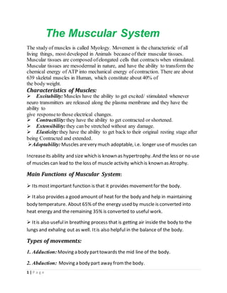
- 1. 1 | P a g e The Muscular System The study of muscles is called Myology. Movement is the characteristic of all living things, most developed in Animals because of their muscular tissues. Muscular tissues are composed ofelongated cells that contracts when stimulated. Muscular tissues are mesodermal in nature, and have the ability to transform the chemical energy of ATP into mechanical energy of contraction. There are about 639 skeletal muscles in Human, which constitute about 40% of the body weight. Characteristics of Muscles: Excitability: Muscles have the ability to get excited/ stimulated whenever neuro transmitters are released along the plasma membrane and they have the ability to give responseto those electrical changes. Contractility: they have the ability to get contracted or shortened. Extensibility: they can be stretched without any damage. Elasticity: they have the ability to get back to their original resting stage after being Contracted and extended. Adoptability: Muscles arevery much adoptable, i.e. longer use of muscles can Increaseits ability and size which is known as hypertrophy. And the less or no use of muscles can lead to the loss of muscle activity which is known as Atrophy. Main Functions of Muscular System: Its mostimportant function is that it provides movementfor the body. Italso provides a good amount of heat for the body and help in maintaining body temperature. About 65% of the energy used by muscle is converted into heat energy and the remaining 35% is converted to useful work. Itis also usefulin breathing process that is getting air inside the body to the lungs and exhaling out as well. Itis also helpful in the balance of the body. Types of movements: 1. Adduction: Moving a body parttowards the mid line of the body. 2. Abduction: Moving a body part away fromthe body.
- 2. 2 | P a g e 3. Flexion: Bending a joint to decreasethe angel. 4. Extension: Straightening a jointto increasethe angel. 5. Rotation: Moving a body part around its own axis. Types of Human Muscles: 1. Skeletal Muscles: ThoseMuscles which is attached to skeleton i.e. Bones or Cartilage are called Skeletal Muscles. They are attached to the skeleton by fibrous connective tissues called tendons. They arevoluntary in function and they are striated,(having alternate light and dark bands. A single Skeletal Muscle Cell is elongated, striated and multinucleated. 2. CardiacMuscles:Heartis the only organ which is not directly controlled by Brain. Heart’s pacemaker controlcardiac muscles contraction in about 0.8 sec/beat. Sometime the Heart beating or contraction shifts into high gear, for a brief period of time controlled by Vegus Nerve. The cardiac Muscles are Involuntary: they are not under conscious control. They have Rhythmicity, continuously contracting and relaxing fromthe start of birth until death. They are striated, having alternate light and dark bands. They are Strongestof the all muscles, i.e. pumps blood to overall 80-thousand- mile Length of blood vessels. They may be uninucleated or sometime binucleated. They havecytoplasmic bridges called Intercalated discs, through which the nervous transmission is conducted fromone cell to another. 3. Smooth Muscles:Those muscles which are found in visceralorgans arecalled smooth muscles. They are not striated, and very slow in contraction and relaxation. They are present in alimentary canal, blood vessels, urinary system and respiratory system etc.
- 3. 3 | P a g e Skeletal muscles: Muscle fiber: Musclefiber is elongated cylindricalmultinucleated cell of the skeletal muscles. Itis unbranched and striated and have a highly organized internal arrangement. For example, they are Multinucleated Their cytoplasm is known as sarcoplasm. There plasma membrane is called Sarcolemma, which is infolded inwards to the sarcoplasmforming a tubule called T-Tubule. T-tubuleis continuous with extra cellular fluid. Mitochondria (Sarcosomes) are present in large number. Sarcoplasmicreticulum storeCa++ions. Myoglobin is also presentas oxygen storagecompounds. there is also some stored glycogen. Structureof skeletal muscles: Skeletal muscles are surrounded by layer of connective tissues called epimysium. The skeletal muscles are divided into many portions called Fascicles, surrounded by perimysium.
- 4. 4 | P a g e A fascicle contains many musclefibers which are surrounded by endomysium. Protein bundles present in the musclefiber are called myofibrils. Each myofibril is a bundle of parallel proteins called Myofilaments. There are two kinds of myofilaments. 1. Thick filament (Myosin):They are fixed. Ithas double globular head or cross bridge and a tail. The head region contains actin binding site and also contain an ATP binding site at hinge point. 2. Thin Filament (Actins):Itis composed of two stands of proteins called Actin. It has an active site which can bind to myosin head. Italso contains another protein called tropomyosin which blocks the active site of Actin, preventing the binding of actin to myosin head. It also has calcium binding protein called troponin. Sarcomere:It is the smallest contractile unit of muscle which contains one dark band (A Band) and one light band (I Band).
- 5. 5 | P a g e Muscle contraction: All kind of movement accepts growth and cytoplasmic streaming are controlled by muscle contraction. The contraction of muscleis explained by a theory called sliding filament theory proposed by Jean Hanson and Hugh Huxley. According to this hypothesis when a muscle contracts, the actin and myosin slide pass each other. Excitation of muscle: A nervefiber and all the muscle fibers innervated by it are called a motor unit. They behave as a single motor unit. Upon receiving nerve impulse fromthe brain, the nervefiber releases neurotransmitters, the action potential pass through T-tubules to the sarcoplasmic reticulum to release the Ca++ ions. Mechanism of Muscle contraction: Due to the neuro transmitter the action potential prolonged down to the nerve fiber through t-tubule and release Ca++ ions from the sarcoplasmic reticulum. The Ca ions get attached to the troponin of the thin filaments. Due to this attachment "structuralorientation" occurs in the thin filament and the tropomyosin move away from actin exposing its active site. At the same time an ATP molecule attach to the myosin head due to which it cocks (moveinto extended high energy position) and it get attached to the active site of the actin forming "cross bride". Now as the myosin useoff the ATP molecule converting to ADP and releasing energy. That energy Exert power on the actin and move it "notch" and the myosin head reduce its angel from90 ° to 45°. This is called power stroke, sliding of thin filament over thick filament. The myosin head remain attached to the actin until it binds a new ATP. Upon binding more ATP, myosin releases the actin. It Is now prepared to repeat the whole process—itwill hydrolyzetheATP, recock (the recovery stroke) attached to the new active sited own on the thin filament and produceanother power stroke.
- 6. 6 | P a g e In the intense exercise a molecule called phosphocreatineis used which provide inorganic phosphate for the used ATPs. Recovery: when the impulse ceases fromthe CNS, the Sarcoplasmic reticulum Reaccumulates /reabsorbed all the Ca++ ions with in milliseconds. As the Ca++ ions are lost from troponin the tropomyosin again come to its original position again blocking the active site of the actin. And when new ATP molecule get attached to the myosin it releases from actin and the whole sarcomereget relaxed so as the whole muscle. CREATED BY: AZIZ KHAN BS ZOOLOGY: 3RD SEMESTER UNIVERSITY OF SWAT