
Recommended
More Related Content
What's hot
What's hot (20)
Similar to A report about the accessory organs to the digestive system
Similar to A report about the accessory organs to the digestive system (20)
Recently uploaded
Recently uploaded (20)
A report about the accessory organs to the digestive system
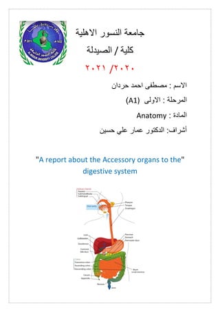
- 1. االهلية النسور جامعة كلية / الصيدلة 0202 / 0202 حردان احمد مصطفى : االسم المرحل ة االولى : A1) ) الماد ة : Anatomy حسين علي عمار الدكتور :أشراف "A report about the Accessory organs to the" digestive system
- 2. " " Accessory organs to the digestive system The salivary glands : are ductal glands in mammals that secrete saliva. They are solid formations made up of millions of secretory cells. Thin channels flow between these cells that collect and carry saliva and direct it to a single channel that in turn carries saliva into the mouth . 1-The parotid glands are the largest salivary glands, and they are located on the side of the lower jaw in front of the ear . 2-The sublingual or lingual glands are the smallest salivary glands and are found under the tongue. What distinguishes them from other salivary glands is that instead of having a single large duct such as the parotid and submandibular glands, it has an entire row of much smaller ducts that open in the mouth along the transverse edge. Minor located in the floor of the mouth under the tongue. 3-submandibular glands, they are located below the base of the tongue, and each submandibular gland has a channel that extends forward, penetrating the tissues at
- 3. the bottom of the mouth and opening through an opening that can be seen easily at the base of the small limb of the tongue. ? Q/What is the function of the salivary glands The salivary glands secrete saliva, which has many benefits for the oral cavity and its overall health. These benefits include: Protection Saliva is made up of proteins that lubricate and protect the soft and hard tissues of the oral cavity. Caching In general the higher the saliva flow rate, the faster the clearance and the greater the buffer capacity, thus better protection from tooth decay is provided. Therefore, people who have a slower rate of salivation
- 4. along with a lower ability to buffer, reduce the protection of saliva against microbes. Membrane formation Saliva forms a thin film on the surface of a tooth. The membrane contains mucus and glycoprotein rich in proline from saliva. The proteins (ethestrin and proline- rich proteins) within the salivary glutes prevent demineralization and encourage remineralization by attracting calcium ions. Maintaining healthy teeth Demineralization occurs when enamel degrades due to the presence of acid. When this happens, the effect of the buffering ability of saliva (increases the rate of saliva flow) prevents demineralization. Saliva can then begin to promote tooth remineralization by hardening the enamel with calcium minerals and phosphates. Antimicrobial Depending on the elements it contains, saliva can inhibit microbial growth. For example, lactoferrin in saliva naturally binds to iron. Since iron is a major component of bacterial cell walls, iron removal breaks down the cell
- 5. wall which in turn breaks down the bacteria. Antimicrobial peptides inhibit the growth of Candida albicans and streptococcus. Salivary immune globulin A collects oral bacteria and prevents dental plaque from forming. Tissue repair Saliva can encourage soft tissue repair by reducing clotting time and increasing wound shrinkage. digestion Saliva contains the enzyme amylase, which breaks down starch into maltose and dextrin. As a result, saliva allows digestion before food reaches the stomach. Tasting Saliva acts as a solvent in which solid particles can dissolve and enter the taste buds through the oral mucosa on the tongue. These taste buds are found within the synthesized loops, where small salivary glands secrete saliva.
- 6. The liver :an organ found only in vertebrates, detoxifies various metabolites, synthesizes proteins, and produces biochemicals needed for digestion. In humans, the liver is located in the upper right quadrant of the abdomen, below the diaphragm. Other roles in metabolism include: regulation of glycogen storage, the breakdown of red blood cells, and hormone production. The liver is an attached digestive gland that produces bile (an alkaline compound that helps break down fats). Bile aids digestion by emulsifying fats. The bile produced by the liver is stored in the gallbladder - a small sac that lies under the liver - and then it travels to the small intestine to complete the digestive process. Functional anatom: Liver hilum, which is the yellow circle par 1-The central area, or hilum of the liver, includes the area known as the porta hepatis, which consists of: the common bile duct, the common hepatic artery, and the portal vein. The duct, vein, and artery divide into left and right branches, and the hepatic regions fed by these branches form the left and right functional lobes . 2-The functional lobes are separated by an imaginary plane (the Cantele line, which extends from the
- 7. gallbladder fossa to the inferior vena cava) into true right and left lobes. 3-The liver is also divided into left and right lobes by the middle hepatic vein. 4-The right lobe is divided into an anterior segment and a posterior segment by the right hepatic vein 5-The left lobe is divided into medial and lateral segments by the left hepatic vein The gallbladder: in vertebrates, is a small, hollow organ in which bile is stored and concentrated before it is released into the small intestine. In humans, the gallbladder piriformis is located below the liver, although the structure and location of the gallbladder may differ greatly between animal species. The gallbladder receives and stores bile produced from the common hepatic duct, then releases it through the common bile duct towards the duodenum, where the juice works to aid in the digestion of fats. Microscopic anatomy: Micrograph of normal gallbladder wall, stain H&E The gallbladder wall consists of a number of layers, where the inner surface of the gallbladder wall is lined by a single layer of columnar cells with a brushy edge of
- 8. the microvilli, much like intestinal absorptive cells. Below the epithelium lies the lamina propria, the muscular layer, the outer layer around the muscle and the serous membrane. Unlike all other places in the intestinal tract, the gallbladder does not contain a mucosal muscle plate, and muscle fibers are not arranged in distinct layers . The mucosa is the inner part of the gallbladder wall, where it consists of a single layer of cephalic cells, with cells that have small hair-like attachments called microvilli, which are located on a thin layer of connective tissue called the lamina propria. The mucosa contracts and collects in small external pockets called rugae. The muscle layer is located below the mucosa, where it is made up of a smooth muscle with fibers that run longitudinally, obliquely and transversely, and are not arranged in separate layers. The function of muscle fibers is to contract to remove bile from the gallbladder. The gallbladder contains Rokitansky-Aschov pockets, which are a distinctive feature of it, which are external pockets of the mucous membrane, and may extend into the muscle layer, indicating the occurrence of adenoma.
- 9. The muscle layer is surrounded by a layer of connective and fatty tissue . The outer layer of the bottom of the gallbladder, and the surfaces not in contact with the liver, are covered by a thick serous membrane, which faces the abdominal serous membrane. The serous membrane contains blood and lymph vessels. Connective tissue covers the surfaces that come into contact with the liver. Q/What are the functions of the gallbladder? 1-The storage of bile is the primary function of the gallbladder, as it is used to digest fats from food. Juice is produced in the liver, then it flows through small vessels into large liver ducts, and eventually it flows through the gallbladder duct (part of the bile tree) to reach the gallbladder, where it is stored. At any given time, 30 to 60 milliliters (1.0 to 2.0 fluid ounces) of juice are stored in the gallbladder. 2-The liver also produces bile, which is stored in the gallbladder, and it differs from the bile secreted by the gallbladder. During the gallbladder storing the bile, it is concentrated 3-10 times by removing some water and electrolytes, as this process takes place through the active transport of sodium and chlorine ions through the epithelium of the gallbladder, which creates an osmotic
- 10. pressure that leads to the reabsorption of water and other electrolytes. The pancreas: or pancreatic organ, or complicated or sweet, is one of the most important glands in the body of mammals and has a dual role: 1-Exocrine gland: It secretes pancreatic juice containing digestive enzymes. 2- An endocrine gland, or endocrine gland, is based on the secretion of a number of hormones, the most important of which is insulin, which is secreted by the Langerhans Islands and which acts to represent sugar and carbohydrates. Its imbalance mainly leads to diabetes, for example. Anatomy of pancreas: The colostrum is a soft, lobular gland that lies on the posterior abdominal wall and extends transversely in the abdomen from the duodenal curve to the navel of the spleen, behind the stomach. They are about 13 cm long, 2-7 cm wide, and weigh 70-80 g. It is located at the level of the first lumbar vertebra, just below the celiac trunk. The transverse colon mesentery is attached to it, separating the supraclone and hypochondral space.
- 11. 1.The bile ducts: 2. Intrahepatic bile ducts, 3. Right and left hepatic ducts, 4. The common hepatic duct, 5. The gallbladder duct, 6. The common bile duct, 7. The ampulla of Vater, 8. The large duodenal papilla 9.Gallbladder, 10--11. The left and right lobes of the liver. 12. The spleen 13.The esophagus. 14. The stomach. 15. The pancreas: 16. The extra pancreatic duct, 17. The pancreatic duct 18.Small intestine: 19. Duodenum, 20. The jejunum 21—22.Right and left kidneys