
Recommended
More Related Content
What's hot
What's hot (20)
Similar to Scottie Dog Sign.pdf
Similar to Scottie Dog Sign.pdf (20)
Recently uploaded
Recently uploaded (20)
Scottie Dog Sign.pdf
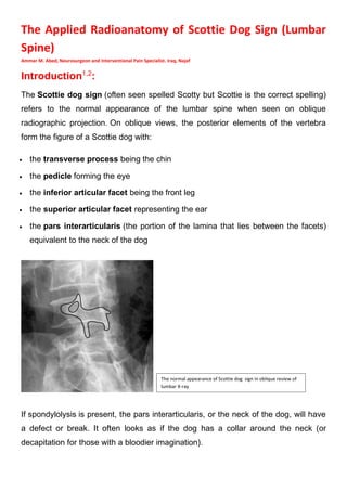
- 1. The Applied Radioanatomy of Scottie Dog Sign (Lumbar Spine) Ammar M. Abed, Neurosurgeon and Interventional Pain Specialist. Iraq, Najaf Introduction1,2 : The Scottie dog sign (often seen spelled Scotty but Scottie is the correct spelling) refers to the normal appearance of the lumbar spine when seen on oblique radiographic projection. On oblique views, the posterior elements of the vertebra form the figure of a Scottie dog with: • the transverse process being the chin • the pedicle forming the eye • the inferior articular facet being the front leg • the superior articular facet representing the ear • the pars interarticularis (the portion of the lamina that lies between the facets) equivalent to the neck of the dog If spondylolysis is present, the pars interarticularis, or the neck of the dog, will have a defect or break. It often looks as if the dog has a collar around the neck (or decapitation for those with a bloodier imagination). The normal appearance of Scottie dog sign in oblique review of lumbar X-ray
- 2. The clinical significant of these parts will be discussed below: The Chin of Scottie Dog: The chin of Scottie dog represents the transverse process of the lumbar vertebra and it is important in minimally invasive spine procedures during the lumbar transforaminal epidural steroid injection, radiofrequency nerve ablation, and endoscopy The approach to the lumbar transforaminal region is by prone position, local anesthesia, and fluoroscopy guided. The site of the injection or procedure is in Kambin's triangle inferior to the transverse process in oblique view (chin of Scottie dog). Kambin's triangle is a safe space because it is devoid of vascular and neural structures of importance. Fluoroscopy3,4,5 : 1. Start with AP view to identify the targeted level. L5 pars defect Kambin's triangle
- 3. 2. Then square the superior endplate of the targeted level (This entails adjusting the fluoroscope in a cranial/caudal orientation until the endplates are aligned and appear as a single straight line). 3. Once the appropriate level is identified, oblique the view 15–30° to the ipsilateral side, producing the Scottie dog view. Procedure3,4,5 : 1. Entry Point: Lateral to the desired foramen. 2. Anesthetize the skin at the desired entry point. 3. Target: On the oblique view, just below the 6 o’clock position of the pedicle or below the chin of the Scottie dog 4. Insert the needle and slowly advance toward the antero/lateral aspect of the epidural space. 5. The needle is advanced until the foraminal ligament is felt. 6. After negative aspiration, injection of contrast should show contrast spread in the anterior epidural space in the lateral view, as well as perineural and epidural spread in the AP view without vascular runoff or intrathecal spread (If aspiration is positive for blood or CSF, the needle should be repositioned). Oblique view for transforaminal approach to an epidural steroid injection at L4–L5. The L4 nerve is identified as yellow color. It appears from the region below the chin of Scottie dog (from: Pain Management and Palliative Care, Kimberly A. Sackheim Editor, Springer)
- 4. The Eye of Scottie Dog: The eye of Scottie dog represents the pedicle and it is an excellent sign for percutaneous screw fixation, percutaneous cementoplasty, and lumbar medial branch block as the medial nerve passes there. Here we will discuss the lumbar medial branch block. Anatomy of lumbar medial branch nerves6-9 : 1. Each lumbar medial branch nerve originates from the dorsal root ganglia of the spinal nerve above and descends to lie in the junction between the superior articular process and transverse process at each level. The site of injection below the chin of Scottie dog (from: Pain Management and Palliative Care, Kimberly A. Sackheim Editor, Springer) The site of injection at the eye of Scottie dog (from: Pain Management and Palliative Care, Kimberly A. Sackheim Editor, Springer)
- 5. 2. The medial branch nerve slopes inferomedially before branching to give innervation to the facet joint above and below. 3. Lumbar facet joints are innervated by the medial branch at the same level and from the level above. Therefore, both of these nerves must be blocked in order to diagnose/treat pain emanating from a single facet joint. (for example, the L3–L4 facet joint is innervated by the L2 and L3 medial branch nerves) 4. The L5 dorsal primary ramus is blocked at the junction of the S1 superior articular process and the sacral ala. Fluoroscopy6-9 : 1. Obtain an AP view to identify the appropriate level. 2. Oblique the fluoroscopic view 25–35° to the ipsilateral side to optimize visualization of the Scottie dog. 3. Cranial or cephalad tilt can be used to square off the endplates of the targeted level. 4. The target is the intersection of the transverse process and the superior articular process (the eye of the Scottie dog). 5. The one variation is the intersection of the S1 superior articular process with the sacral ala for the L5 dorsal primary ramus block. Procedure: 1. Entry Point: lateral and slightly superior to the targeted pedicle. 2. Anesthetize the skin at the entry point. 3. Advance a 22 gauge curved spinal needle toward the target site. 4. Target is the periosteum at the junction of the superior articular process and transverse process. 5. Once the needle makes contact with periosteum, a lateral fluoroscopic image can be used to confirm appropriate depth (posterior to the neural foramen). 6. Once needle position is confirmed and aspiration is negative for blood, medications are administered at each level.
- 6. The Leg and Ear of Scottie Dog: The meeting of the front leg (inferior facet) with the ear (superior facet) of the below Scottie dog represents the facet joint. And this meeting is the site of the steroid injection of intra-facet injection. Anatomy of the Lumbar Facet Joint6-9 : 1. Facet joints are formed from the superior articular process (ear of Scottie dog) of the inferior vertebra and the inferior articular process (front leg of Scottie dog) of the superior vertebra. 2. Each lumbar facet joint is innervated by two medial branch nerves: the first branch arises at the level of each joint and the Second branch originates from the level directly above. 3. The facet joint is a true synovial joint with a limited volume that is typically <1.5 mL in the lumbar region. Fluoroscopy6-9 : 1. AP view is used to identify the targeted level. 2. Then oblique 25–35° to the ipsilateral side to visualize the joint space formed by the junction of the superior and inferior articular processes. The front leg meets the below ear and the result is facet joint
- 7. Procedure6-9 : 1. Entry Point: inferior one-third of the targeted facet joint. 2. The skin is anesthetized at the entry point. 3. A 22 gauge spinal needle is advanced in a coaxial view into the inferior aspect of the posterior joint space. 4. Lateral fluoroscopy is used to confirm appropriate depth with the needle tip in the posterior aspect of the joint. 5. Some resistance should be felt when the needle tip enters the posterior capsule of the facet joint. The neck of Scottie Dog: Acute pars fracture describes a defect of the lumbar spine due to recurring mechanical stress and is commonly seen in athletic individuals. Low back pain that is worse with extension may be seen on presentation. X-ray with an oblique view is usually the first test to order. oblique X-ray view show lateral a deformity of the neck or collar of the Scottie dog. The neck or collar of Scottie dog indicates the pars fracture
- 8. Summary the Scottie sign is an excellent sign in the minimally invasive lumbar procedures by which the surgeon can do these invasive procedure as accurate, easy, and rapid by fluoroscopy guided imaging. References: 1. Mellado JM, Larrosa R, Martín J et-al. MDCT of variations and anomalies of the neural arch and its processes: part 1--pedicles, pars interarticularis, laminae, and spinous process. AJR Am J Roentgenol. 2011;197 (1): W104-13. doi:10.2214/AJR.10.5803 - Pubmed citation 2. 2. Yochum TR, Rowe LJ. Essentials of Skeletal Radiology. Lippincott Williams & Wilkins. (2004) ISBN:0781739462. Read it at Google Books - Find it at Amazon 3. Stizman BT, Stizman BT. Epidural injections. In: Fenton DS, Czervionke LF, editors. Image-guided spine intervention. Philadelphia: Saunders; 2003 4. Benzon HT. Selective nerve root blocks and transforaminal epidural steroid injections for back pain and sciatica. In: Benzon HT, Raja SN, Molloy RE, Liu SS, Fishman SM, editors. Essentials of pain medicine and regional anesthesia. 2nd ed. Philadelphia: Elsevier; 2005 5. Rathmell JP. Atlas of image-guided intervention in regional anesthesia and pain medicine. 2nd ed. Philadelphia: Lippincott Williams & Wilkins; 2012. p. 64–79. Chapter 6: Transforaminal and selective spinal nerve injection 6. Rathmell JP. Atlas of image-guided intervention in regional anesthesia and pain medicine. 2nd ed. Philadelphia: Lippincott Williams & Wilkins; 2012. p. 80–117. Chapter 7: Facet injection: intraarticular injection, medial branch block, and radiofrequency treatment 7. Czervionke LF, Fenton DS. Facet joint injection and medial branch block. In: Fenton DS, Czervionke LF, editors. Image-guided spine intervention. Philadelphia: Saunders; 2003 8. Raj PP, Lou L, Erdine S, Staats PS, Waldman SD. Radiographic imaging for regional anesthesia and pain management. Philadelphia: Churchill Livingstone; 2003. p. 185–96. Chapter 30: Lumbar facet block and median branch blocks 9. Clemons RR, Benzon HT. Facet syndrome: facet joint injections and facet nerve blocks. In: Benzon HT, Raja SN, Molloy RE, Liu SS, Fishman SM, editors. Essentials of pain medicine and regional anesthesia. 2nd ed. Philadelphia: Elsevier; 2005