Call Girls Hsr Layout Just Call 7001305949 Top Class Call Girl Service Available
Endo perio 2020 (1).pdf
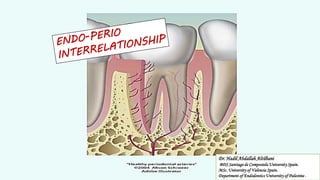
1. Dr. Hadil Abdallah Altilbani
BDS Santiago de Compostela University Spain.
MSc. University of Valencia Spain.
Department of Endodontics University of Palestine .
2. The tooth, the pulp tissue within it and its supporting
structures should be viewed as
One Biologic Unit
The interrelationship of these structures
influences each other during health, function
and disease.
It's the spread of inflammation and infection from one
component to the other.
Periodontic _ Endodontic
Relationship
DEFINITION
The interrelationships between pulpal and
periodontal disease primarily occur by way of the
intimate anatomic and vascular connections between
the pulp and the periodontium ,also they have the
same embryonic origin and they share the same
function protect, and provide nourishment to the
teeth.
3. The various pathways of communication between the pulp and the periodontium may be classified as:
I) DEVELOPMENTAL
I. Apical Foramen
II. Lateral or Accessory Canals
III. Dentinal Tubules
IV. Developmental or Lingual Grooves
V. Tooth / Root Anomalies
II) PATHOLOGICAL
I. Empty spaces created by destroyed Sharpey’s fibres
II. Root fractures following trauma
III. Idiopathic Resorption (Internal or External)
IV. Cemental Agenesis or Hypoplasia
III) IATROGENIC
I. Exposure of dentinal tubules following root planing
II. Accidental lateral perforations during endodontic treatment
Pathways of Communication
The similarity between the endodontic and periodontal microflora suggests that a cross infection can occur between the root canals and
periodontal pocket.
communication between the pulp and the
periodontal tissues.
Pulpal and periodontal problems are responsible for more than 50% of tooth mortality.
J Conserv Dent. 2008 Apr-Jun; 11(2): 54–62.
PATHWAYS OF COMMUNICATION
The microbial findings, similarities in the composition of cellular infiltrates also suggest the
existence of
4. Anatomical pathways
Apical Foramen:
Total disintegration of the pulp is only a certainty if bacterial plaque involves the main apical foramen,
compromising the vascular supply.
The pulp has good defense capacity as long as the blood supply through
the apical holes is intact.
Following Necrosis Of The Pulp, various bacterial products like enzymes, metabolites, antigens etc. reach the
periodontium through the apical foramen, initiating and perpetuating an inflammatory response there.
This results in destruction of periodontal tissue fibers and resorption of the adjacent alveolar bone.
External resorption of the cementum can also occur concurrently.
Lateral canals:
These ramifications are now currently termed as ‘accessory canals’.
The term accessory canal is now used to describe any ramification that
connects the root canal system to the periodontal ligament.
30-40% of canals have lateral or accessory canals
The frequency of these ramifications on the root surface are as follows:
❑ apical third 17%,
❑ coronal third 1.6%
❑ body of the root 8.8%.
Bender et al., stated that periodontal endodontic problems were much more frequent in the molars than
in the anterior teeth
The majority of the accessory canals are found in The Apical Part of the root and lateral canals in the
Molar Furcation Region.
5. The radiographic indications of the presence of lateral canals
before obturation are:
1. Localized Thickening of periodontal
ligament on the lateral root surface
2. A frank Lateral Lesion It is essential that the
dentist recognizes and is familiar with canal
ramifications and variations.
6. Passage of microorganisms between the pulp and periodontal
tissues is possible through these tubules, when the dentinal
tubules are exposed in areas of
denuded cementum.
Tubular pathways
The tubules may be denuded of their
cementum coverage as a result of :
❑ Perio Disease
❑ Surgical Procedures
❑ Developmentally
when the cementum and enamel do not meet at the
CEJ thus leaving areas of exposed dentin
8. A. Exposure of dentinal tubules following
root planning
Iatrogenic origin
Nonphysiological pathways
b. Accdental lateral perforation during
endodontic procedure.
c. Root fracture due to endodontic
procedure.
The incidence of root fractures is
more in the roots that are filled
with lateral condensation technique
and the teeth restored with
intracanal posts.
9. • Two basic questions have been raised and continue to be a matter of
dispute :
ENDODONTIC PERIODONTAL-CONTROVERSY
1) Is periodontal disease a cause of pulp necrosis?
2) Can a pulpless tooth be the cause of periodontal disease?
Etiopathogenesis
Of
Perio-endo Lesions
Effect of periodontal lesions on the pulp
The effects of pulpal disease on the periodontium are well documented , a clear –cut
relationship between periodontitis and pulpal involvement is less evident .
One may possible that bacteria and the inflammatory products of periodontitis could
gain access to the pulp through accessory canals , apical foramen , or dentinal tubules .
This process , the reverse of the effects
of a necrotic pulp on the periodontum
has been referred to As
if it exists ,it is Rare .
10. The effect of periodontal lesions on the pulp can Result in atrophic and other degenerative changes like:
CAUSE:
Disruption of blood flow through the lateral canals localized areas of Coagulation Necrosis in the
pulp.
Effect of periodontal lesions on the pulp
1. Reduction In The Number Of Pulp Cells,
2. Dystrophic Mineralization,
3. Fibrosis,
4. Reparative Dentin Formation,
5. Inflammation And Resorption
With slowly advancing periodontal disease,
cementum deposition may act to obliterate
lateral canals before pulpal irritation occurs.
11. The most common periodontal lesion produced by the pulp disease is The
Localized Apical Granuloma.
The effects of pulpal disease on the periodontium
As long as the pulp remains vital, it is unlikely that significant changes will occur in
the periodontium..
Pulpal tissue inflamed may be little or no effect on the periodontium .
Necrosis , can result in bone resorption and rediolucency at the apex , In the furcation
, or at points along the root .
The resulting lesion may be an acute apical lesion , or abscess , a more chronic
periradicular Lesion ( cyst or granuloma ), or lesion associated with a lateral or
accesory canals .
Resorption:
Resorption of the sides of the roots is frequently found adjacent to the granulation tissue
overlying the roots.
When the periodontal lesions are deep, resorption may also be found within the root canals,
often opposite lateral canals, and at the apical foramen.
Since this resorptive process extends into the dentin peripherally towards the pulp, and the
activating factors are produced from the periodontal lesion, a name which reflects the
etiology of this phenomenon,
Peripheral Inflammatory Root Resorption (PIRR) Was Proposed.
13. A close relationship exists between disease of the dental pulp and
periodontal disease, and it expresses itself in several ways.
The most commonly used classification was given by Simon, Glick
and Frank in 1972 According to this classification, perio-endo
lesions can be classified into:
1. Primary endodontic lesion
2. Primary periodontal lesion
3. Primary endodontic lesion with secondary periodontal involvement
4. Primary periodontal lesion with secondary endodontic involvement
5. True combined lesion
Based on etiology Diagnosis, Prognosis and Treatment
CLASSIFICATION OF PERIO-ENDO
LESIONS
14. Based on the primary source of infection.
Chronic apical lesion on a tooth with a necrotic pulp may Drian coronally
through the periodontal ligament into the gingival sulcus.
Pulpal infection may cause a tissue-destructive process that proceeds from the
apical region of a tooth toward the gingival margin.
“Retrograde Periodontitis”
PRIMARY ENDODONTIC LESION
Extraosseous fistulation
Drainage of endodontic abscess into the
sulcus follows one or two routes:
15. This condition may clinically mimic the presence of a Periodontal
Abscess.
In reality, however, it would be a sinus tract originating from the pulp
that opens into the periodontal ligament.
The sinus tract extending into the gingival sulcus or furcation area
disappears at an early stage, if the necrotic pulp has been removed
and the root canals are well sealed.
PRIMARY ENDODONTIC LESION
Pre-op - periapical and furcal RL + a deep
narrow perio defect
Periodontal ligament fistulation
16. PRIMARY PERIODONTAL LESION
These lesions are caused primarily by periodontal pathogens.
In this process, chronic periodontitis progresses apically along the root surface.
In most cases, pulpal tests indicate a clinically Normal Pulpal Reaction.
There is frequently an accumulation of plaque and calculus and the presence of deep
pockets may be detected.
Pre-op: alveolar bone loss + a periapical lesion, a deep narrow
pocket was traced on the mesial aspect of the root, The Tooth
Tested Vital
17. Primary endodontic lesion with
secondary periodontal involvement
If a primary endodontic lesion remains Untreated, it may become secondarily involved with
periodontal breakdown.
Plaque accumulation at the gingival margin of the sinus tract leads to plaque induced
periodontitis in this area.
When plaque and calculus are detected, the Treatment And Prognosis of the teeth are
different from those of the teeth involved with only endodontic disease.
The tooth now requires both Endodontic And Periodontal Treatment.
COMBINED DISEASES
Root fractures may also present as primary endodontic lesions with
secondary periodontal involvement.
Thes typically occur in root canal treated teeth, often with posts and
crowns.
The signs may range from a local deepening of periodontal pocket to
a more acute periodontal abscess formation.
18. Primary periodontal disease with secondary
endodontic involvement
The apical progression of a periodontal pocket may continue until the apical tissues are
involved.
In this case, the Pulp May become necrotic as a result of infection entering through lateral
canals or the apical foramen.
In single-rooted teeth, the prognosis is usually poor.
In molar teeth, the prognosis may be better.
Since not all the roots may suffer the same loss of supporting tissue, root resection can be
considered as a treatment alternative.
At initial presentation shows evidence of horizontal bone loss as well as a periapical radiolucency. The
crown was intact, but Vitality Tests Were Negative. The post-op radiograph shows that a lateral canal was
exposed to the oral environment due to bone loss. That lateral canal could serve as a potential pathway
for bacteria.
19. It is possible for a blood vessel within a lateral canal to be severed by a curette and for the microorganisms to be
pushed into the area during treatment, resulting in pulp inflammation and necrosis.
If the blood supply circulating through the apex is intact, the pulp has good prospects for survival.
Lateral canals and dentinal tubules may be opened to the oral environment by scaling and root planning or surgical
flap procedures.
20. True combined lesion
True combined endodontic periodontal disease occurs less frequently than other
endodontic-periodontal problems.
It is formed when an endodontic lesion progressing coronally joins an infected periodontal
pocket progressing apically.
The degree of attachment loss in this type of lesion is invariably large and the prognosis
guarded.
This is particularly true in single rooted teeth. In molar teeth, root resection can be an
alternative treatment.
The radiographic appearance of combined endodontic periodontal disease may be similar to
that of a vertically fractured tooth.
If a sinus tract is present, it may be necessary to raise a flap to determine the etiology of the
lesion.
Radiograph shows separate progression of endodontic disease and
periodontal disease. The tooth remained untreated and
consequently the two lesions joined together
21. True Combined Disease
Radiograph shows bone loss in 2/3 of the root with calculus
present and a separate periapical radiolucency. Clinical
exam revealed coronal color change and pus exuding from
the gingival crevice. Pulp vitality tests were negative
22. Concomitant Endo-perio Lesion
Is An Additional Classification
That Has Been Proposed To
Describe
The Presence Of Endo
And Perio Disease As Two
Separate And Distinct Entities
Concomitant Lesion
23. Diagnosis of primary endodontic disease and primary
periodontal disease usually present no clinical
difficulty.
In primary periodontal
disease, the pulp is vital and
responsive to testing.
In primary endodontic
disease, the pulp is infected
and nonvital.
DIAGNOSIS OF PERIODONTALENDODONTIC
LESIONS
24. The following steps in diagnosis, aids in deciding an appropriate treatment plan:
However, primary endodontic disease with secondary periodontal involvement, primary periodontal disease with secondary
endodontic involvement, or true combined diseases are clinically and radiographically Very Similar.
Accurate diagnosis can be achieved by careful history taking, examination and the use of special tests .
The extra oral and intra oral tissues are examined for the presence of any abnormality or disease.
Differences between periodontal and pulpal lesion
Signs and symptoms of periodontitis :
Teeth With Chronic Periodontal Lesions are typically free of acute symptoms, the patient may be unware of the condition ,except for bleeding
on brushing and flossing , or bad breath .
Increased tooth mobility may occur if sufficient attachment has been lost .
dental radiographs usually disclose the extent of attachment loss , which should correlate with clinical probing data .
Differences between periodontal and pulpal lesion
Signs and symptoms of pulpal disease:
The Teeth With Pulpal Inflammation ,respond normally to percussion and palpation .
Thermal stimuli or percussion applied to teeth with irreversible pulpitis can provoke sever Pain .
This pain may be intense and is often described as bright or throbbing .
If The Inflammatory Process Extends to involve the periodontal ligament , the affected tooth can become
tender to pressure , biting ,or light tapping with an instrument .
Dental radiograghs usally document the presence of apical or lateral lesions .
The clinician should remember that some inflamed and Necrotic Pulps are asymptomatic and that the patient is
unaware of their existence .
26. ✓ When the pulp is nonvital and infected, conventional endodontic therapy
alone will resolve the lesion.
✓ Surgical endodontic therapy is Not necessary, even in the presence
of large periradicular radiolucencies and periodontal abscesses.
✓ If primary endodontic lesions persist, despite extensive endodontic
treatment, the lesion may have secondary periodontal involvement
or it may be a true combined lesion.factors
Before the commencement of any kind of advanced restorative work to treat a perio-endo lesion
The Prognosis
of the tooth should be considered carefully.
❑ Whether there is a Functional need for the tooth
❑ whether the tooth is Restorable after the lesion has been treated
❑ whether the Patient is suitable for a lengthy, costly and invasive treatment
27. TREATEMENT
PRIMARY ENDODONTIC LESION
Appropriate treatment varies with the presence , nature , and extent of Involvement of the disease .
Are sufficient to result in healing of the lesion .
Periodontal treatment is not required in the absence of any periodontal involvement .
CONVENTIONAL ENDODONTIC THERAPY
In case of secondary periodontal involvement
root canal therapy is instituted immediately and the cleaned and shaped root canal is filled with Calcium Hydroxide paste, which has
bactericidal, anti inflammatory and proteolytic property, inhibiting resorption and favoring repair.
It also inhibits periodontal contamination of Instrumented canals via patent channels connecting the pulp and periodontium before
periodontal treatment removes the contaminants.
Treatment results should be evaluated after Two To Three Months and only then should periodontal treatment be considered.
Prognosis of primary endodontic disease with secondary periodontal involvement depends on periodontal
treatment and patient response.
28. 1. Periodontal Therapy
2. Guided Tissue Regeneration
3. Root Amputation & Hemisection
4. Pulp Therapy
Treatement
Primary Periodontal Lesion
Should Be Treated First By Proper Hygiene Phase Therapy.
Poor Restorations And Any Developmental Grooves That Are Difficult To Be Altered And Make Oral Hygiene
Maintenance Problematic For The Patient Should Be Removed.
Periodontal Surgery Is Performed After The Completion Of Hygiene Phase Therapy.
Re-evaluation must be performed periodically after therapy to check for possible retrograde endodontic problems.
The prognosis is entirely dependent on the periodontal therapy, in such cases.
Early stage periodontal lesions with secondary endodontic involvement may present as reversible pulpal hypersensitivity, which can be
treated purely by periodontal therapy.
Periodontal treatment removes noxious stimuli, and secondary mineralization of dentinal tubules allows the resolution of
hypersensitivity.
The prognosis of periodontal lesions is poorer than endodontic lesions and is dependent on the apical extensions of the lesion.
29. True combined lesions are treated initially as for Primary Endodontic lesions with secondary periodontal involvement.
The prognosis of a true combined perio-endo lesion is often poor or even hopeless, especially when periodontal lesions are chronic, with extensive
loss of attachment.
Root amputation, hemisection or bicuspidization may allow the root configurations to be changed sufficiently for a part of the root structure to be saved.
The prognosis of an affected tooth can also be improved by increasing bony support, which can be achieved by bone grafting and guided tissue regeneration.
These advanced treatment options are based on responses to conventional periodontal and endodontic treatment over an extended time period.