SlideShare a Scribd company logo
1 of 22
Download to read offline
Chapter 3
Simple cloning
3.1 The basic experiment
3.1.1 Cloning using a simple plasmid
Now that we have surveyed the enzymes and techniques that are used
in cloning, we can take an easy example and see how they are applied.
One of the simplest exercises is inserting DNA into E. coli in a plasmid
such as pUC18 (Figure 3.1). Bacterial plasmids are circular dsDNA
molecules capable of replicating in bacterial cells. Insertion of
DNA into the plasmid allows the DNA to be propagated in a host
cell. Molecules that are used for propagating DNA in this way are
called vectors. The pUC18 plasmid is one of a series of similar vectors
and carries two genes of particular importance. One confers
resistance to the antibiotic ampicillin (Figure 3.2), which blocks the
cross-linking of the polysaccharide chains in the bacterial cell
walls. The lack of cross-linking weakens the bacterial cell wall and
the cells eventually lyse. The resistance gene (bla or ampR
) encodes
a beta-lactamase enzyme that hydrolyses the beta-lactam ring of
the antibiotic, inactivating it. The other gene (lacZ’) encodes part of
Fig 3.1 Generalized pUC
plasmid. It contains an ampicillin
resistance gene (ampR), part of a
beta-galactosidase gene (lacZ’),
the Lac repressor gene (lacI) and
an orgin of replication (ori).
The multiple cloning site (MCS)
is located in the beta-galactosidase
gene.
https://www.cambridge.org/core/terms. https://doi.org/10.1017/CBO9780511807343.005
Downloaded from https://www.cambridge.org/core. The Librarian-Seeley Historical Library, on 13 Dec 2019 at 14:57:54, subject to the Cambridge Core terms of use, available at
a beta-galactosidase enzyme, which normally cleaves disaccharides,
such as lactose, into monosaccharides. It can also cleave an artificial
substrate 5-bromo-4-chloro-3-indolyl-b-D-galactoside (also known as
X-gal), shown in Figure 3.2, to liberate a blue dye. For this reason, the
substrate is said to be chromogenic. We will see later why the gene for
the whole beta-galactosidase enzyme is not used, but for the moment
we can treat the plasmid as though it contained the entire gene.
As well as the ampR
and lacZ’ genes, the plasmid has some less
immediately obvious features that we will look at in more detail
later, such as an origin of replication. It also has a number of
recognition sites for restriction endonucleases, and many of these are
located in a region of the lacZ’ gene known as the multiple cloning
site or polylinker.
Suppose we wanted to clone genomic DNA from the organism we
were studying into the BamHI site of pUC18, located in the multiple
cloning site in the lacZ’ gene. (We do not have to use the BamHI
site. We could use any of the other restriction sites in the multiple
cloning site.) The procedure would be as follows. The first stage is
to purify genomic DNA from the organism of interest and cut it with
the enzyme BamHI. We would also purify some pUC18 and cut it
with the same enzyme. Then the BamHI-digested plasmid and the
BamHI-digested genomic DNA are mixed and DNA ligase in a suit-
able buffer is added. BamHI ends will anneal to BamHI ends and
the ligase will seal the nicks. That will generate a large number of
types of molecule, which may be linear or (topologically) circular.
Fig 3.2 Structure of ampicillin
(a), X-gal or 5-bromo-4-
chloro-3-indolyl-b-D-galactoside
(b), and IPTG or isopropyl-b-
D-thiogalactoside (c).
53
3.1 THE BASIC EXPERIMENT
https://www.cambridge.org/core/terms. https://doi.org/10.1017/CBO9780511807343.005
Downloaded from https://www.cambridge.org/core. The Librarian-Seeley Historical Library, on 13 Dec 2019 at 14:57:54, subject to the Cambridge Core terms of use, available at
Linear molecules will not be stably propagated in later stages of
the experiment, so we will not consider them further. Different
circular molecules that may be formed are shown in Figure 3.3.
Intramolecular ligation of plasmid DNA will regenerate the original
circular pUC18 plasmid. Intramolecular ligation of genomic DNA
will produce circular molecules of genomic DNA, with no plasmid
DNA sequences present. A wide range of intermolecular ligations can
take place. Two plasmid DNA molecules can be ligated, in a head-to-
tail or head-to-head configuration. Two genomic DNA molecules could
be ligated, again in either orientation. Most important, a genomic
DNA molecule can be ligated to a plasmid DNA molecule. (Of course,
a huge range of ligations involving three or more molecules can also
take place, but they will be rarer than unimolecular or bimolecular
ligations. How rare depends on the concentrations of the species
involved.)
The molecules that contain new combinations of sequences, not
present before, are termed recombinants. Note that the insertion of
DNA into the lacZ’ gene will destroy the function of that gene, as it
disrupts the coding region for the LacZ’ protein; therefore, it is an
easy matter to work out what properties each of the plasmid types
shown in Figure 3.3 will confer. It is possible that insertion of a short
piece of DNA into the lacZ’ gene might only partially inactivate the
gene function (or not affect it at all) if the reading frame were not
terminated or shifted. This is relatively uncommon, but the pos-
sibility should always be kept in mind.
The next stage is to put the ligation products back into E. coli. For
an experiment such as this, it would usually be done by chemically
induced transformation, or electroporation (Sections 1.5.2 and 1.5.3).
An important point is that the uptake of DNA by E. coli is rather
inefficient, in that the probability of any individual bacterial cell
taking up a piece of DNA is very low. For simple transformation
procedures, of 109
cells treated with 1 mg of supercoiled plasmid
DNA, only 105
or so will successfully take up a molecule. We say there
is a transformation frequency of 105
colonies/microgram of DNA. This
is low, given that 1 mg of pUC18 contains of the order of 1011
molecules. The cells are now plated on agar containing, in this
example, ampicillin, together with an inducer of the lacZ’ gene
(isopropyl-thiogalactoside (IPTG), Figure 3.2) and the chromogenic
substrate X-gal. The cells that have not taken up a plasmid will be
sensitive to the ampicillin and die after a few generations. Those that
took up genomic DNA only will also die. Those that took up pUC18
DNA will have acquired an ampicillin resistance gene and be able to
grow on the selective medium. They will, therefore, form isolated
colonies on the agar if plated at a suitable dilution. Thus, all the
members of a colony are derived from one cell and should have
acquired a single plasmid. (Because the probability of a cell taking up
one DNA molecule is low, the probability of a cell taking up two might
be expected to be very low indeed. Actually, that is likely not to be
the case. Although only a tiny fraction of cells is able to take up DNA,
54 SIMPLE CLONING
https://www.cambridge.org/core/terms. https://doi.org/10.1017/CBO9780511807343.005
Downloaded from https://www.cambridge.org/core. The Librarian-Seeley Historical Library, on 13 Dec 2019 at 14:57:54, subject to the Cambridge Core terms of use, available at
Fig 3.3 Ligation of BamHI-cut genomic DNA into a BamHI-cut vector.The figure shows the regeneration of vector, the
circularization of genomic DNA and the ways in which two separate molecules (vector or genomic DNA) can be joined.
The functional genes present on each molecule after ligation are indicated.
55
3.1 THE BASIC EXPERIMENT
https://www.cambridge.org/core/terms. https://doi.org/10.1017/CBO9780511807343.005
Downloaded from https://www.cambridge.org/core. The Librarian-Seeley Historical Library, on 13 Dec 2019 at 14:57:54, subject to the Cambridge Core terms of use, available at
those cells that do are quite efficient at DNA uptake and may take up
more than one piece. So a low, but significant fraction of colonies
may contain two different plasmids. Over time, these may segregate,
with some cells containing one plasmid and the rest containing
the other.)
The problem now is to distinguish the colonies that have acquired
genomic DNA from all the others. That can be done in this example
by using the lacZ’ gene. Colonies that have acquired pUC18 molecules
without an insert of genomic DNA will have an intact lacZ’ gene
(which will be induced by the IPTG). They will, therefore, be able to
break down the X-gal and be coloured blue. The colonies that have
acquired molecules with a genomic DNA insert will have a lacZ’ gene
that has been inactivated by the insertion. Thus, these colonies will be
unable to metabolize the X-gal and will be the usual white (actually
off-white) colour. The head-to-tail dimeric pUC18 molecules will also
give blue colonies, and the head-to-head dimers will not replicate
stably in the host cell. The relative abundances of blue and white
colonies will depend on factors such as the relative proportions of
genomic and plasmid DNA in the initial ligation. We are interested
in the white colonies. We can confirm that plasmids in the white
colonies contain an insert by growing up the bacteria, purifying
plasmid DNA and cutting it with a restriction enzyme. BamHI would
be the easiest in this case, and should generate two sizes of molecule:
plasmid and inserted genomic DNA. Plasmids from blue colonies,
where there is no insert, will give a single size of restriction frag-
ment, corresponding to the plasmid DNA. The same would be seen
for the head-to-tail dimeric plasmids. (An alternative way to dif-
ferentiate whether plasmids have an insert, and to measure the
size of the insert, is to do PCR on DNA from the colonies using
primers that anneal on each side of the cloning site. This can be done
directly with cells, without needing to purify the DNA, and is called
colony PCR.)
The outcome of this experiment is a large number of E. coli
colonies, each carrying a plasmid containing a BamHI fragment of
genomic DNA. This is useful in a number of ways. For example, to get
a lot more of each piece of genomic DNA, it is just a matter of
growing up a larger quantity of E. coli, extracting the plasmid and,
if necessary, separating the insert from the plasmid. That is much
easier than extracting the DNA from more of the organism(s) we used
in the first place. Also, each plasmid insert is a single, defined piece
of DNA. To separate a single, defined piece out of the total genomic
DNA each time one wanted it would be too laborious. This
propagation of individual, defined pieces of DNA in a suitable
organism is cloning. The generation of recombinants in this way by
randomly cloning fragments of the total DNA from an organism is
called shotgun cloning, and the random collection of E. coli colonies
containing the recombinant DNA molecules is called a library.
Because total genomic DNA was used, it is called a genomic library.
An ideal genomic library would contain all the sequences present
56 SIMPLE CLONING
https://www.cambridge.org/core/terms. https://doi.org/10.1017/CBO9780511807343.005
Downloaded from https://www.cambridge.org/core. The Librarian-Seeley Historical Library, on 13 Dec 2019 at 14:57:54, subject to the Cambridge Core terms of use, available at
in the original organism’s genome, and would then be described as
representative. A library constructed in the way we have just
described would probably not be very representative. For example,
small DNA fragments tend to be cloned more efficiently than large
ones, so our library would be underrepresentative of the large
fragments. Another problem with the library in our example is that
any gene that contained a BamHI site could never be rescued intact,
because BamHI digestion was used to generate the fragments to be
cloned. It would be better to use some other way of generating the
fragments (see Section 5.2).
3.1.2 Use of the lacZ’ indicator system
(alpha-complementation)
In this example, we said that the pUC plasmid contained only a part
of the lacZ beta-galactosidase gene. Beta-galactosidase is rather a large
protein (116 kDa), and it is more convenient to have just a portion
of the gene on the plasmid to keep the plasmid as small as possible.
This portion encodes the first 146 amino acids and is sometimes
called the lacZ’ minigene. In addition to the lacZ’ gene and the
ampicillin resistance gene, the plasmid also contains a lacI gene for
the Lac repressor protein (Figure 3.1). This keeps the minigene
repressed, except in the presence of an inducer such as IPTG. This may
be particularly important if the sequences inserted into the minigene
are toxic if expressed at high levels. The rest of the lacZ gene is
contained within the host, often on an F’ plasmid. In fact it is a
version of the gene called the M15 deletion, which lacks codons
1141. The polypeptide produced from this fails to tetramerize, and
this tetramerization is needed for enzymatic activity. However, in
the presence of the minigene product (amino acids 1146), assembly
can take place to produce a molecule with low but detectable
beta-galactosidase activity. This intragenic complementation is
called alpha-complementation. The principle of alpha-complementa-
tion is summarized in Table 3.1. In essence, it is just a way of reducing
the size of the vector needed. The pUC vectors were developed at the
University of California (hence their name). We will now go back
to the basic procedure we have described and look in a little more
detail at the vectors and hosts.
Table 3.1 Alpha-complementation
LacZresidues
1146 present?
M15-deleted
LacZpresent?
Result(þIPTGþX-galþAmp)?
Host No Yes AmpS
, no LacZ; no colonies
Host þpUCplasmid Yes Yes AmpR
,LacZ; blue colonies
Host þpUCplasmid
(withinsert)
No Yes AmpR
, no LacZ; white colonies
57
3.1 THE BASIC EXPERIMENT
https://www.cambridge.org/core/terms. https://doi.org/10.1017/CBO9780511807343.005
Downloaded from https://www.cambridge.org/core. The Librarian-Seeley Historical Library, on 13 Dec 2019 at 14:57:54, subject to the Cambridge Core terms of use, available at
3.2 Vectors, transformation and hosts
3.2.1 Vectors
As outlined above, vector is the name given to DNA molecules into
which foreign DNA is inserted for subsequent propagation in a host
cell. A vector should have several features:
1. An origin of replication. Without an origin of replication, the
vector could not replicate, and when the cells divide after taking
up the vector molecule, only one of the daughter cells would
retain it. Therefore, we would never get a colony of transformed
cells. The pUC18 origin of replication came ultimately from a
plasmid in a clinical bacterial isolate denoted as pMB1.
2. A selectable marker. This is needed to distinguish cells that have
taken up the vector from those that have not. In the example we
have just looked at, the selectable marker was ampicillin resis-
tance. The ampicillin resistance gene derives from a transposon
(a piece of DNA able to move, or transpose, to different places in
the genome) from another plasmid, pRSF2124.
3. Suitable single restriction sites. In the example we considered,
pUC18 had just one recognition site for the enzyme BamHI, which
was used in the cloning. Had there been more than one BamHI
site in pUC18, the vector would not have been suitable, because
cutting with BamHI would have cut it into more than one piece.
Reassembly of the vector together with an insert during the
ligation would have required a trimolecular reaction at least,
which would have been very unlikely. Note, though, that the same
enzyme does not necessarily have to be used for cutting the vector
and insert (see Section 1.2.1). If the insert molecules had been
generated by Sau3A digestion, we could still have used BamHI
to cut the vector, as BamHI ends are compatible with Sau3A
ends. Cutting the vector with Sau3A would not have been
feasible, as there are too many sites for Sau3A in the vector.
Note also that restriction sites located in indispensable genes in
the vector are unsuitable for cloning, because insertion of
DNA there would be likely to destroy gene function and,
therefore, vector viability. We will see that this is particularly
important when we consider vectors based on bacterial viruses
in Chapter 4.
4. Suitable size. To some extent, having a suitable size is a corollary
of having suitable single restriction sites. A restriction enzyme
cleavage site that comprises six nucleotides will occur on average
approximately once every 46
bp (i.e. every 4 kbp or so). So a
vector that was much larger than 4 kbp might be expected to have
several sites for a given enzyme, and cutting the vector would
reduce it into several pieces that would be unlikely to be correctly
assembled in a ligation reaction. It is possible to remove restric-
tion sites, and methods for doing this are described in Chapter 7.
Simply removing excess restriction sites may not eliminate
58 SIMPLE CLONING
https://www.cambridge.org/core/terms. https://doi.org/10.1017/CBO9780511807343.005
Downloaded from https://www.cambridge.org/core. The Librarian-Seeley Historical Library, on 13 Dec 2019 at 14:57:54, subject to the Cambridge Core terms of use, available at
the problem, though. Large DNA molecules are very susceptible
to physical shearing, even in the simple act of pipetting, so
they are always difficult to handle. Note that the use of the alpha-
complementation system allowed the size of the pUC plasmids to
be reduced from what would otherwise be necessary to encode
beta-galactosidase.
5. Markers for DNA insertion. In the example we studied, the
insertion of DNA into the vector can be detected by inactivation of
the lacZ’ gene, which in turn could easily be assayed by plating
cells onto medium containing an inducer and a chromogenic
substrate as well as ampicillin. In some older vectors, an
additional round of plating was needed to distinguish recombi-
nants from non-recombinants. An example of this is the vector
pBR322, which contains an ampicillin resistance gene and a
tetracycline resistance gene (Figure 3.4). There are cloning sites in
both genes. Suppose we had used the BamHI site in the tetracycline
resistance gene for insertion of DNA. After transformation, we
would plate the E. coli cells on medium containing ampicillin to
select for the acquisition of a plasmid. We would then plate small
samples from ampicillin-resistant colonies onto medium contain-
ing tetracycline. If DNA had been inserted into the BamHI site,
then the tetracycline resistance gene would be inactivated, and
cells containing recombinant plasmids would be sensitive to
tetracycline. Cells that had acquired a plasmid without inserted
DNA would be resistant to tetracycline as well as ampicillin. This
need for two rounds of plating was one of the reasons for the
pBR322 plasmid being superseded by the pUC and other related
plasmids as a routine cloning vector. In some of the first plasmids
used for cloning, the cloning site was not in a functional sequence
at all. The only way to detect insertion, therefore, was to isolate
plasmid DNAs, digest with a suitable enzyme and examine the
Fig 3.4 The plasmid pBR322
(4.3 kb).The figure shows genes
for ampicillin resistance (ampR),
tetracycline resistance, (tetR), the
origin of replication (ori), and
a selection of unique
restriction sites.
59
3.2 VECTORS, TRANSFORMATION AND HOSTS
https://www.cambridge.org/core/terms. https://doi.org/10.1017/CBO9780511807343.005
Downloaded from https://www.cambridge.org/core. The Librarian-Seeley Historical Library, on 13 Dec 2019 at 14:57:54, subject to the Cambridge Core terms of use, available at
fragments produced electrophoretically. That was potentially very
tedious, especially if intramolecular ligation had been favoured
over the intermolecular reactions (the tendency for which depends
on the relative concentrations of plasmid and genomic DNA), in
which case most of the plasmids in transformed cells would not be
recombinant.
6. High copy number. Having a high copy number is desirable but
not essential, like having markers for DNA insertion. To
maximize the yield of plasmid from transformed cells, the copy
number in each cell should be as high as possible. Different
plasmids have different copy numbers. For some, such as the
F factor, the copy number is low, perhaps one or two per cell.
The replication of the F factor is quite closely tied to replication
of the chromosome, and such control of replication is said to be
stringent. The plasmid pSC101 (one of the first plasmids used for
cloning) is also under stringent control and is usually present at
no more than five copies per cell. Other plasmids with different
origins of replication have less tightly controlled replication, and
they are said to be relaxed. Here, the copy number can range
widely. The copy number of pBR322 is typically 1520 per cell,
whereas vectors such as the pUC plasmids may be present in
500700 copies per cell. The origin of replication for both pBR322
and the pUC plasmids came ultimately from the plasmid pMB1.
The reason for the difference in copy number between these
plasmids with, in theory, the same replication origin may lie in a
mutation in the pUC plasmids in the region encoding the RNA
molecules that regulate replication by interacting with the
origin. This mutation makes the repression system that controls
plasmid replication less effective. The copy number of many low
copy-number plasmids containing the pMB1 origin can be
increased by chloramphenicol amplification. The host cell culture
is treated with chloramphenicol, which inhibits bacterial protein
synthesis. The inhibition of protein synthesis blocks chromosomal
DNA replication, because particular proteins, such as DnaA, need
to be synthesized each time chromosomal replication is initiated.
The inhibition also blocks cell division, which is closely tied to
chromosomal DNA replication. Plasmid replication, however,
requires only proteins that are more long-lived. It can, therefore,
continue when chromosomal DNA replication and cell division
have stopped. Eventually, plasmid DNA replication will stop too,
as the supply of general replication proteins, such as DNA
polymerase, runs out; but the average copy number will have
increased greatly.
7. Disablement. Ever since the earliest experiments in genetic
manipulation, there has been concern over the possibility of
recombinant DNA molecules ‘escaping’ into the environment and
spreading. The likelihood of this can be reduced if the plasmid
is in some way disabled so that it cannot spread to other bacteria
by processes such as conjugation. Many plasmids, such as pBR322,
60 SIMPLE CLONING
https://www.cambridge.org/core/terms. https://doi.org/10.1017/CBO9780511807343.005
Downloaded from https://www.cambridge.org/core. The Librarian-Seeley Historical Library, on 13 Dec 2019 at 14:57:54, subject to the Cambridge Core terms of use, available at
have been disabled by removal of the mob gene, which is required
for them to mobilize themselves by conjugation. However, such
plasmids can still be transmitted from cells containing other
plasmids that can provide the necessary functions for mobiliza-
tion. This transmission can be blocked by removing a region
containing sites called nic and bom from the plasmid that might
be mobilized. This region is where the proteins provided by the
other plasmids act. The pUC vectors are among those that have
had this region removed.
3.2.2 Transformation
The transformation method chosen will depend on the efficiency
required. The more complex chemical methods for inducing com-
petence described in Section 1.5.2 can generate up to 109
transfor-
mants per microgram of plasmid DNA, with up to 5% of viable cells
competent for transformation. If the recombinant molecules are
abundant or of just one (or a few) types, as when reintroducing a
plasmid DNA stock into cells to prepare more of the same plasmid,
then simple treatment with calcium chloride will be sufficient. If the
number of recombinant molecules available is very small and as
many different members of the collection as possible need to be
recovered (e.g. in cDNA cloning, see Section 5.3), then a more efficient
system such as electroporation may be required. When optimized,
this approach can yield a higher number of transformants per
microgram of DNA than any of the chemical treatments (up to
5  1010
per microgram have been claimed). Typically, a field strength
of the order of 15 kV cm1
is used, with a decay time constant of
about 5 ms. These conditions will usually result in most of the cells
remaining viable, and a reasonable balance between cell killing and
the induction of competence.
3.2.3 Hosts
The host is the cell in which the recombinant molecules are to
be propagated. Choosing the right host is as important as choosing
the right vector. Essential or desirable characters include the
following:
1. Efficient transformation. This depends on two main features.
One is the ability actually to take DNA into the cell, and this pro-
cess is poorly understood. Different genotypes respond differently
to different transformation systems. Some mutations appear to
enhance the efficiency of transformation itself. These include the
deoR mutation, which seems to assist particularly in the uptake of
larger DNA molecules. DeoR is a transcriptional regulator with
DNA-binding activity that controls expression of a set of genes
involved in deoxyribonucleoside catabolism.
The main feature determining transformation efficiency is
the presence or absence of endogenous DNA-degrading systems.
61
3.2 VECTORS, TRANSFORMATION AND HOSTS
https://www.cambridge.org/core/terms. https://doi.org/10.1017/CBO9780511807343.005
Downloaded from https://www.cambridge.org/core. The Librarian-Seeley Historical Library, on 13 Dec 2019 at 14:57:54, subject to the Cambridge Core terms of use, available at
Many hosts used for cloning are derived from E. coli strain K,
which contains the K restrictionmodification system encoded by
the hsdRMS locus. The hsdR gene encodes an endonuclease that
cleaves DNA containing the sequence -AACNNNNNNGTGC-, unless
the second of the two adenine residues and the adenine residue
on the other strand opposite the thymine are methylated. Many
hosts, therefore, have an hsdR mutation (or a larger deletion) to
avoid cleavage of incoming unprotected DNA. The hsdM gene
encodes the methylase that protects against degradation so
passaging of DNA through an hsdR
Mþ
strain can be used to
allow methylation if it is subsequently necessary to propagate in
an hsdRþ
strain.
There are other proteins in E. coli strain K that will degrade
incoming DNA if it is methylated, belonging to the methylation-
dependent restriction systems (MDRS). These are not as well
understood as the Classes IIII restrictionmodification systems.
They include the endonucleases that are the products of the
mcrA, mcrB and mrr loci, which will degrade DNA containing
methylcytosine (mcrA, mcrB) or methyladenine (mrr). Using strains
mutant in these loci, therefore, is desirable, particularly when
cloning highly methylated DNA.
The extent of methylation of DNA in the host may also affect
the efficiency with which other restriction enzymes will subse-
quently cleave the DNA in vitro. Methylation can be carried out by
the enzymes mentioned already, and also by the products of the
dam and dcm genes. The Dam protein methylates adenines at the
sequence -GATC-, and Dcm methylates cytosines at the sequences
-CCAGG- and -CCTGG-. Some frequently used restriction enzymes
have recognition sites that overlap with these and are inhibited by
methylation, so DNA prepared from strains that are wild type for
these loci will not be efficiently restricted. Use of dam
or dcm
strains has the disadvantage, however, that they have high
mutation rates. This is because newly replicated dsDNA is hemi-
methylated (as the newly synthesized material has not yet been
methylated). If the polymerase has introduced any errors during
synthesis, they will result in a mismatch; these mismatches are
normally resolved in the cell by correction to the methylated
strand. In a dam
or similar strain, neither strand is methylated,
and the mismatch correction is as likely to be to the incorrect
(newly synthesized) strand as to the correct one.
2. Stable maintenance of plasmid. Once a recombinant plasmid has
entered the cell (assuming it has escaped any endogenous restric-
tion enzyme activity), it is still not guaranteed to replicate indefi-
nitely and stably even if it has a suitable origin of replication.
Rearrangement of the recombinant may occur, and the most
frequent manifestation of this is partial deletion (i.e. loss of
part of the molecule). This usually occurs by recombination
across directly repeated sequences, as shown in Figure 3.5. Not
all the plasmid molecules in one cell need undergo the same
62 SIMPLE CLONING
https://www.cambridge.org/core/terms. https://doi.org/10.1017/CBO9780511807343.005
Downloaded from https://www.cambridge.org/core. The Librarian-Seeley Historical Library, on 13 Dec 2019 at 14:57:54, subject to the Cambridge Core terms of use, available at
recombination at the same time, so recombination across a pair of
directly repeated sequences in a plasmid will leave three types of
molecule in the cell: the original, unrecombined one; and the two
smaller recombination products. One of the products will lack
an origin of replication and be lost from the population. The
other will still be able to replicate, and will do so faster than the
original molecule because it is smaller. Over a number of rounds
of replication, even a small difference in the time required for
replication can result in a large difference in the number of
molecules present. Generating 1 mg of DNA from a single copy of
a 4 kbp plasmid takes about 40 cycles of replication. A plasmid
that could replicate 10% faster would, over this period, generate
approximately 16 times as much DNA and, therefore, be by far the
most abundant molecule in the total population. The problem will
be exacerbated if the original plasmid is deleterious to the host,
perhaps by expression of a protein that is toxic to the cell, and
the partial deletion caused by recombination abolishes production
of that protein. The possibility of preferential propagation of
altered molecules during cloning is, therefore, one that should
always be borne in mind. Recombination need not always result
in deletion. It can also cause inversion, if it takes place across
inverted repeats (Figure 3.5).
Fig 3.5 Recombination across repeated sequences.If the repeats are in a direct configuration, then the material between them is
lost (a).If they are inverted, then the materialbetween them is inverted on recombination (b).1and 2 represent arbitrary points on
the molecule.
63
3.2 VECTORS, TRANSFORMATION AND HOSTS
https://www.cambridge.org/core/terms. https://doi.org/10.1017/CBO9780511807343.005
Downloaded from https://www.cambridge.org/core. The Librarian-Seeley Historical Library, on 13 Dec 2019 at 14:57:54, subject to the Cambridge Core terms of use, available at
Since deletions and inversions usually depend upon recombi-
nation, mutations in the host that suppress recombination will
help to ensure the stability of transforming molecules. There are
three main recombination systems in E. coli, using the products of
the recBCD, recE and recF genes. However, these systems depend
largely on the product of the recA gene, so strains mutant in this
will have a greatly reduced recombination frequency. Some
sequences, particularly those containing inverted repeats, may
be subject to rearrangement in a recA-independent way. This may
be due to the recF pathway functioning in the absence of (or low
levels of ) recA, so an recF mutation may be desirable in addition to
recA. It is also reported that deficiency of the DNA gyrase encoded
by the gyrA and gyrB genes can lead to enhanced stability of
molecules containing repeated sequences.
3. Disablement. As with vectors, it is necessary to take precautions
to ensure that strains carrying recombinant plasmids are
unlikely to escape and propagate outside the laboratory (i.e. to
enhance containment). For this reason, the preferred strains
usually carry mutations that reduce their viability in the wild.
These are often mutations conferring auxotrophy (i.e. the require-
ment for a particular metabolite to be supplied in the medium,
resulting from an inability to synthesize the metabolite).
4. Features allowing use of the lacZ’ minigene. We saw earlier that
the use of the lacZ’ minigene requires the use of a host producing
a suitable LacZ fragment, and that this is sometimes encoded on
an F’ plasmid. In these cases, therefore, it is necessary to be able to
ensure retention of the F’ plasmid. For this reason, the F’ plasmid
also contains genes proA and proB for two of the enzymes of
proline biosynthesis (encoding 5-glutamyl phosphate reductase
and glutamate-5-kinase respectively), and these are deleted from
the chromosome of the host. Propagation of the host on medium
lacking proline ensures that only cells carrying the resident F’
plasmid will grow.
5. Other markers. In any work with bacteria, it is very useful to have
strains that are genetically marked, so the correct strains can be
recognized as such. The recombination and nutritional deficiency
markers mentioned above may be useful in this respect, although
it may also be convenient to have other markers that can be
selected more easily. Antibiotic resistance can be useful, although
of course it should be different from any of the antibiotic
resistances conferred by the vectors to be used. Another mutation
that is often incorporated into hosts is endA, inactivating the gene
for a DNA-specific endonuclease. This mutation enhances both
the yield and quality of plasmid DNA preparations. The relA
mutation overrides controls on RNA synthesis, improving the rate
of synthesis in the absence of protein synthesis. The tonA mutation
causes loss of an outer membrane protein, to which phages T1
and T5 adsorb. Infection by these phages can be a problem in the
laboratory, so the tonA mutation is useful in conferring resistance
64 SIMPLE CLONING
https://www.cambridge.org/core/terms. https://doi.org/10.1017/CBO9780511807343.005
Downloaded from https://www.cambridge.org/core. The Librarian-Seeley Historical Library, on 13 Dec 2019 at 14:57:54, subject to the Cambridge Core terms of use, available at
to phage infection. Host strains used for particular purposes, such
as the expression of cloned genes, have additional features, as
discussed in Chapter 8.
It may be helpful to look at the genotype of a common host strain,
and see how it fulfils the considerations above. The strain DH5a is
a common host for the pUC plasmids, and is a derivative of E. coli
strain K. Its genotype is endA1 hsdR17 supE44 thi-1 recA1 gyrA (nalR
) relA1
D(lacZYA-argF)U169 F80lacZDM15
endA1 is discussed above.
hsdR17 inactivates the host restriction system, allowing DNA to
be protected by methylation, but not digested, which is sometimes
abbreviated to rK

mK
þ
.
supE44 is an ‘amber’ chain termination suppressor mutation
that allows readthrough of UAG codons in translation. This is
used because the same host (or derivatives of it) can also be used
for propagation of phages, a number of which have amber chain
termination mutations as a biological containment measure.
Growth of such phages is possible only in a suppressor host.
thi-1 is a nutritional requirement (for thiamine) that gives some
containment.
recA1 gyrA (nalR
) relA1 are discussed above. (nalR
) is resistance
to the DNA gyrase inhibitor nalidixic acid, conferred by the gyrA
mutation.
D(lacZYA-argF)U169 is a deletion of the lac operon (extending
into an arginine biosynthesis gene) that is required for the blue-
white selection.
U80lacZDM15 describes a U80lacZ prophage that directs
synthesis of the partially deleted LacZ, required for bluewhite
selection.
Another example of a widely used host is JM109, also a derivative
of E. coli strain K. It is endA1 hsdR17 supE44 thi-1 recA1 gyrA (nalR
) relA1
mcrA D(lacZYA-proAB) F’ traD36 proAþ
proBþ
lacIq
lacZDM15.
Many of the mutations are similar to those in DH5a. JM109 also
carries the mcrA mutation, described earlier, and a different lac
deletion, extending into the proline biosynthesis operon. The symbol
F’ indicates that the markers following it are on an F’ plasmid.
traD36 reduces the efficiency of conjugation by a factor of 105
,
contributing to biological containment.
proAþ
proBþ
restores the ability to synthesize proline that was lost
by the deletion into proAB. This means that growth of the strain on
a medium that lacks added proline selects for the presence of F’, as
it requires the cells to synthesize their own proline.
lacIq
is a mutant form of the Lac repressor gene, causing increased
levels of repressor in the cell, and allowing tighter control of the lacZ
gene. This is important for the use of this host for vectors that repli-
cate to a high copy number. Such vectors might titrate out all the
repressor produced by a wild-type lacI gene, especially if the gene was
present only on the chromosome or on a low-copy-number plasmid
65
3.2 VECTORS, TRANSFORMATION AND HOSTS
https://www.cambridge.org/core/terms. https://doi.org/10.1017/CBO9780511807343.005
Downloaded from https://www.cambridge.org/core. The Librarian-Seeley Historical Library, on 13 Dec 2019 at 14:57:54, subject to the Cambridge Core terms of use, available at
such as F’. Titration of the repressor might lead to uncontrolled
transcription of sequences inserted within the lacZ region of the
vector.
lacZDM15 is the partially deleted lacZ needed for alpha-
complementation.
Notice that JM109 allows selection for retention of the F’ plasmid,
as described. This is important for two reasons. The partially deleted
lacZ needed for alpha-complementation is encoded on the F’. Also,
the host is sometimes used for growing phages (Section 4.3) that
propagate only in a host carrying the F (or F’) plasmid.
3.3 Modifications
3.3.1 Avoiding self-ligation
Although the bluewhite screening that is available with the pUC
vectors allows us to see which colonies on a plate contain recom-
binant plasmids, these are sometimes very few in number compared
with the colonies containing vector that religated without any insert.
If we need to recover a large number of different recombinant mole-
cules, then the large number of non-recombinant colonies may be
a problem. There are various approaches to minimizing this. One
approach is to increase the ratio of insert DNA to vector DNA. A more
reliable solution is to use alkaline phosphatase (Section 1.3.1). This
enzyme is capable of removing 5’-terminal phosphate groups from
nucleic acid molecules (and nucleotides). However, these phosphate
groups are required for ligation to take place. So if the vector is
treated with alkaline phosphatase to remove its terminal phosphate
groups, then self-ligation is no longer possible. Ligation of vector to
insert, however, is still possible because each insert molecule carries
two phosphate groups, one at each end. Note though, that sealing
each vectorinsert boundary requires two phosphate groups, one for
each strand. Phosphatase treatment of the vector, therefore, means
that each boundary can be covalently sealed by ligase on one strand
only. The other strand will remain nicked, as shown in Figure 3.6.
However, these nicks are not a serious problem. Transformation is
still possible, and the normal cellular replication and repair processes
will produce molecules that are no longer nicked.
In practical terms, the alkaline phosphatase treatment is carried
out after the vector has been cut, and before vector and insert DNA
are mixed. All traces of phosphatase activity must be removed before
the insert DNA is added, otherwise the insert would be dephos-
phorylated, too, and intermolecular ligation will also be blocked.
Commonly used alkaline phosphatases are inactivated by heating for
a few minutes. This treatment can be followed by removal of protein
with phenol and chloroform to minimize any carry over of active
enzyme into the ligation reactions. If the enzyme used is sufficiently
heat labile, purification of the DNA prior to ligation can be omitted.
66 SIMPLE CLONING
https://www.cambridge.org/core/terms. https://doi.org/10.1017/CBO9780511807343.005
Downloaded from https://www.cambridge.org/core. The Librarian-Seeley Historical Library, on 13 Dec 2019 at 14:57:54, subject to the Cambridge Core terms of use, available at
A third approach to increasing the fraction of recombinants
recovered is to use more complex vectors that, if self-ligated,
direct the synthesis of a molecule that kills the host cell. One
example uses the ccdB (control of cell death) gene. The coding region
for this is arranged in the vector so that self-ligation results in the
expression of the CcdB protein and consequent death of cells
containing the molecule. This is the basis of ‘Zero BackgroundTM
’
vectors.
3.3.2 Forced cloning
In the example we considered, the insert DNA could have been
ligated into the vector DNA in either of two orientations. In some
circumstances it is desirable to control the orientation in which
a particular restriction fragment is inserted. This is sometimes
called forced cloning. This is possible with the pUC vectors, as
there are many different restriction sites clustered in the multiple
cloning site. The region is also sometimes called the polylinker.
The consequence of having this collection of sites is that, if the insert
DNA has different restriction sites at each end, we can readily use
a vector cut with the two corresponding enzymes. There will then
be only a single orientation in which the insert can be ligated to the
vector.
There is a series of pUC vectors with different combinations and
arrangements of restriction sites in the multiple cloning site. They
generally come in pairs whose multiple cloning sites are mirror
images of one another, as shown in Figure 3.7. They were generated
Fig 3.6 Phosphatase treatment
of vector.Removal of the
phosphate groups (P) from the
vector means that self-ligation is
notpossible.Ligation to a molecule
with 5’-phosphates is still possible,
although one strand will be nicked
at each junction.
67
3.3 MODIFICATIONS
https://www.cambridge.org/core/terms. https://doi.org/10.1017/CBO9780511807343.005
Downloaded from https://www.cambridge.org/core. The Librarian-Seeley Historical Library, on 13 Dec 2019 at 14:57:54, subject to the Cambridge Core terms of use, available at
by the ligation of different synthetic DNA molecules containing the
appropriate restriction sites into an EcoRI site close to the beginning
of the lacZ’ minigene.
This wide combination of restriction sites, combined with the
bluewhite screening, makes the pUC vectors very versatile. There
are many other series of vectors based on the same principle, but with
additional features (e.g. to drive transcription of the insert DNA).
We shall deal with some examples of these later.
3.3.3 Cloning PCR products
The basic cloning method can be applied to the cloning of PCR
products. In the simplest case, we can treat the PCR products as blunt-
ended DNA molecules and ligate them into a vector that has been
cut to give blunt ends. Very often, this blunt-end ligation is not very
efficient. A better approach is to incorporate restriction sites into
the primers used for PCR. The PCR products will, therefore, have
these restriction sites located at each end and they can be cut with
the appropriate enzyme(s) prior to a sticky-ended ligation into an
appropriately cut vector. It can be even more efficient to exploit the
fact that many polymerase preparations used for PCR incorporate an
additional A residue that is not template encoded onto the end of the
molecules they synthesize. The products can, therefore, be ligated
directly into vector molecules that have an overhanging T residue.
A suitable vector DNA preparation can be made by cutting vector
with an enzyme that generates blunt ends and then enzymatically
adding a 3’ T residue to the ends. This is the basis of the pGEM-T
vector series, which are available commercially as linearized plasmids
with the T residue added.
Fig 3.7 Multiple cloning sites of pUC19 and pUC18.Note that they contain the same restriction sites but in the opposite
orientation. So an EcoRIHindIII fragment, say, could be cloned into either vector, but would have opposite orientations.
68 SIMPLE CLONING
https://www.cambridge.org/core/terms. https://doi.org/10.1017/CBO9780511807343.005
Downloaded from https://www.cambridge.org/core. The Librarian-Seeley Historical Library, on 13 Dec 2019 at 14:57:54, subject to the Cambridge Core terms of use, available at
3.3.4 Methods that do not use DNA ligase
In the basic experiment, we used restriction enzymes to cut DNA
molecules and ligase to rejoin them. There are other systems
available that have different ways of cutting and ligating molecules.
They are often faster than approaches based on DNA ligase, but they
are newer and less widely used.
1. Topoisomerase. This approach uses the Vaccinia virus
topoisomerase I. In vivo, it cleaves one strand of a DNA molecule
at the recognition sequence -C/TCCTT- and becomes covalently
attached to one end of the cleaved molecule. The supercoiling of
the DNA template can then be altered by swivelling around the
uncleaved strand, and the enzyme then reseals the original strand
and dissociates from the DNA. Commercially available topo-
isomerase-based vectors (‘TOPOÕ
Cloning’ systems) are supplied
in linear form, with the topoisomerase enzyme attached. The
topoisomerase will rapidly ligate the vector to suitable substrate
molecules. For cloning PCR products with overhanging A residues,
the vector has a single overhanging T residue with the topoisom-
erase attached. The PCR product can anneal to the vector and
similarly be rapidly ligated.
2. Recombinase. Site-specific recombination reactions can be used
to clone PCR products or transfer inserted pieces of DNA
rapidly from one vector to another. This is the basis of the
‘GatewayÕ
’ system, based on bacteriophage lambda (Section 4.4).
This bacteriophage can integrate its DNA by site-specific
recombination between a site on the phage (attP) and a site on
the host genome (attB). Recombination across att sites is exploited
in the Gateway system to transfer inserts directly between vectors
carrying att sites. For cloning PCR products, for example, the
primers contain the attB site. The recipient vector (called an
entry vector) contains attP sequences flanking the insertion site.
Addition of recombinase protein catalyses recombination
between attB and attP sites, integrating the PCR product into
the vector, as shown in Figure 3.8. The insert can then be
transferred, if required, into different vectors that have suitable
att sites by similar site-specific recombination. Alternatively,
restriction fragments can be inserted into an entry vector by
conventional ligation and then transferred into other vectors by
recombination.
3.4 Linkers, adaptors and cassettes
3.4.1 Linkers
The importance of the pUC and similar vectors owes a great deal to
the presence of the multiple cloning site, with its ability to accept
a wide range of different fragments. A similar feature is offered by
69
3.4 LINKERS, ADAPTORS AND CASSETTES
https://www.cambridge.org/core/terms. https://doi.org/10.1017/CBO9780511807343.005
Downloaded from https://www.cambridge.org/core. The Librarian-Seeley Historical Library, on 13 Dec 2019 at 14:57:54, subject to the Cambridge Core terms of use, available at
molecules called linkers. These are short, chemically synthesized
molecules that contain a particular restriction enzyme recognition
site within their sequence. An example of these, an EcoRI linker, is
shown in Figure 3.9. Using EcoRI linkers, it is possible to clone a blunt-
ended insert molecule into a vector carrying an EcoRI cloning site.
The linkers are themselves blunt-ended molecules, and can be joined
onto the blunt-ended insert molecule using T4 DNA ligase. Although
the reaction (being a blunt-ended ligation) is relatively inefficient,
the use of an excess of linker helps to ensure that a large proportion
of the insert molecules have linkers on the ends. Some molecules
end up with more than one linker attached to each end, but that
is not a problem. The next step is to treat the insert-linker molecules
with the appropriate restriction enzyme, which cuts within the
linkers to leave a single cut linker attached to each end of the insert.
So the insert now has sticky ends, which can be used for insertion
into a restriction site in the usual way. This is summarized in
Figure 3.9.
Fig 3.8 ‘GATEWAYÕ
’cloning
system. In the first stage a PCR
product (PCR) is transferred into
an entry vector by site-specific
recombination across att sites.
In the second stage, the product
is transferred into a
specialized vector by
recombination.
70 SIMPLE CLONING
https://www.cambridge.org/core/terms. https://doi.org/10.1017/CBO9780511807343.005
Downloaded from https://www.cambridge.org/core. The Librarian-Seeley Historical Library, on 13 Dec 2019 at 14:57:54, subject to the Cambridge Core terms of use, available at
There is a potential problem, though. If the insert itself contains
a recognition site for the linker restriction enzyme, then the second
step of the process in Figure 3.9 will cut the insert as well as the
linkers. This is likely to be undesirable. It can be avoided by treatment
of the insert, prior to the addition of linkers, with the appropriate
methylase: EcoRI methylase if EcoRI linkers are being used. This
renders the molecule insensitive to the restriction enzyme, which
will still be able to cut the linker.
Of course, an alternative way of cloning blunt-ended DNA into a
vector with EcoRI ends is to avoid using linkers and instead polish up
the ends of the cut vector to make them blunt. That could be followed
by a blunt-ended ligation of insert DNA into vector. What are the
advantages of using linkers? There are three main advantages. One is
that it makes better use of the insert and vector DNA, which may
be in short supply. Ligation of blunt-ended insert into blunt-ended
vector is an inefficient reaction, with much of the insert being
wasted. Ligation of blunt-ended linker onto blunt-ended insert
can also be inefficient. However, we use a large excess of linkers
Fig 3.9 An EcoRI linker. An
example of a linker is shown in (a).
The use of linkers is shown in (b).
The linkers are first ligated onto a
target molecule in a blunt-ended
reaction and are then cleaved with
EcoRI.
71
3.4 LINKERS, ADAPTORS AND CASSETTES
https://www.cambridge.org/core/terms. https://doi.org/10.1017/CBO9780511807343.005
Downloaded from https://www.cambridge.org/core. The Librarian-Seeley Historical Library, on 13 Dec 2019 at 14:57:54, subject to the Cambridge Core terms of use, available at
(which are in abundant supply, being synthesized on a chemical
scale), so that a large proportion of insert acquires linkers and can be
efficiently ligated into the vector. The second advantage, which also
comes with using an excess of linkers, is that the likelihood of two
insert DNA molecules being ligated to each other (concatenated) is
reduced. The third advantage comes when it is necessary to cut the
insert out of the vector at a later stage. It is unlikely that ligation of a
blunt-ended insert into vector that has been cut with EcoRI and then
polished will regenerate any restriction sites. It is difficult, therefore,
to excise the insert precisely from the vector. However, insert cloned
into EcoRI-cut vector using EcoRI linkers can readily be excised simply
by redigestion with EcoRI.
There is no reason why a linker should have only one restriction
site within it. Linkers that contain a number of sites are available.
These are often called polylinkers, and resemble the multiple cloning
site of the pUC vectors.
3.4.2 Adaptors
One or both ends of a linker may be single stranded. These are
sometimes called adaptors. Examples are given in Figure 3.10. The
first shown is an adaptor that is blunt at one end (like a conventional
linker) and sticky at the other. Therefore, it can be ligated, without
further digestion, to a blunt-ended molecule to leave a sticky end.
Note that in this example the 5’ overhanging end is not phosphory-
lated; this lack of a phosphate group prevents the concatenation of
adaptors in the ligation reaction (which would obscure the sticky
ends to be used for cloning). Some adaptors (such as the second
example in Figure 3.10) are sticky at both ends and can be attached to
a molecule that is already sticky-ended. Adaptors may also have extra
restriction sites within their sequence.
3.4.3 Cassettes (cloning cartridges)
Cassettes (cloning cartridges) are a combination of linkers with other
features, such as selectable markers. In their simplest form they
consist of an antibiotic resistance gene flanked by DNA that contains
multiple cloning sites (Figure 3.11). Consequently, they can be used
as an easy way of incorporating selectable markers or other
features into DNA molecules. This can be particularly useful as a
way of inactivating a cloned gene in the technique of gene disruption
Fig 3.10 Adaptors.The upper
molecule has a blunt end (left-hand
side) and an EcoRI sticky end. It can
be used to convert a blunt-ended
molecule into an EcoRI-ended
molecule. The lower molecule has
a BamHI end (left-hand side) and a
PstI end (right-hand side).
Therefore, it can be ligated onto a
molecule with BamHI ends to
produce one that has PstI ends
(or vice versa, depending on the
availability of phosphates on the
target molecule).
72 SIMPLE CLONING
https://www.cambridge.org/core/terms. https://doi.org/10.1017/CBO9780511807343.005
Downloaded from https://www.cambridge.org/core. The Librarian-Seeley Historical Library, on 13 Dec 2019 at 14:57:54, subject to the Cambridge Core terms of use, available at
(see Section 7.6). Cassettes may contain gene expression signals
(such as promoters, terminators, etc.) rather than selectable markers.
These cassettes are called promoter cassettes, terminator cassettes,
and so on.
Fig 3.11 Cassettes.The diagram
shows examples of an antibiotic
resistance cassette (upper) and an
expression cassette (lower).
73
3.4 LINKERS, ADAPTORS AND CASSETTES
https://www.cambridge.org/core/terms. https://doi.org/10.1017/CBO9780511807343.005
Downloaded from https://www.cambridge.org/core. The Librarian-Seeley Historical Library, on 13 Dec 2019 at 14:57:54, subject to the Cambridge Core terms of use, available at

More Related Content

Similar to Simple gene cloning and manipulation_Howe (TUGAS PEMBUATAN PETA KONSEP).pdf

Cloning vectors based on m13 and lambda bacteriophage
Cloning vectors based on m13 and lambda bacteriophageCloning vectors based on m13 and lambda bacteriophage
Cloning vectors based on m13 and lambda bacteriophageRashmi Rawat
 
The Efficiency and Ethics of the CRISPR System in Human Embryos
The Efficiency and Ethics of the CRISPR System in Human EmbryosThe Efficiency and Ethics of the CRISPR System in Human Embryos
The Efficiency and Ethics of the CRISPR System in Human EmbryosStephen Cranwell
 
Gene Cloning Vectors - Plasmids, Bacteriophages and Phagemids.
Gene Cloning Vectors - Plasmids, Bacteriophages and Phagemids.Gene Cloning Vectors - Plasmids, Bacteriophages and Phagemids.
Gene Cloning Vectors - Plasmids, Bacteriophages and Phagemids.Ambika Prajapati
 
Synthesis of c dna
Synthesis of c dnaSynthesis of c dna
Synthesis of c dnaDUVASU
 
Restriction mapping of bacterial dna
Restriction mapping of bacterial dnaRestriction mapping of bacterial dna
Restriction mapping of bacterial dnaAtai Rabby
 
Recombinant DNA Technology
Recombinant DNA TechnologyRecombinant DNA Technology
Recombinant DNA TechnologyFaisal Hussain
 
Chapter 3 cloning vectors
Chapter 3  cloning vectorsChapter 3  cloning vectors
Chapter 3 cloning vectorsHikmetGeckil1
 
The complete guide to learn gene cloning
The complete guide to learn gene cloningThe complete guide to learn gene cloning
The complete guide to learn gene cloningCreative Biogene
 
transfection and invitro packaging of phage genome
transfection and invitro packaging of phage genometransfection and invitro packaging of phage genome
transfection and invitro packaging of phage genomeNOMI KhanS
 
Screening and selection of recombinants
Screening and selection of recombinants Screening and selection of recombinants
Screening and selection of recombinants Kristu Jayanti College
 
Recombinant DNA Technology- Part 1.pdf
Recombinant DNA Technology- Part 1.pdfRecombinant DNA Technology- Part 1.pdf
Recombinant DNA Technology- Part 1.pdfNamrata Chhabra
 
Enzymes used in RDT
Enzymes used in RDTEnzymes used in RDT
Enzymes used in RDTRashmi Rawat
 
Recombinant DNA Technology.pptx
Recombinant DNA Technology.pptxRecombinant DNA Technology.pptx
Recombinant DNA Technology.pptxIbrahim A. Zahran
 
Different types of vectors.pptx
Different types of vectors.pptxDifferent types of vectors.pptx
Different types of vectors.pptxAlisha Shaikh
 

Similar to Simple gene cloning and manipulation_Howe (TUGAS PEMBUATAN PETA KONSEP).pdf (20)

Gene Cloning
Gene CloningGene Cloning
Gene Cloning
 
Cloning vectors based on m13 and lambda bacteriophage
Cloning vectors based on m13 and lambda bacteriophageCloning vectors based on m13 and lambda bacteriophage
Cloning vectors based on m13 and lambda bacteriophage
 
The Efficiency and Ethics of the CRISPR System in Human Embryos
The Efficiency and Ethics of the CRISPR System in Human EmbryosThe Efficiency and Ethics of the CRISPR System in Human Embryos
The Efficiency and Ethics of the CRISPR System in Human Embryos
 
Gene Cloning Vectors - Plasmids, Bacteriophages and Phagemids.
Gene Cloning Vectors - Plasmids, Bacteriophages and Phagemids.Gene Cloning Vectors - Plasmids, Bacteriophages and Phagemids.
Gene Cloning Vectors - Plasmids, Bacteriophages and Phagemids.
 
Synthesis of c dna
Synthesis of c dnaSynthesis of c dna
Synthesis of c dna
 
Restriction mapping of bacterial dna
Restriction mapping of bacterial dnaRestriction mapping of bacterial dna
Restriction mapping of bacterial dna
 
Recombinant DNA Technology
Recombinant DNA TechnologyRecombinant DNA Technology
Recombinant DNA Technology
 
Chapter 3 cloning vectors
Chapter 3  cloning vectorsChapter 3  cloning vectors
Chapter 3 cloning vectors
 
The complete guide to learn gene cloning
The complete guide to learn gene cloningThe complete guide to learn gene cloning
The complete guide to learn gene cloning
 
transfection and invitro packaging of phage genome
transfection and invitro packaging of phage genometransfection and invitro packaging of phage genome
transfection and invitro packaging of phage genome
 
Screening and selection of recombinants
Screening and selection of recombinants Screening and selection of recombinants
Screening and selection of recombinants
 
Cloning vectors
Cloning vectorsCloning vectors
Cloning vectors
 
Microbiology Assignment Help
Microbiology Assignment HelpMicrobiology Assignment Help
Microbiology Assignment Help
 
Recombinant DNA Technology- Part 1.pdf
Recombinant DNA Technology- Part 1.pdfRecombinant DNA Technology- Part 1.pdf
Recombinant DNA Technology- Part 1.pdf
 
Vectors.pptx
 Vectors.pptx Vectors.pptx
Vectors.pptx
 
Samim
SamimSamim
Samim
 
Enzymes used in RDT
Enzymes used in RDTEnzymes used in RDT
Enzymes used in RDT
 
Recombinant DNA Technology.pptx
Recombinant DNA Technology.pptxRecombinant DNA Technology.pptx
Recombinant DNA Technology.pptx
 
Different types of vectors.pptx
Different types of vectors.pptxDifferent types of vectors.pptx
Different types of vectors.pptx
 
Gene Cloning
Gene CloningGene Cloning
Gene Cloning
 

Recently uploaded

Introduction to ArtificiaI Intelligence in Higher Education
Introduction to ArtificiaI Intelligence in Higher EducationIntroduction to ArtificiaI Intelligence in Higher Education
Introduction to ArtificiaI Intelligence in Higher Educationpboyjonauth
 
Influencing policy (training slides from Fast Track Impact)
Influencing policy (training slides from Fast Track Impact)Influencing policy (training slides from Fast Track Impact)
Influencing policy (training slides from Fast Track Impact)Mark Reed
 
AmericanHighSchoolsprezentacijaoskolama.
AmericanHighSchoolsprezentacijaoskolama.AmericanHighSchoolsprezentacijaoskolama.
AmericanHighSchoolsprezentacijaoskolama.arsicmarija21
 
Types of Journalistic Writing Grade 8.pptx
Types of Journalistic Writing Grade 8.pptxTypes of Journalistic Writing Grade 8.pptx
Types of Journalistic Writing Grade 8.pptxEyham Joco
 
Proudly South Africa powerpoint Thorisha.pptx
Proudly South Africa powerpoint Thorisha.pptxProudly South Africa powerpoint Thorisha.pptx
Proudly South Africa powerpoint Thorisha.pptxthorishapillay1
 
How to do quick user assign in kanban in Odoo 17 ERP
How to do quick user assign in kanban in Odoo 17 ERPHow to do quick user assign in kanban in Odoo 17 ERP
How to do quick user assign in kanban in Odoo 17 ERPCeline George
 
EPANDING THE CONTENT OF AN OUTLINE using notes.pptx
EPANDING THE CONTENT OF AN OUTLINE using notes.pptxEPANDING THE CONTENT OF AN OUTLINE using notes.pptx
EPANDING THE CONTENT OF AN OUTLINE using notes.pptxRaymartEstabillo3
 
call girls in Kamla Market (DELHI) 🔝 >༒9953330565🔝 genuine Escort Service 🔝✔️✔️
call girls in Kamla Market (DELHI) 🔝 >༒9953330565🔝 genuine Escort Service 🔝✔️✔️call girls in Kamla Market (DELHI) 🔝 >༒9953330565🔝 genuine Escort Service 🔝✔️✔️
call girls in Kamla Market (DELHI) 🔝 >༒9953330565🔝 genuine Escort Service 🔝✔️✔️9953056974 Low Rate Call Girls In Saket, Delhi NCR
 
ENGLISH 7_Q4_LESSON 2_ Employing a Variety of Strategies for Effective Interp...
ENGLISH 7_Q4_LESSON 2_ Employing a Variety of Strategies for Effective Interp...ENGLISH 7_Q4_LESSON 2_ Employing a Variety of Strategies for Effective Interp...
ENGLISH 7_Q4_LESSON 2_ Employing a Variety of Strategies for Effective Interp...JhezDiaz1
 
Employee wellbeing at the workplace.pptx
Employee wellbeing at the workplace.pptxEmployee wellbeing at the workplace.pptx
Employee wellbeing at the workplace.pptxNirmalaLoungPoorunde1
 
Earth Day Presentation wow hello nice great
Earth Day Presentation wow hello nice greatEarth Day Presentation wow hello nice great
Earth Day Presentation wow hello nice greatYousafMalik24
 
Grade 9 Q4-MELC1-Active and Passive Voice.pptx
Grade 9 Q4-MELC1-Active and Passive Voice.pptxGrade 9 Q4-MELC1-Active and Passive Voice.pptx
Grade 9 Q4-MELC1-Active and Passive Voice.pptxChelloAnnAsuncion2
 
Introduction to AI in Higher Education_draft.pptx
Introduction to AI in Higher Education_draft.pptxIntroduction to AI in Higher Education_draft.pptx
Introduction to AI in Higher Education_draft.pptxpboyjonauth
 
Solving Puzzles Benefits Everyone (English).pptx
Solving Puzzles Benefits Everyone (English).pptxSolving Puzzles Benefits Everyone (English).pptx
Solving Puzzles Benefits Everyone (English).pptxOH TEIK BIN
 
ACC 2024 Chronicles. Cardiology. Exam.pdf
ACC 2024 Chronicles. Cardiology. Exam.pdfACC 2024 Chronicles. Cardiology. Exam.pdf
ACC 2024 Chronicles. Cardiology. Exam.pdfSpandanaRallapalli
 
ECONOMIC CONTEXT - LONG FORM TV DRAMA - PPT
ECONOMIC CONTEXT - LONG FORM TV DRAMA - PPTECONOMIC CONTEXT - LONG FORM TV DRAMA - PPT
ECONOMIC CONTEXT - LONG FORM TV DRAMA - PPTiammrhaywood
 
Computed Fields and api Depends in the Odoo 17
Computed Fields and api Depends in the Odoo 17Computed Fields and api Depends in the Odoo 17
Computed Fields and api Depends in the Odoo 17Celine George
 

Recently uploaded (20)

Introduction to ArtificiaI Intelligence in Higher Education
Introduction to ArtificiaI Intelligence in Higher EducationIntroduction to ArtificiaI Intelligence in Higher Education
Introduction to ArtificiaI Intelligence in Higher Education
 
Influencing policy (training slides from Fast Track Impact)
Influencing policy (training slides from Fast Track Impact)Influencing policy (training slides from Fast Track Impact)
Influencing policy (training slides from Fast Track Impact)
 
AmericanHighSchoolsprezentacijaoskolama.
AmericanHighSchoolsprezentacijaoskolama.AmericanHighSchoolsprezentacijaoskolama.
AmericanHighSchoolsprezentacijaoskolama.
 
Types of Journalistic Writing Grade 8.pptx
Types of Journalistic Writing Grade 8.pptxTypes of Journalistic Writing Grade 8.pptx
Types of Journalistic Writing Grade 8.pptx
 
Proudly South Africa powerpoint Thorisha.pptx
Proudly South Africa powerpoint Thorisha.pptxProudly South Africa powerpoint Thorisha.pptx
Proudly South Africa powerpoint Thorisha.pptx
 
How to do quick user assign in kanban in Odoo 17 ERP
How to do quick user assign in kanban in Odoo 17 ERPHow to do quick user assign in kanban in Odoo 17 ERP
How to do quick user assign in kanban in Odoo 17 ERP
 
EPANDING THE CONTENT OF AN OUTLINE using notes.pptx
EPANDING THE CONTENT OF AN OUTLINE using notes.pptxEPANDING THE CONTENT OF AN OUTLINE using notes.pptx
EPANDING THE CONTENT OF AN OUTLINE using notes.pptx
 
call girls in Kamla Market (DELHI) 🔝 >༒9953330565🔝 genuine Escort Service 🔝✔️✔️
call girls in Kamla Market (DELHI) 🔝 >༒9953330565🔝 genuine Escort Service 🔝✔️✔️call girls in Kamla Market (DELHI) 🔝 >༒9953330565🔝 genuine Escort Service 🔝✔️✔️
call girls in Kamla Market (DELHI) 🔝 >༒9953330565🔝 genuine Escort Service 🔝✔️✔️
 
ENGLISH 7_Q4_LESSON 2_ Employing a Variety of Strategies for Effective Interp...
ENGLISH 7_Q4_LESSON 2_ Employing a Variety of Strategies for Effective Interp...ENGLISH 7_Q4_LESSON 2_ Employing a Variety of Strategies for Effective Interp...
ENGLISH 7_Q4_LESSON 2_ Employing a Variety of Strategies for Effective Interp...
 
Employee wellbeing at the workplace.pptx
Employee wellbeing at the workplace.pptxEmployee wellbeing at the workplace.pptx
Employee wellbeing at the workplace.pptx
 
Earth Day Presentation wow hello nice great
Earth Day Presentation wow hello nice greatEarth Day Presentation wow hello nice great
Earth Day Presentation wow hello nice great
 
Grade 9 Q4-MELC1-Active and Passive Voice.pptx
Grade 9 Q4-MELC1-Active and Passive Voice.pptxGrade 9 Q4-MELC1-Active and Passive Voice.pptx
Grade 9 Q4-MELC1-Active and Passive Voice.pptx
 
Introduction to AI in Higher Education_draft.pptx
Introduction to AI in Higher Education_draft.pptxIntroduction to AI in Higher Education_draft.pptx
Introduction to AI in Higher Education_draft.pptx
 
OS-operating systems- ch04 (Threads) ...
OS-operating systems- ch04 (Threads) ...OS-operating systems- ch04 (Threads) ...
OS-operating systems- ch04 (Threads) ...
 
Raw materials used in Herbal Cosmetics.pptx
Raw materials used in Herbal Cosmetics.pptxRaw materials used in Herbal Cosmetics.pptx
Raw materials used in Herbal Cosmetics.pptx
 
Solving Puzzles Benefits Everyone (English).pptx
Solving Puzzles Benefits Everyone (English).pptxSolving Puzzles Benefits Everyone (English).pptx
Solving Puzzles Benefits Everyone (English).pptx
 
ACC 2024 Chronicles. Cardiology. Exam.pdf
ACC 2024 Chronicles. Cardiology. Exam.pdfACC 2024 Chronicles. Cardiology. Exam.pdf
ACC 2024 Chronicles. Cardiology. Exam.pdf
 
ECONOMIC CONTEXT - LONG FORM TV DRAMA - PPT
ECONOMIC CONTEXT - LONG FORM TV DRAMA - PPTECONOMIC CONTEXT - LONG FORM TV DRAMA - PPT
ECONOMIC CONTEXT - LONG FORM TV DRAMA - PPT
 
Computed Fields and api Depends in the Odoo 17
Computed Fields and api Depends in the Odoo 17Computed Fields and api Depends in the Odoo 17
Computed Fields and api Depends in the Odoo 17
 
Rapple "Scholarly Communications and the Sustainable Development Goals"
Rapple "Scholarly Communications and the Sustainable Development Goals"Rapple "Scholarly Communications and the Sustainable Development Goals"
Rapple "Scholarly Communications and the Sustainable Development Goals"
 

Simple gene cloning and manipulation_Howe (TUGAS PEMBUATAN PETA KONSEP).pdf

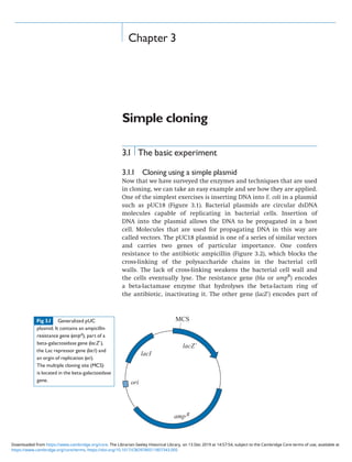
  • 1. Chapter 3 Simple cloning 3.1 The basic experiment 3.1.1 Cloning using a simple plasmid Now that we have surveyed the enzymes and techniques that are used in cloning, we can take an easy example and see how they are applied. One of the simplest exercises is inserting DNA into E. coli in a plasmid such as pUC18 (Figure 3.1). Bacterial plasmids are circular dsDNA molecules capable of replicating in bacterial cells. Insertion of DNA into the plasmid allows the DNA to be propagated in a host cell. Molecules that are used for propagating DNA in this way are called vectors. The pUC18 plasmid is one of a series of similar vectors and carries two genes of particular importance. One confers resistance to the antibiotic ampicillin (Figure 3.2), which blocks the cross-linking of the polysaccharide chains in the bacterial cell walls. The lack of cross-linking weakens the bacterial cell wall and the cells eventually lyse. The resistance gene (bla or ampR ) encodes a beta-lactamase enzyme that hydrolyses the beta-lactam ring of the antibiotic, inactivating it. The other gene (lacZ’) encodes part of Fig 3.1 Generalized pUC plasmid. It contains an ampicillin resistance gene (ampR), part of a beta-galactosidase gene (lacZ’), the Lac repressor gene (lacI) and an orgin of replication (ori). The multiple cloning site (MCS) is located in the beta-galactosidase gene. https://www.cambridge.org/core/terms. https://doi.org/10.1017/CBO9780511807343.005 Downloaded from https://www.cambridge.org/core. The Librarian-Seeley Historical Library, on 13 Dec 2019 at 14:57:54, subject to the Cambridge Core terms of use, available at
  • 2. a beta-galactosidase enzyme, which normally cleaves disaccharides, such as lactose, into monosaccharides. It can also cleave an artificial substrate 5-bromo-4-chloro-3-indolyl-b-D-galactoside (also known as X-gal), shown in Figure 3.2, to liberate a blue dye. For this reason, the substrate is said to be chromogenic. We will see later why the gene for the whole beta-galactosidase enzyme is not used, but for the moment we can treat the plasmid as though it contained the entire gene. As well as the ampR and lacZ’ genes, the plasmid has some less immediately obvious features that we will look at in more detail later, such as an origin of replication. It also has a number of recognition sites for restriction endonucleases, and many of these are located in a region of the lacZ’ gene known as the multiple cloning site or polylinker. Suppose we wanted to clone genomic DNA from the organism we were studying into the BamHI site of pUC18, located in the multiple cloning site in the lacZ’ gene. (We do not have to use the BamHI site. We could use any of the other restriction sites in the multiple cloning site.) The procedure would be as follows. The first stage is to purify genomic DNA from the organism of interest and cut it with the enzyme BamHI. We would also purify some pUC18 and cut it with the same enzyme. Then the BamHI-digested plasmid and the BamHI-digested genomic DNA are mixed and DNA ligase in a suit- able buffer is added. BamHI ends will anneal to BamHI ends and the ligase will seal the nicks. That will generate a large number of types of molecule, which may be linear or (topologically) circular. Fig 3.2 Structure of ampicillin (a), X-gal or 5-bromo-4- chloro-3-indolyl-b-D-galactoside (b), and IPTG or isopropyl-b- D-thiogalactoside (c). 53 3.1 THE BASIC EXPERIMENT https://www.cambridge.org/core/terms. https://doi.org/10.1017/CBO9780511807343.005 Downloaded from https://www.cambridge.org/core. The Librarian-Seeley Historical Library, on 13 Dec 2019 at 14:57:54, subject to the Cambridge Core terms of use, available at
  • 3. Linear molecules will not be stably propagated in later stages of the experiment, so we will not consider them further. Different circular molecules that may be formed are shown in Figure 3.3. Intramolecular ligation of plasmid DNA will regenerate the original circular pUC18 plasmid. Intramolecular ligation of genomic DNA will produce circular molecules of genomic DNA, with no plasmid DNA sequences present. A wide range of intermolecular ligations can take place. Two plasmid DNA molecules can be ligated, in a head-to- tail or head-to-head configuration. Two genomic DNA molecules could be ligated, again in either orientation. Most important, a genomic DNA molecule can be ligated to a plasmid DNA molecule. (Of course, a huge range of ligations involving three or more molecules can also take place, but they will be rarer than unimolecular or bimolecular ligations. How rare depends on the concentrations of the species involved.) The molecules that contain new combinations of sequences, not present before, are termed recombinants. Note that the insertion of DNA into the lacZ’ gene will destroy the function of that gene, as it disrupts the coding region for the LacZ’ protein; therefore, it is an easy matter to work out what properties each of the plasmid types shown in Figure 3.3 will confer. It is possible that insertion of a short piece of DNA into the lacZ’ gene might only partially inactivate the gene function (or not affect it at all) if the reading frame were not terminated or shifted. This is relatively uncommon, but the pos- sibility should always be kept in mind. The next stage is to put the ligation products back into E. coli. For an experiment such as this, it would usually be done by chemically induced transformation, or electroporation (Sections 1.5.2 and 1.5.3). An important point is that the uptake of DNA by E. coli is rather inefficient, in that the probability of any individual bacterial cell taking up a piece of DNA is very low. For simple transformation procedures, of 109 cells treated with 1 mg of supercoiled plasmid DNA, only 105 or so will successfully take up a molecule. We say there is a transformation frequency of 105 colonies/microgram of DNA. This is low, given that 1 mg of pUC18 contains of the order of 1011 molecules. The cells are now plated on agar containing, in this example, ampicillin, together with an inducer of the lacZ’ gene (isopropyl-thiogalactoside (IPTG), Figure 3.2) and the chromogenic substrate X-gal. The cells that have not taken up a plasmid will be sensitive to the ampicillin and die after a few generations. Those that took up genomic DNA only will also die. Those that took up pUC18 DNA will have acquired an ampicillin resistance gene and be able to grow on the selective medium. They will, therefore, form isolated colonies on the agar if plated at a suitable dilution. Thus, all the members of a colony are derived from one cell and should have acquired a single plasmid. (Because the probability of a cell taking up one DNA molecule is low, the probability of a cell taking up two might be expected to be very low indeed. Actually, that is likely not to be the case. Although only a tiny fraction of cells is able to take up DNA, 54 SIMPLE CLONING https://www.cambridge.org/core/terms. https://doi.org/10.1017/CBO9780511807343.005 Downloaded from https://www.cambridge.org/core. The Librarian-Seeley Historical Library, on 13 Dec 2019 at 14:57:54, subject to the Cambridge Core terms of use, available at
  • 4. Fig 3.3 Ligation of BamHI-cut genomic DNA into a BamHI-cut vector.The figure shows the regeneration of vector, the circularization of genomic DNA and the ways in which two separate molecules (vector or genomic DNA) can be joined. The functional genes present on each molecule after ligation are indicated. 55 3.1 THE BASIC EXPERIMENT https://www.cambridge.org/core/terms. https://doi.org/10.1017/CBO9780511807343.005 Downloaded from https://www.cambridge.org/core. The Librarian-Seeley Historical Library, on 13 Dec 2019 at 14:57:54, subject to the Cambridge Core terms of use, available at
  • 5. those cells that do are quite efficient at DNA uptake and may take up more than one piece. So a low, but significant fraction of colonies may contain two different plasmids. Over time, these may segregate, with some cells containing one plasmid and the rest containing the other.) The problem now is to distinguish the colonies that have acquired genomic DNA from all the others. That can be done in this example by using the lacZ’ gene. Colonies that have acquired pUC18 molecules without an insert of genomic DNA will have an intact lacZ’ gene (which will be induced by the IPTG). They will, therefore, be able to break down the X-gal and be coloured blue. The colonies that have acquired molecules with a genomic DNA insert will have a lacZ’ gene that has been inactivated by the insertion. Thus, these colonies will be unable to metabolize the X-gal and will be the usual white (actually off-white) colour. The head-to-tail dimeric pUC18 molecules will also give blue colonies, and the head-to-head dimers will not replicate stably in the host cell. The relative abundances of blue and white colonies will depend on factors such as the relative proportions of genomic and plasmid DNA in the initial ligation. We are interested in the white colonies. We can confirm that plasmids in the white colonies contain an insert by growing up the bacteria, purifying plasmid DNA and cutting it with a restriction enzyme. BamHI would be the easiest in this case, and should generate two sizes of molecule: plasmid and inserted genomic DNA. Plasmids from blue colonies, where there is no insert, will give a single size of restriction frag- ment, corresponding to the plasmid DNA. The same would be seen for the head-to-tail dimeric plasmids. (An alternative way to dif- ferentiate whether plasmids have an insert, and to measure the size of the insert, is to do PCR on DNA from the colonies using primers that anneal on each side of the cloning site. This can be done directly with cells, without needing to purify the DNA, and is called colony PCR.) The outcome of this experiment is a large number of E. coli colonies, each carrying a plasmid containing a BamHI fragment of genomic DNA. This is useful in a number of ways. For example, to get a lot more of each piece of genomic DNA, it is just a matter of growing up a larger quantity of E. coli, extracting the plasmid and, if necessary, separating the insert from the plasmid. That is much easier than extracting the DNA from more of the organism(s) we used in the first place. Also, each plasmid insert is a single, defined piece of DNA. To separate a single, defined piece out of the total genomic DNA each time one wanted it would be too laborious. This propagation of individual, defined pieces of DNA in a suitable organism is cloning. The generation of recombinants in this way by randomly cloning fragments of the total DNA from an organism is called shotgun cloning, and the random collection of E. coli colonies containing the recombinant DNA molecules is called a library. Because total genomic DNA was used, it is called a genomic library. An ideal genomic library would contain all the sequences present 56 SIMPLE CLONING https://www.cambridge.org/core/terms. https://doi.org/10.1017/CBO9780511807343.005 Downloaded from https://www.cambridge.org/core. The Librarian-Seeley Historical Library, on 13 Dec 2019 at 14:57:54, subject to the Cambridge Core terms of use, available at
  • 6. in the original organism’s genome, and would then be described as representative. A library constructed in the way we have just described would probably not be very representative. For example, small DNA fragments tend to be cloned more efficiently than large ones, so our library would be underrepresentative of the large fragments. Another problem with the library in our example is that any gene that contained a BamHI site could never be rescued intact, because BamHI digestion was used to generate the fragments to be cloned. It would be better to use some other way of generating the fragments (see Section 5.2). 3.1.2 Use of the lacZ’ indicator system (alpha-complementation) In this example, we said that the pUC plasmid contained only a part of the lacZ beta-galactosidase gene. Beta-galactosidase is rather a large protein (116 kDa), and it is more convenient to have just a portion of the gene on the plasmid to keep the plasmid as small as possible. This portion encodes the first 146 amino acids and is sometimes called the lacZ’ minigene. In addition to the lacZ’ gene and the ampicillin resistance gene, the plasmid also contains a lacI gene for the Lac repressor protein (Figure 3.1). This keeps the minigene repressed, except in the presence of an inducer such as IPTG. This may be particularly important if the sequences inserted into the minigene are toxic if expressed at high levels. The rest of the lacZ gene is contained within the host, often on an F’ plasmid. In fact it is a version of the gene called the M15 deletion, which lacks codons 1141. The polypeptide produced from this fails to tetramerize, and this tetramerization is needed for enzymatic activity. However, in the presence of the minigene product (amino acids 1146), assembly can take place to produce a molecule with low but detectable beta-galactosidase activity. This intragenic complementation is called alpha-complementation. The principle of alpha-complementa- tion is summarized in Table 3.1. In essence, it is just a way of reducing the size of the vector needed. The pUC vectors were developed at the University of California (hence their name). We will now go back to the basic procedure we have described and look in a little more detail at the vectors and hosts. Table 3.1 Alpha-complementation LacZresidues 1146 present? M15-deleted LacZpresent? Result(þIPTGþX-galþAmp)? Host No Yes AmpS , no LacZ; no colonies Host þpUCplasmid Yes Yes AmpR ,LacZ; blue colonies Host þpUCplasmid (withinsert) No Yes AmpR , no LacZ; white colonies 57 3.1 THE BASIC EXPERIMENT https://www.cambridge.org/core/terms. https://doi.org/10.1017/CBO9780511807343.005 Downloaded from https://www.cambridge.org/core. The Librarian-Seeley Historical Library, on 13 Dec 2019 at 14:57:54, subject to the Cambridge Core terms of use, available at
  • 7. 3.2 Vectors, transformation and hosts 3.2.1 Vectors As outlined above, vector is the name given to DNA molecules into which foreign DNA is inserted for subsequent propagation in a host cell. A vector should have several features: 1. An origin of replication. Without an origin of replication, the vector could not replicate, and when the cells divide after taking up the vector molecule, only one of the daughter cells would retain it. Therefore, we would never get a colony of transformed cells. The pUC18 origin of replication came ultimately from a plasmid in a clinical bacterial isolate denoted as pMB1. 2. A selectable marker. This is needed to distinguish cells that have taken up the vector from those that have not. In the example we have just looked at, the selectable marker was ampicillin resis- tance. The ampicillin resistance gene derives from a transposon (a piece of DNA able to move, or transpose, to different places in the genome) from another plasmid, pRSF2124. 3. Suitable single restriction sites. In the example we considered, pUC18 had just one recognition site for the enzyme BamHI, which was used in the cloning. Had there been more than one BamHI site in pUC18, the vector would not have been suitable, because cutting with BamHI would have cut it into more than one piece. Reassembly of the vector together with an insert during the ligation would have required a trimolecular reaction at least, which would have been very unlikely. Note, though, that the same enzyme does not necessarily have to be used for cutting the vector and insert (see Section 1.2.1). If the insert molecules had been generated by Sau3A digestion, we could still have used BamHI to cut the vector, as BamHI ends are compatible with Sau3A ends. Cutting the vector with Sau3A would not have been feasible, as there are too many sites for Sau3A in the vector. Note also that restriction sites located in indispensable genes in the vector are unsuitable for cloning, because insertion of DNA there would be likely to destroy gene function and, therefore, vector viability. We will see that this is particularly important when we consider vectors based on bacterial viruses in Chapter 4. 4. Suitable size. To some extent, having a suitable size is a corollary of having suitable single restriction sites. A restriction enzyme cleavage site that comprises six nucleotides will occur on average approximately once every 46 bp (i.e. every 4 kbp or so). So a vector that was much larger than 4 kbp might be expected to have several sites for a given enzyme, and cutting the vector would reduce it into several pieces that would be unlikely to be correctly assembled in a ligation reaction. It is possible to remove restric- tion sites, and methods for doing this are described in Chapter 7. Simply removing excess restriction sites may not eliminate 58 SIMPLE CLONING https://www.cambridge.org/core/terms. https://doi.org/10.1017/CBO9780511807343.005 Downloaded from https://www.cambridge.org/core. The Librarian-Seeley Historical Library, on 13 Dec 2019 at 14:57:54, subject to the Cambridge Core terms of use, available at
  • 8. the problem, though. Large DNA molecules are very susceptible to physical shearing, even in the simple act of pipetting, so they are always difficult to handle. Note that the use of the alpha- complementation system allowed the size of the pUC plasmids to be reduced from what would otherwise be necessary to encode beta-galactosidase. 5. Markers for DNA insertion. In the example we studied, the insertion of DNA into the vector can be detected by inactivation of the lacZ’ gene, which in turn could easily be assayed by plating cells onto medium containing an inducer and a chromogenic substrate as well as ampicillin. In some older vectors, an additional round of plating was needed to distinguish recombi- nants from non-recombinants. An example of this is the vector pBR322, which contains an ampicillin resistance gene and a tetracycline resistance gene (Figure 3.4). There are cloning sites in both genes. Suppose we had used the BamHI site in the tetracycline resistance gene for insertion of DNA. After transformation, we would plate the E. coli cells on medium containing ampicillin to select for the acquisition of a plasmid. We would then plate small samples from ampicillin-resistant colonies onto medium contain- ing tetracycline. If DNA had been inserted into the BamHI site, then the tetracycline resistance gene would be inactivated, and cells containing recombinant plasmids would be sensitive to tetracycline. Cells that had acquired a plasmid without inserted DNA would be resistant to tetracycline as well as ampicillin. This need for two rounds of plating was one of the reasons for the pBR322 plasmid being superseded by the pUC and other related plasmids as a routine cloning vector. In some of the first plasmids used for cloning, the cloning site was not in a functional sequence at all. The only way to detect insertion, therefore, was to isolate plasmid DNAs, digest with a suitable enzyme and examine the Fig 3.4 The plasmid pBR322 (4.3 kb).The figure shows genes for ampicillin resistance (ampR), tetracycline resistance, (tetR), the origin of replication (ori), and a selection of unique restriction sites. 59 3.2 VECTORS, TRANSFORMATION AND HOSTS https://www.cambridge.org/core/terms. https://doi.org/10.1017/CBO9780511807343.005 Downloaded from https://www.cambridge.org/core. The Librarian-Seeley Historical Library, on 13 Dec 2019 at 14:57:54, subject to the Cambridge Core terms of use, available at
  • 9. fragments produced electrophoretically. That was potentially very tedious, especially if intramolecular ligation had been favoured over the intermolecular reactions (the tendency for which depends on the relative concentrations of plasmid and genomic DNA), in which case most of the plasmids in transformed cells would not be recombinant. 6. High copy number. Having a high copy number is desirable but not essential, like having markers for DNA insertion. To maximize the yield of plasmid from transformed cells, the copy number in each cell should be as high as possible. Different plasmids have different copy numbers. For some, such as the F factor, the copy number is low, perhaps one or two per cell. The replication of the F factor is quite closely tied to replication of the chromosome, and such control of replication is said to be stringent. The plasmid pSC101 (one of the first plasmids used for cloning) is also under stringent control and is usually present at no more than five copies per cell. Other plasmids with different origins of replication have less tightly controlled replication, and they are said to be relaxed. Here, the copy number can range widely. The copy number of pBR322 is typically 1520 per cell, whereas vectors such as the pUC plasmids may be present in 500700 copies per cell. The origin of replication for both pBR322 and the pUC plasmids came ultimately from the plasmid pMB1. The reason for the difference in copy number between these plasmids with, in theory, the same replication origin may lie in a mutation in the pUC plasmids in the region encoding the RNA molecules that regulate replication by interacting with the origin. This mutation makes the repression system that controls plasmid replication less effective. The copy number of many low copy-number plasmids containing the pMB1 origin can be increased by chloramphenicol amplification. The host cell culture is treated with chloramphenicol, which inhibits bacterial protein synthesis. The inhibition of protein synthesis blocks chromosomal DNA replication, because particular proteins, such as DnaA, need to be synthesized each time chromosomal replication is initiated. The inhibition also blocks cell division, which is closely tied to chromosomal DNA replication. Plasmid replication, however, requires only proteins that are more long-lived. It can, therefore, continue when chromosomal DNA replication and cell division have stopped. Eventually, plasmid DNA replication will stop too, as the supply of general replication proteins, such as DNA polymerase, runs out; but the average copy number will have increased greatly. 7. Disablement. Ever since the earliest experiments in genetic manipulation, there has been concern over the possibility of recombinant DNA molecules ‘escaping’ into the environment and spreading. The likelihood of this can be reduced if the plasmid is in some way disabled so that it cannot spread to other bacteria by processes such as conjugation. Many plasmids, such as pBR322, 60 SIMPLE CLONING https://www.cambridge.org/core/terms. https://doi.org/10.1017/CBO9780511807343.005 Downloaded from https://www.cambridge.org/core. The Librarian-Seeley Historical Library, on 13 Dec 2019 at 14:57:54, subject to the Cambridge Core terms of use, available at
  • 10. have been disabled by removal of the mob gene, which is required for them to mobilize themselves by conjugation. However, such plasmids can still be transmitted from cells containing other plasmids that can provide the necessary functions for mobiliza- tion. This transmission can be blocked by removing a region containing sites called nic and bom from the plasmid that might be mobilized. This region is where the proteins provided by the other plasmids act. The pUC vectors are among those that have had this region removed. 3.2.2 Transformation The transformation method chosen will depend on the efficiency required. The more complex chemical methods for inducing com- petence described in Section 1.5.2 can generate up to 109 transfor- mants per microgram of plasmid DNA, with up to 5% of viable cells competent for transformation. If the recombinant molecules are abundant or of just one (or a few) types, as when reintroducing a plasmid DNA stock into cells to prepare more of the same plasmid, then simple treatment with calcium chloride will be sufficient. If the number of recombinant molecules available is very small and as many different members of the collection as possible need to be recovered (e.g. in cDNA cloning, see Section 5.3), then a more efficient system such as electroporation may be required. When optimized, this approach can yield a higher number of transformants per microgram of DNA than any of the chemical treatments (up to 5 1010 per microgram have been claimed). Typically, a field strength of the order of 15 kV cm1 is used, with a decay time constant of about 5 ms. These conditions will usually result in most of the cells remaining viable, and a reasonable balance between cell killing and the induction of competence. 3.2.3 Hosts The host is the cell in which the recombinant molecules are to be propagated. Choosing the right host is as important as choosing the right vector. Essential or desirable characters include the following: 1. Efficient transformation. This depends on two main features. One is the ability actually to take DNA into the cell, and this pro- cess is poorly understood. Different genotypes respond differently to different transformation systems. Some mutations appear to enhance the efficiency of transformation itself. These include the deoR mutation, which seems to assist particularly in the uptake of larger DNA molecules. DeoR is a transcriptional regulator with DNA-binding activity that controls expression of a set of genes involved in deoxyribonucleoside catabolism. The main feature determining transformation efficiency is the presence or absence of endogenous DNA-degrading systems. 61 3.2 VECTORS, TRANSFORMATION AND HOSTS https://www.cambridge.org/core/terms. https://doi.org/10.1017/CBO9780511807343.005 Downloaded from https://www.cambridge.org/core. The Librarian-Seeley Historical Library, on 13 Dec 2019 at 14:57:54, subject to the Cambridge Core terms of use, available at
  • 11. Many hosts used for cloning are derived from E. coli strain K, which contains the K restrictionmodification system encoded by the hsdRMS locus. The hsdR gene encodes an endonuclease that cleaves DNA containing the sequence -AACNNNNNNGTGC-, unless the second of the two adenine residues and the adenine residue on the other strand opposite the thymine are methylated. Many hosts, therefore, have an hsdR mutation (or a larger deletion) to avoid cleavage of incoming unprotected DNA. The hsdM gene encodes the methylase that protects against degradation so passaging of DNA through an hsdR Mþ strain can be used to allow methylation if it is subsequently necessary to propagate in an hsdRþ strain. There are other proteins in E. coli strain K that will degrade incoming DNA if it is methylated, belonging to the methylation- dependent restriction systems (MDRS). These are not as well understood as the Classes IIII restrictionmodification systems. They include the endonucleases that are the products of the mcrA, mcrB and mrr loci, which will degrade DNA containing methylcytosine (mcrA, mcrB) or methyladenine (mrr). Using strains mutant in these loci, therefore, is desirable, particularly when cloning highly methylated DNA. The extent of methylation of DNA in the host may also affect the efficiency with which other restriction enzymes will subse- quently cleave the DNA in vitro. Methylation can be carried out by the enzymes mentioned already, and also by the products of the dam and dcm genes. The Dam protein methylates adenines at the sequence -GATC-, and Dcm methylates cytosines at the sequences -CCAGG- and -CCTGG-. Some frequently used restriction enzymes have recognition sites that overlap with these and are inhibited by methylation, so DNA prepared from strains that are wild type for these loci will not be efficiently restricted. Use of dam or dcm strains has the disadvantage, however, that they have high mutation rates. This is because newly replicated dsDNA is hemi- methylated (as the newly synthesized material has not yet been methylated). If the polymerase has introduced any errors during synthesis, they will result in a mismatch; these mismatches are normally resolved in the cell by correction to the methylated strand. In a dam or similar strain, neither strand is methylated, and the mismatch correction is as likely to be to the incorrect (newly synthesized) strand as to the correct one. 2. Stable maintenance of plasmid. Once a recombinant plasmid has entered the cell (assuming it has escaped any endogenous restric- tion enzyme activity), it is still not guaranteed to replicate indefi- nitely and stably even if it has a suitable origin of replication. Rearrangement of the recombinant may occur, and the most frequent manifestation of this is partial deletion (i.e. loss of part of the molecule). This usually occurs by recombination across directly repeated sequences, as shown in Figure 3.5. Not all the plasmid molecules in one cell need undergo the same 62 SIMPLE CLONING https://www.cambridge.org/core/terms. https://doi.org/10.1017/CBO9780511807343.005 Downloaded from https://www.cambridge.org/core. The Librarian-Seeley Historical Library, on 13 Dec 2019 at 14:57:54, subject to the Cambridge Core terms of use, available at
  • 12. recombination at the same time, so recombination across a pair of directly repeated sequences in a plasmid will leave three types of molecule in the cell: the original, unrecombined one; and the two smaller recombination products. One of the products will lack an origin of replication and be lost from the population. The other will still be able to replicate, and will do so faster than the original molecule because it is smaller. Over a number of rounds of replication, even a small difference in the time required for replication can result in a large difference in the number of molecules present. Generating 1 mg of DNA from a single copy of a 4 kbp plasmid takes about 40 cycles of replication. A plasmid that could replicate 10% faster would, over this period, generate approximately 16 times as much DNA and, therefore, be by far the most abundant molecule in the total population. The problem will be exacerbated if the original plasmid is deleterious to the host, perhaps by expression of a protein that is toxic to the cell, and the partial deletion caused by recombination abolishes production of that protein. The possibility of preferential propagation of altered molecules during cloning is, therefore, one that should always be borne in mind. Recombination need not always result in deletion. It can also cause inversion, if it takes place across inverted repeats (Figure 3.5). Fig 3.5 Recombination across repeated sequences.If the repeats are in a direct configuration, then the material between them is lost (a).If they are inverted, then the materialbetween them is inverted on recombination (b).1and 2 represent arbitrary points on the molecule. 63 3.2 VECTORS, TRANSFORMATION AND HOSTS https://www.cambridge.org/core/terms. https://doi.org/10.1017/CBO9780511807343.005 Downloaded from https://www.cambridge.org/core. The Librarian-Seeley Historical Library, on 13 Dec 2019 at 14:57:54, subject to the Cambridge Core terms of use, available at
  • 13. Since deletions and inversions usually depend upon recombi- nation, mutations in the host that suppress recombination will help to ensure the stability of transforming molecules. There are three main recombination systems in E. coli, using the products of the recBCD, recE and recF genes. However, these systems depend largely on the product of the recA gene, so strains mutant in this will have a greatly reduced recombination frequency. Some sequences, particularly those containing inverted repeats, may be subject to rearrangement in a recA-independent way. This may be due to the recF pathway functioning in the absence of (or low levels of ) recA, so an recF mutation may be desirable in addition to recA. It is also reported that deficiency of the DNA gyrase encoded by the gyrA and gyrB genes can lead to enhanced stability of molecules containing repeated sequences. 3. Disablement. As with vectors, it is necessary to take precautions to ensure that strains carrying recombinant plasmids are unlikely to escape and propagate outside the laboratory (i.e. to enhance containment). For this reason, the preferred strains usually carry mutations that reduce their viability in the wild. These are often mutations conferring auxotrophy (i.e. the require- ment for a particular metabolite to be supplied in the medium, resulting from an inability to synthesize the metabolite). 4. Features allowing use of the lacZ’ minigene. We saw earlier that the use of the lacZ’ minigene requires the use of a host producing a suitable LacZ fragment, and that this is sometimes encoded on an F’ plasmid. In these cases, therefore, it is necessary to be able to ensure retention of the F’ plasmid. For this reason, the F’ plasmid also contains genes proA and proB for two of the enzymes of proline biosynthesis (encoding 5-glutamyl phosphate reductase and glutamate-5-kinase respectively), and these are deleted from the chromosome of the host. Propagation of the host on medium lacking proline ensures that only cells carrying the resident F’ plasmid will grow. 5. Other markers. In any work with bacteria, it is very useful to have strains that are genetically marked, so the correct strains can be recognized as such. The recombination and nutritional deficiency markers mentioned above may be useful in this respect, although it may also be convenient to have other markers that can be selected more easily. Antibiotic resistance can be useful, although of course it should be different from any of the antibiotic resistances conferred by the vectors to be used. Another mutation that is often incorporated into hosts is endA, inactivating the gene for a DNA-specific endonuclease. This mutation enhances both the yield and quality of plasmid DNA preparations. The relA mutation overrides controls on RNA synthesis, improving the rate of synthesis in the absence of protein synthesis. The tonA mutation causes loss of an outer membrane protein, to which phages T1 and T5 adsorb. Infection by these phages can be a problem in the laboratory, so the tonA mutation is useful in conferring resistance 64 SIMPLE CLONING https://www.cambridge.org/core/terms. https://doi.org/10.1017/CBO9780511807343.005 Downloaded from https://www.cambridge.org/core. The Librarian-Seeley Historical Library, on 13 Dec 2019 at 14:57:54, subject to the Cambridge Core terms of use, available at
  • 14. to phage infection. Host strains used for particular purposes, such as the expression of cloned genes, have additional features, as discussed in Chapter 8. It may be helpful to look at the genotype of a common host strain, and see how it fulfils the considerations above. The strain DH5a is a common host for the pUC plasmids, and is a derivative of E. coli strain K. Its genotype is endA1 hsdR17 supE44 thi-1 recA1 gyrA (nalR ) relA1 D(lacZYA-argF)U169 F80lacZDM15 endA1 is discussed above. hsdR17 inactivates the host restriction system, allowing DNA to be protected by methylation, but not digested, which is sometimes abbreviated to rK mK þ . supE44 is an ‘amber’ chain termination suppressor mutation that allows readthrough of UAG codons in translation. This is used because the same host (or derivatives of it) can also be used for propagation of phages, a number of which have amber chain termination mutations as a biological containment measure. Growth of such phages is possible only in a suppressor host. thi-1 is a nutritional requirement (for thiamine) that gives some containment. recA1 gyrA (nalR ) relA1 are discussed above. (nalR ) is resistance to the DNA gyrase inhibitor nalidixic acid, conferred by the gyrA mutation. D(lacZYA-argF)U169 is a deletion of the lac operon (extending into an arginine biosynthesis gene) that is required for the blue- white selection. U80lacZDM15 describes a U80lacZ prophage that directs synthesis of the partially deleted LacZ, required for bluewhite selection. Another example of a widely used host is JM109, also a derivative of E. coli strain K. It is endA1 hsdR17 supE44 thi-1 recA1 gyrA (nalR ) relA1 mcrA D(lacZYA-proAB) F’ traD36 proAþ proBþ lacIq lacZDM15. Many of the mutations are similar to those in DH5a. JM109 also carries the mcrA mutation, described earlier, and a different lac deletion, extending into the proline biosynthesis operon. The symbol F’ indicates that the markers following it are on an F’ plasmid. traD36 reduces the efficiency of conjugation by a factor of 105 , contributing to biological containment. proAþ proBþ restores the ability to synthesize proline that was lost by the deletion into proAB. This means that growth of the strain on a medium that lacks added proline selects for the presence of F’, as it requires the cells to synthesize their own proline. lacIq is a mutant form of the Lac repressor gene, causing increased levels of repressor in the cell, and allowing tighter control of the lacZ gene. This is important for the use of this host for vectors that repli- cate to a high copy number. Such vectors might titrate out all the repressor produced by a wild-type lacI gene, especially if the gene was present only on the chromosome or on a low-copy-number plasmid 65 3.2 VECTORS, TRANSFORMATION AND HOSTS https://www.cambridge.org/core/terms. https://doi.org/10.1017/CBO9780511807343.005 Downloaded from https://www.cambridge.org/core. The Librarian-Seeley Historical Library, on 13 Dec 2019 at 14:57:54, subject to the Cambridge Core terms of use, available at
  • 15. such as F’. Titration of the repressor might lead to uncontrolled transcription of sequences inserted within the lacZ region of the vector. lacZDM15 is the partially deleted lacZ needed for alpha- complementation. Notice that JM109 allows selection for retention of the F’ plasmid, as described. This is important for two reasons. The partially deleted lacZ needed for alpha-complementation is encoded on the F’. Also, the host is sometimes used for growing phages (Section 4.3) that propagate only in a host carrying the F (or F’) plasmid. 3.3 Modifications 3.3.1 Avoiding self-ligation Although the bluewhite screening that is available with the pUC vectors allows us to see which colonies on a plate contain recom- binant plasmids, these are sometimes very few in number compared with the colonies containing vector that religated without any insert. If we need to recover a large number of different recombinant mole- cules, then the large number of non-recombinant colonies may be a problem. There are various approaches to minimizing this. One approach is to increase the ratio of insert DNA to vector DNA. A more reliable solution is to use alkaline phosphatase (Section 1.3.1). This enzyme is capable of removing 5’-terminal phosphate groups from nucleic acid molecules (and nucleotides). However, these phosphate groups are required for ligation to take place. So if the vector is treated with alkaline phosphatase to remove its terminal phosphate groups, then self-ligation is no longer possible. Ligation of vector to insert, however, is still possible because each insert molecule carries two phosphate groups, one at each end. Note though, that sealing each vectorinsert boundary requires two phosphate groups, one for each strand. Phosphatase treatment of the vector, therefore, means that each boundary can be covalently sealed by ligase on one strand only. The other strand will remain nicked, as shown in Figure 3.6. However, these nicks are not a serious problem. Transformation is still possible, and the normal cellular replication and repair processes will produce molecules that are no longer nicked. In practical terms, the alkaline phosphatase treatment is carried out after the vector has been cut, and before vector and insert DNA are mixed. All traces of phosphatase activity must be removed before the insert DNA is added, otherwise the insert would be dephos- phorylated, too, and intermolecular ligation will also be blocked. Commonly used alkaline phosphatases are inactivated by heating for a few minutes. This treatment can be followed by removal of protein with phenol and chloroform to minimize any carry over of active enzyme into the ligation reactions. If the enzyme used is sufficiently heat labile, purification of the DNA prior to ligation can be omitted. 66 SIMPLE CLONING https://www.cambridge.org/core/terms. https://doi.org/10.1017/CBO9780511807343.005 Downloaded from https://www.cambridge.org/core. The Librarian-Seeley Historical Library, on 13 Dec 2019 at 14:57:54, subject to the Cambridge Core terms of use, available at
  • 16. A third approach to increasing the fraction of recombinants recovered is to use more complex vectors that, if self-ligated, direct the synthesis of a molecule that kills the host cell. One example uses the ccdB (control of cell death) gene. The coding region for this is arranged in the vector so that self-ligation results in the expression of the CcdB protein and consequent death of cells containing the molecule. This is the basis of ‘Zero BackgroundTM ’ vectors. 3.3.2 Forced cloning In the example we considered, the insert DNA could have been ligated into the vector DNA in either of two orientations. In some circumstances it is desirable to control the orientation in which a particular restriction fragment is inserted. This is sometimes called forced cloning. This is possible with the pUC vectors, as there are many different restriction sites clustered in the multiple cloning site. The region is also sometimes called the polylinker. The consequence of having this collection of sites is that, if the insert DNA has different restriction sites at each end, we can readily use a vector cut with the two corresponding enzymes. There will then be only a single orientation in which the insert can be ligated to the vector. There is a series of pUC vectors with different combinations and arrangements of restriction sites in the multiple cloning site. They generally come in pairs whose multiple cloning sites are mirror images of one another, as shown in Figure 3.7. They were generated Fig 3.6 Phosphatase treatment of vector.Removal of the phosphate groups (P) from the vector means that self-ligation is notpossible.Ligation to a molecule with 5’-phosphates is still possible, although one strand will be nicked at each junction. 67 3.3 MODIFICATIONS https://www.cambridge.org/core/terms. https://doi.org/10.1017/CBO9780511807343.005 Downloaded from https://www.cambridge.org/core. The Librarian-Seeley Historical Library, on 13 Dec 2019 at 14:57:54, subject to the Cambridge Core terms of use, available at
  • 17. by the ligation of different synthetic DNA molecules containing the appropriate restriction sites into an EcoRI site close to the beginning of the lacZ’ minigene. This wide combination of restriction sites, combined with the bluewhite screening, makes the pUC vectors very versatile. There are many other series of vectors based on the same principle, but with additional features (e.g. to drive transcription of the insert DNA). We shall deal with some examples of these later. 3.3.3 Cloning PCR products The basic cloning method can be applied to the cloning of PCR products. In the simplest case, we can treat the PCR products as blunt- ended DNA molecules and ligate them into a vector that has been cut to give blunt ends. Very often, this blunt-end ligation is not very efficient. A better approach is to incorporate restriction sites into the primers used for PCR. The PCR products will, therefore, have these restriction sites located at each end and they can be cut with the appropriate enzyme(s) prior to a sticky-ended ligation into an appropriately cut vector. It can be even more efficient to exploit the fact that many polymerase preparations used for PCR incorporate an additional A residue that is not template encoded onto the end of the molecules they synthesize. The products can, therefore, be ligated directly into vector molecules that have an overhanging T residue. A suitable vector DNA preparation can be made by cutting vector with an enzyme that generates blunt ends and then enzymatically adding a 3’ T residue to the ends. This is the basis of the pGEM-T vector series, which are available commercially as linearized plasmids with the T residue added. Fig 3.7 Multiple cloning sites of pUC19 and pUC18.Note that they contain the same restriction sites but in the opposite orientation. So an EcoRIHindIII fragment, say, could be cloned into either vector, but would have opposite orientations. 68 SIMPLE CLONING https://www.cambridge.org/core/terms. https://doi.org/10.1017/CBO9780511807343.005 Downloaded from https://www.cambridge.org/core. The Librarian-Seeley Historical Library, on 13 Dec 2019 at 14:57:54, subject to the Cambridge Core terms of use, available at
  • 18. 3.3.4 Methods that do not use DNA ligase In the basic experiment, we used restriction enzymes to cut DNA molecules and ligase to rejoin them. There are other systems available that have different ways of cutting and ligating molecules. They are often faster than approaches based on DNA ligase, but they are newer and less widely used. 1. Topoisomerase. This approach uses the Vaccinia virus topoisomerase I. In vivo, it cleaves one strand of a DNA molecule at the recognition sequence -C/TCCTT- and becomes covalently attached to one end of the cleaved molecule. The supercoiling of the DNA template can then be altered by swivelling around the uncleaved strand, and the enzyme then reseals the original strand and dissociates from the DNA. Commercially available topo- isomerase-based vectors (‘TOPOÕ Cloning’ systems) are supplied in linear form, with the topoisomerase enzyme attached. The topoisomerase will rapidly ligate the vector to suitable substrate molecules. For cloning PCR products with overhanging A residues, the vector has a single overhanging T residue with the topoisom- erase attached. The PCR product can anneal to the vector and similarly be rapidly ligated. 2. Recombinase. Site-specific recombination reactions can be used to clone PCR products or transfer inserted pieces of DNA rapidly from one vector to another. This is the basis of the ‘GatewayÕ ’ system, based on bacteriophage lambda (Section 4.4). This bacteriophage can integrate its DNA by site-specific recombination between a site on the phage (attP) and a site on the host genome (attB). Recombination across att sites is exploited in the Gateway system to transfer inserts directly between vectors carrying att sites. For cloning PCR products, for example, the primers contain the attB site. The recipient vector (called an entry vector) contains attP sequences flanking the insertion site. Addition of recombinase protein catalyses recombination between attB and attP sites, integrating the PCR product into the vector, as shown in Figure 3.8. The insert can then be transferred, if required, into different vectors that have suitable att sites by similar site-specific recombination. Alternatively, restriction fragments can be inserted into an entry vector by conventional ligation and then transferred into other vectors by recombination. 3.4 Linkers, adaptors and cassettes 3.4.1 Linkers The importance of the pUC and similar vectors owes a great deal to the presence of the multiple cloning site, with its ability to accept a wide range of different fragments. A similar feature is offered by 69 3.4 LINKERS, ADAPTORS AND CASSETTES https://www.cambridge.org/core/terms. https://doi.org/10.1017/CBO9780511807343.005 Downloaded from https://www.cambridge.org/core. The Librarian-Seeley Historical Library, on 13 Dec 2019 at 14:57:54, subject to the Cambridge Core terms of use, available at
  • 19. molecules called linkers. These are short, chemically synthesized molecules that contain a particular restriction enzyme recognition site within their sequence. An example of these, an EcoRI linker, is shown in Figure 3.9. Using EcoRI linkers, it is possible to clone a blunt- ended insert molecule into a vector carrying an EcoRI cloning site. The linkers are themselves blunt-ended molecules, and can be joined onto the blunt-ended insert molecule using T4 DNA ligase. Although the reaction (being a blunt-ended ligation) is relatively inefficient, the use of an excess of linker helps to ensure that a large proportion of the insert molecules have linkers on the ends. Some molecules end up with more than one linker attached to each end, but that is not a problem. The next step is to treat the insert-linker molecules with the appropriate restriction enzyme, which cuts within the linkers to leave a single cut linker attached to each end of the insert. So the insert now has sticky ends, which can be used for insertion into a restriction site in the usual way. This is summarized in Figure 3.9. Fig 3.8 ‘GATEWAYÕ ’cloning system. In the first stage a PCR product (PCR) is transferred into an entry vector by site-specific recombination across att sites. In the second stage, the product is transferred into a specialized vector by recombination. 70 SIMPLE CLONING https://www.cambridge.org/core/terms. https://doi.org/10.1017/CBO9780511807343.005 Downloaded from https://www.cambridge.org/core. The Librarian-Seeley Historical Library, on 13 Dec 2019 at 14:57:54, subject to the Cambridge Core terms of use, available at
  • 20. There is a potential problem, though. If the insert itself contains a recognition site for the linker restriction enzyme, then the second step of the process in Figure 3.9 will cut the insert as well as the linkers. This is likely to be undesirable. It can be avoided by treatment of the insert, prior to the addition of linkers, with the appropriate methylase: EcoRI methylase if EcoRI linkers are being used. This renders the molecule insensitive to the restriction enzyme, which will still be able to cut the linker. Of course, an alternative way of cloning blunt-ended DNA into a vector with EcoRI ends is to avoid using linkers and instead polish up the ends of the cut vector to make them blunt. That could be followed by a blunt-ended ligation of insert DNA into vector. What are the advantages of using linkers? There are three main advantages. One is that it makes better use of the insert and vector DNA, which may be in short supply. Ligation of blunt-ended insert into blunt-ended vector is an inefficient reaction, with much of the insert being wasted. Ligation of blunt-ended linker onto blunt-ended insert can also be inefficient. However, we use a large excess of linkers Fig 3.9 An EcoRI linker. An example of a linker is shown in (a). The use of linkers is shown in (b). The linkers are first ligated onto a target molecule in a blunt-ended reaction and are then cleaved with EcoRI. 71 3.4 LINKERS, ADAPTORS AND CASSETTES https://www.cambridge.org/core/terms. https://doi.org/10.1017/CBO9780511807343.005 Downloaded from https://www.cambridge.org/core. The Librarian-Seeley Historical Library, on 13 Dec 2019 at 14:57:54, subject to the Cambridge Core terms of use, available at
  • 21. (which are in abundant supply, being synthesized on a chemical scale), so that a large proportion of insert acquires linkers and can be efficiently ligated into the vector. The second advantage, which also comes with using an excess of linkers, is that the likelihood of two insert DNA molecules being ligated to each other (concatenated) is reduced. The third advantage comes when it is necessary to cut the insert out of the vector at a later stage. It is unlikely that ligation of a blunt-ended insert into vector that has been cut with EcoRI and then polished will regenerate any restriction sites. It is difficult, therefore, to excise the insert precisely from the vector. However, insert cloned into EcoRI-cut vector using EcoRI linkers can readily be excised simply by redigestion with EcoRI. There is no reason why a linker should have only one restriction site within it. Linkers that contain a number of sites are available. These are often called polylinkers, and resemble the multiple cloning site of the pUC vectors. 3.4.2 Adaptors One or both ends of a linker may be single stranded. These are sometimes called adaptors. Examples are given in Figure 3.10. The first shown is an adaptor that is blunt at one end (like a conventional linker) and sticky at the other. Therefore, it can be ligated, without further digestion, to a blunt-ended molecule to leave a sticky end. Note that in this example the 5’ overhanging end is not phosphory- lated; this lack of a phosphate group prevents the concatenation of adaptors in the ligation reaction (which would obscure the sticky ends to be used for cloning). Some adaptors (such as the second example in Figure 3.10) are sticky at both ends and can be attached to a molecule that is already sticky-ended. Adaptors may also have extra restriction sites within their sequence. 3.4.3 Cassettes (cloning cartridges) Cassettes (cloning cartridges) are a combination of linkers with other features, such as selectable markers. In their simplest form they consist of an antibiotic resistance gene flanked by DNA that contains multiple cloning sites (Figure 3.11). Consequently, they can be used as an easy way of incorporating selectable markers or other features into DNA molecules. This can be particularly useful as a way of inactivating a cloned gene in the technique of gene disruption Fig 3.10 Adaptors.The upper molecule has a blunt end (left-hand side) and an EcoRI sticky end. It can be used to convert a blunt-ended molecule into an EcoRI-ended molecule. The lower molecule has a BamHI end (left-hand side) and a PstI end (right-hand side). Therefore, it can be ligated onto a molecule with BamHI ends to produce one that has PstI ends (or vice versa, depending on the availability of phosphates on the target molecule). 72 SIMPLE CLONING https://www.cambridge.org/core/terms. https://doi.org/10.1017/CBO9780511807343.005 Downloaded from https://www.cambridge.org/core. The Librarian-Seeley Historical Library, on 13 Dec 2019 at 14:57:54, subject to the Cambridge Core terms of use, available at
  • 22. (see Section 7.6). Cassettes may contain gene expression signals (such as promoters, terminators, etc.) rather than selectable markers. These cassettes are called promoter cassettes, terminator cassettes, and so on. Fig 3.11 Cassettes.The diagram shows examples of an antibiotic resistance cassette (upper) and an expression cassette (lower). 73 3.4 LINKERS, ADAPTORS AND CASSETTES https://www.cambridge.org/core/terms. https://doi.org/10.1017/CBO9780511807343.005 Downloaded from https://www.cambridge.org/core. The Librarian-Seeley Historical Library, on 13 Dec 2019 at 14:57:54, subject to the Cambridge Core terms of use, available at