1. CD47, a 50kDa protein receptor for Thrombospondin-1 (TSP-1), plays an
important role in cancer by promoting tumor growth through a process
known as angiogenesis. David Roberts and coworkers showed that this
process requires the interaction of CD47 with vascular endothelial growth
factor receptor-2 (VEGFR2). In addition to angiogenesis, CD47 also
plays a role in Nitric Oxide (NO) signaling by activating the NO signaling
cascade through an unknown mechanism. Dysregulation of NO
signaling contributes to difficulties with wound healing, angiogenesis, and
cardiovascular disease. Soluble guanylyl cyclase (sGC), a 150kDa
heterodimeric enzyme that converts GTP to cGMP, is the receptor for NO
and is a downstream target of CD47. Thrombospondin-1 (TSP-1), a
450kDa homotrimeric protein, binds to CD47 and inhibits NO binding to
sGC, causing an increase in cytosolic calcium. Previous data from
Saumya Ramanathan suggests that an unknown protein interacts with
CD47 in order to bind TSP-1 and cause this cytosolic calcium increase.
BirA, a biotin ligase tag, will be attached to the C-terminus of CD47,
which will biotinylate proximal proteins, helping to identify the unknown
protein involved in TSP-1 binding to CD47. Our goals are to activate
CD47 by adding truncated TSP-1, E3CaG1 to cells stained with
fluorescent calcium indicator Fluo-3AM and measuring increases in
calcium. Preliminary results suggest that our calcium-based assay needs
further investigation due to the lack of calcium response to ligand.
Western blots showed expression of E3CaG1 from SF-9 cells, however
after purifying E3CaG1 on a nickel column, our protein sample was not
pure as evident by coomassie staining. Cloning of CD47-BirA into the
pCMV vector is underway for transient expression in HEK293T cells.
Abstract
Key Players in Nitric Oxide Signaling
Regulation
Alexander Ollerton1, Sarah Young2, William Montfort2
Department of Chemistry & Biochemistry, Northern Arizona University1
Department of Chemistry & Biochemistry, University of Arizona2
Figure 4: Overview of NO signaling
Nitric Oxide synthase converts L-arginine to L-citrulline, producing
NO as a byproduct. NO binds to the ferrous heme of soluble guanylyl
cyclase, decreasing cytosolic calcium concentrations. TSP-1 binds to
CD47 causing an increase in calcium, which in turn inhibits sGC and
NO signaling.
Our
Focus
Figure 3: Domain structure of CD47
CD47, a 50kDa protein receptor for TSP-1,
has 5 glycosylation sites located in the
extracellular domain, 5 transmembrane
helices, and a c-terminus with 4 splice
variants of unknown function. CD47 has a
“don’t eat me” signal which allows it to
escape detection from the immune system.
Additionally, CD47 plays a role in NO
signaling. Its direct role has not been well
characterized.
Figure 1: Diagram of soluble guanylyl
cyclase
Soluble guanylyl cyclase, a 150 kDa
heterodimeric enzyme, has two
subunits, α and β, which contain a
H-NOX, PAS, coiled-coil domain and
catalytic domain. NO signaling occurs
when NO binds to the ferrous heme of
the beta strand of sGC and converts
GTP to cGMP.
Figure 2: Domain structure of Thrombospondin-1 and E3CaG1
(A) Thrombospondin-1, a 450 kDa homotrimeric protein, consists of an N-terminal domain, a
procollagen domain, three TSP repeats, three EGF-like repeats, seven calcium binding
repeats and a C-terminal binding domain. TSP-1 binds to the N-terminus of CD47, through
its carboxy domain inhibiting angiogenesis through the NO signaling by sGC inhibition.(B)
E3CaG1, 63kDa truncated TSP-1, is the C-terminus of TSP-1 and is expressed in SF9 cells
using the baculovirus system.
Introduction
Results
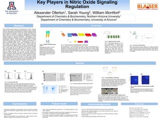
Figure 5: Flow Cytometry Data
(A) Baseline histogram of Jurkat T-cells stained with 5μM fluo-3AM. Cells
washed three times in Krebs buffer. (B) 1μM Angiotensin-II and 5μM of fluo-
3AM was added to Jurkat T-cells and after 15 minutes, flow cytometric
measurments were obtained. Unexpectedly, we did not see calcium increase as
previously described so further optimization of assay is needed. (C) Addition of
1
μ 𝑔
𝑚𝐿
Ionomycin, 400nM Thapsigargin, 2mM Calcium Chloride and 5μM of fluo-
3AM were added to the Jurkat T-cells as a positive control. (A,B,C)
Fluorescence measurements were obtained using a 488nm laser.
(A) (B) (C) (A)
1 2 3 4 5 6 7 8 9 10
(B)
5000
4000
3000
1500
1000
700
5000
4000
1500
1000
700
(A) (B)
Conclusions Future work
Jurkat T-cells will be treated with phorbol ester or T-cell receptor antibody to induce
calcium signaling
Once E3CaG1 is prepared, its ability to induce calcium signaling will be examined by
flow cytometry. This will allow for unraveling signaling mechanism.
Once cloning is complete and transfection optimized, proximity labeling by CD47-BirA
will be used to isolate and identify co-receptors and signaling partners by mass
spectrometry.
Acknowledgments References
Kaur, S.et al. 2013, Sci. Rep. 3,1673
Kim, D.I et al. 2014, PNAS, 10.1073, E2453-E2461
Lawler,J., et al. 1992, Biochemistry., 31, 1173-1180
Ramanathan,S. et al. 2011, Biochemistry, 50, 7787-7799
Rogers, N.M. et al. 2012, AJP-renal ,303, F1117-1125
Roux,K. et al. 2012, JCB, 196, 801-810
Willingham, S.B., et al. 2012, PNAS, 109, 6662-6667
Figure 7: Cloning Strategy of CD47-BirA
Cloning of CD47-BirA into the pCMV-3Tag-3A (destination)
vector was a two step process. First, CD47 was cloned out
of the pEGFP-N3 vector using NotI/BamHI restriction sites
and inserted into the destination vector. BirA-HA was cloned
out of the pcDNA3.1 vector using BamHI/XhoI restriction
sites and inserted at the C-terminus of CD47 in the
destination vector using a GGSG linker.
Figure 6: E3CaG1 Purification from 200-mL Prep of SF-9 Cells
(A)(left) Western Blot: E3CaG1 eluted with 30 mM imidazole
against a 15-40 mM imidazole concentration gradient; this
indicated weak binding of His-tagged E3CaG1 to the Ni
column. (right) E3CaG1 eluted in fractions 1-3. These
fractions were pooled and concentrated (lane 8); anti-His
monoclonal antibody was very specific to the His tag of
E3CaG1. (B) The coomassie gel shows low purity or
degradation of E3CaG1.
Figure 8: PCR of CD47 and Double Digestion of pCMV-
3Tag-3A
(A) Inserting NotI at the N-terminus and BamHI at the C-
terminus of CD47 (976 bp) by PCR. (B) Double digest
product of pCMV-3tag 3A (4214 bp) with NotI and BamHI
restriction enzymes.
Figure 9: LB/Agar plates after transformation of ligated product
NotI-CD47-BamHI was ligated into the pCMV-3Tag-3A vector in a 3:1 ratio.
Ligated product was transformed into DH5α E. coli cells. Plates were
incubated at 37°C and after 13 hours of growth a single colony grew. We
inoculated an overnight culture and isolated the DNA yielding a concentration
of 10 ng/uL.
Thank you to the Montfort lab for allowing me to be a part of their research lab.
Thank you Sarah Young for teaching me new scientific techniques and for giving me
great advice throughout this process.
Thank you to the BLAISER program for giving me this wonderful opportunity to research
over the summer.
Funding: American Heart Association, NIH R01 GM117357
• Vasoconstrictor angiotensin-II, which signals via calcium, did not elicit a response
in preliminary experiments, suggesting receptor may be missing in these Jurkat
cells
• E3CaG1 is expressing in Sf9 cells; however, expression levels and purification
need optimization.
• Ligation and transformation led to a possible clone. Greater quantity of DNA is
needed for sequencing and conformation of correct cloning.