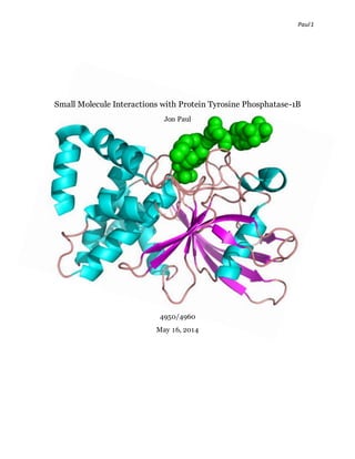
2. Paul2
INTRODUCTION
Protein Tyrosine Phophatase-1Bis a class ofenzymes playing an important part in cell-signaling among
and between cells.1 Protein Tyrosine Kinases are necessary to the functioning ofproteinsin the body.
These PTPs work antagonistically with PTKs; this alteration in protein function is accomplished through
phosphorylation and dephosphorylation ofthe phosphotyrosine residues by PTKs and PTPs respectively.
As well as its aid towards cell differentiation and proliferation, PTP-1Bhas shown involvement in
diabetes, hypertension, rheumatoid arthritis, and cancer.2 As a negativeregulator ofinsulin, PTP-1B
inhibition proves critical in the developmentofdrugs treating type two diabetes. This examination of
PTP-1B will begin with its structureand signaturemotifs, we will then move towards a molecular
modeling approach wherewe examine a ligand docking to several ofPTP-1Bcrystal structure –both wild
type and mutant. A list ofimportant residues toward binding will be discussed - the RMSD of each ligand
will be measured and analyzed against each PTP-1Bcrystal structure. A qualitative structure activity-
relationship will be performed to validatethe method and test whether future studies can be used with
such parameters. Finally, a pharmacophore model will then be performed on a NCBI list ofcompounds
containing carboxylate and amino bi-functional groups. Among thesecompounds, 50 will be selectedand
docked to a wild type protein, 1PXH, in the NCBI protein database.A further discussion will conclude
with the docking scoreofthe top two ligands and their docking score;an analysis as to their usefulness in
inhibiting PTP-1Bwill encourage the development offurtherresearch.An outline ofthis paper can be
seen below:
Structure
PTP-1B Proteins
Ligand Properties
Ligand Docking
QSAR Studies
Docking and QSAR Discussion
1 Silva, Nathan, and David Marcey."Protein TyrosinePhosphatase."Protein Tyrosine Phosphatase. N.p., 1
Jan. 2001. Web. 12 May 2014. <http://www.callutheran.edu/BioDev/omm/ptp1b/molmast.htm>.
2 http://ruchir.myweb.uga.edu/bcmb8010/Enzyme%20report.pdf
3. Paul3
Pharmacophore Model
Selection ofLigands
Docking of Ligands
Conclusion
STRUCTURE
PTP-1B’s structure contains three loops proving important in catalysis; these are the WDP-loop, the PTP-
loop, and the recognition loop.The secondary structure ofPTP-1Bcontains 9 alpha helices and 1 main
beta sheet – 8 strands. The PTP-loop is highly conserved containing the signature motif, H-C-X-X-G-X-X-
R.3 This 1 letter codecorresponds to theirrespective amino acids. Xstand for the stop codon ty pically
addressed as TAA,TAG, or TGA. Cys215 and Arg221 ofthe PTP-loop are the mostcrucial for catalysis.
Val49 and Tyr46 help aid the substrate into the activesite. Ser216 forms a hydrogen bond with the
recognition loop further stabilizing the active site. The WDP-loop, Tryptophan, aspartic acid, and proline
residues account for the name ofthis loop. Asp181 and Gln262 are also important in catalysis.Of the PTP-
loop, Ser222 stabilizes the thiolatewhile arginine helps bind and stabilize the transition state. The WDP-
loop is commonly acid-basemechanics with Asp181 activity relying on the closed conformation ofthis
loop. Other loops include the Q-loop and the lysine loop. The Q-loop contains Gln262 which serves in the
second step ofcatalysis.The lysine loop contains Lys120 –this interacts with the WDP-loop. Below is an
image marking the important residues and loops.
3 http://www.auburn.edu/academic/classes/biol/6190/CellSignalingBiology/csb005.pdf
4. Paul4
Figure 1 - Important Residues and Loops of PTP-1 B
PTP-1B Proteins
The crystal structure ofseveral PTP-1Bwere lookedat. The PDB contained all ofthese structures and can
be found using the following ID entries:1I57, 1PXH, 1PA1, 2B4S, 3CV2, 3I80, 3A5K, and 2CM2. PTP-1B
crystal structures were downloaded from the NCBI database and imported to Maestro and MOE; this step
reviewed missing residues and water molecules;accomplished by fellow classmate, John Martinez.
Several versionswithout water interactions were studied;however,these were not extensively focused on
during the research. Ofthe proteins, 1PXH, 2B4S, 3I80, and 2CM2 are wild-type. 1I57 is a mutant
showing C215S – Cysteine to Serine. 1PA1 shows C215D– Cysteine to Aspartic Acid. 3A5K shows a
mutation at C121W– Cysteine to Tryptophan. 3ZV2contains mutation C215A –Cysteine to Alanine;
S216A –Serine to Alanine. An image below shows the proteins superposed. Mutations in the crystal
structure accountfor the differences seen in the loop structurenext to the binding pocketing. One thing to
note is that only 1PXH, 2B4S, 3A5K, 3I80, and 3ZV2are shown in the image below.
5. Paul5
Figure 2 - PTP-1 B Superposition
LIGAND PROPERTIES
63 ligands were used for the docking used in this research study. The ligands were downloaded from the
protein databank. Minimization was performedand superposition before docking occurred.The ligand
properties included the KD/IC50 (nM) and resolution. Below is a list ofthe 63 ligands with their respective
activity concentration and resolution. Theseproperties were all found from the RCSB protein data bank
and their respectivevalues can be confirmed using thesesearch engines. Any ligand that had an ac tivity
below 1000 nMwas considered to be a strong binder. The ligands that exhibitedthis feature are
highlighted in yellow. The root mean squared distance was also evaluated and can be seen using the
following graphs. In addition, this shows the numberofligands that achieved a distance ofless than < 2.0
or 2.5 Å.
9. Paul9
LIGAND DOCKING
Docking was carried outusing sixty-two ligands from the NCBI database. The ligands were incorporated
to MOE with energy minimization. The above PTP-1Bcrystal structures wereused as the docking protein
for these ligands – these are mutant/wild-typevariants ofPTP-1B. A table ofthe sixty-two ligands with
their entry id and their docking scorecan be found in appendix A1. After the ligand docking was
completed,images ofthe ligand interactions with the proteins wereanalyzed to find similarities and
differences. Below is an image ofthe interactions between the top two ligands with the highest docking
scores. Further interactions ofthe top ten ligands can be found in appendix A2. The ligands below are
2CNF and 1Q6S with a docking score of -9.512and -9.433 respectively. These weredocked to the IPXH
wild-type protein.
A table was compiled to list the ligand and their corresponding residue interactions. It shouldbe noted
that Arg221 is a common trend among most ofthe ligands, followed by Ser216. The least occurring
interaction was Arg24.
10. Paul10
The common Arg221 is seen in these ligands as it is crucial in catalysis.The interaction is formed
generally from the deprotonated sulfonamideor phosphate group.As noted earlier Ser216 will form a
hydrogen bondto stabilizethe active site;a common feature in the ligands above. Gln262 is a
characteristic amideamino acid and will interact with the hydrogens attached to the amino gro ups.
Arginine is present at residue 24. Only one interaction with Arg24 is seen in 1Q6S;this is likely due to the
fact that there are no deprotonated carboxyl groups present in the given radius. Alanine is a small amino
acid that interacts with a free ketone usually located on a phosphategroup;this is not seen in 2CNF as the
orientation ofthe ligand to the activesite does not allow for Arg217 to interact –instead, Arg221 is more
important in catalysis than Arg217. 2NTA has the third highest docking score but contains only Arg221 as
an interaction. The moleculeis small in nature with only a sulfonamide and a ketone. 2CNHhas the
fourth highest docking score featuring a benzenering that interacts with Tyr46 and a sulfonamide group
interacting with Asp48.A benefit ofadding more benzenerings along with other sulfonamidegroups may
allow for a better docking score for 2CNF and 1Q6S as this would allow for more interactions. Among all
of these ligands we see characteristic function groups ofketones,sulfonamides, amines,phosphates, and
some ions such as chlorineor fluorine. We could further expand this study in the future by incorporating
more ligands and running a quantitativestructure-activity relationship (QSAR) study with these
compounds;however, this is out ofthe aim of this current research analysis. A list ofthe docking scoreof
the ten ligands can be seen below:
11. Paul11
2CNF -9.512
1Q6S -9.433
2NTA -9.404
2BGD -9.189
2CNH -9.134
2VEW -9.077
1Q6N -8.867
2FJM -8.821
2VEV -8.817
1Q6J -8.791
The aboveligands were selected for pharmacophoremodeling based on their docking score.The selection
was chosen based on the fact that 1PXHis a wild-typecrystal structureofPTP-1B. Other wild-type
proteins could havebeen used.A similar study would incorporate pharmacophore modeling ofthe ligands
docked to other PTP-1Bprotein structures. Protein 2B4s was able to obtain the highest docking scores
with the ligands. Ligands 2CMB and 2CNI had docking scores of -12.509 and -12.381 respectively. An
image of their interactions can be seen below. These two ligands contained the notableArg221 and Asp48.
Other interactions includedArg45, Arg47, and Phe182. A list ofthe top ten ligands docked to 2B4Sand
their interactions can be seen below:
2CMB -12.509 ASP48, ARG45, ARG221
2CNI -12.381 ARG24, ASP48, PHE182, GLN266, ALA217
2CNH -12.376 ARG47 , ASP48, ALA217, GLY220,GLN266, PHE182, ARG221
2VEY -11.514 ALA217, ARG47, PHE182, ILE219, ASP48, GLN266, ARG221,GLY 220
2CM8 -11.441 ARG221, GLN262
1Q6S -11.401 ARG24, SER216, ALA217, GLY218,ILE219, GLY 220, ARG221
2CMA -11.161 GLN266, PHE182, ARG47 , ASP48, I:LE219, GLY 220,GLY 218, ALA217, ARG24
2VEU -10.857 ALA217, GLY 220,GLN266, PHE182, ARG221
2VEV -10.743 GLN266, PHE182, ALA217, GLY 220,ASP48
2VEW -10.739 ARG47 , ASP48, ALA217, GLY220,ARG221, PHE182, GLN266
13. Paul13
Another wild-typeprotein 3I80 was examined.The top two ligands had a docking score of -10.832 and -
9.223. A list ofthe docking scores and interactions can be found below. Two images of1NWE and 1Q6T
interactions will follow.
1NWE -10.832 ARG47 ,45, ASP48, GLN262, PHE182, ARG221,ALA217
1Q6T -9.223 ARG47 , GLY 259
2CNI -8.339 ARG47 , ARG45
3EB1 -8.337 TY R46, ARG47, GLN262, ARG221,ALA217, PHE182
1Q6N -8.195 ARG47
1Q1M -8.120 TY R46, LY S120, PHE182, ALA217, ARG221,ARG45
1T49 -7 .979 ASP48, TY R46, ARG47
2CNH -7 .932 N/A
3EAX -7 .845 LY S41, ASP48, ARG47, TYR46
1Q6S -7 .844 PHE182, ARG47
INWE Interactions 1 Q6TInteractions
QSAR STUDIES
The qualitativestructure-activity relationship was performed on this using a training set and test set. The
ligands were chosen at random and to achieve a better RMSD the highest ranges were removed. Whe n
using four descriptors, partition coefficient, SMR, TPSA, and weight, a RMSE of 0.86344 and A R2 of
0.67188 was obtained. Using three descriptors:partition coefficient, SMR, and Weight, achieved a RMSE
14. Paul14
of 0.86537 and a R2 of 0.67041. Using two descriptors:partition coefficient and SMR gave RMSE 1.13430
and R2 0.43373. It is important to note that when we removedweight as an importantdescriptor we found
that the QSAR study was not as reproducible;this means weight is an importantdescriptor.The best
model was found using one descriptor, the partition coefficient which achieved an RMSE of 0.63531 and a
R2 of 7 .4466.While I did not include graphs that show the line equation or outliers, the general purpose of
the QSAR study was accomplished.
DOCKING AND QSAR DISCUSSION
Successful completion ofthe docking and ligand properties was performed. A careful analysis shows that
these ligands contain many ofthe importantresidues interactions necessary for catalysis. From the QSAR
study it would be important to look at other ligands with different logP(o/w) coefficients. A further
research would includea set ofcompounds that have low/high logP(o/w) and low/high weight. Since
these were the most important descriptors it is possible to obtain a bettermethod for testing these
compounds ifwe can perform docking over a broader rangeofligands.
PHARMACOPHORE MODEL
Pharmacophore modeling allows the user to select different areas ofa moleculeand search them against a
known NCBI database. The ten ligands that were docked to the 1PXHcrystal structure weresuperimposed
and a pharmacophoremodel was selected. A list of20,465 molecules were foundfitting the 7 descriptors
ranging from aromatic hydrogen acceptor to hydrogen donor. Running the list with 6 of the 7 descriptors
found 7 28,754 molecules. A final run finding 5 out of the 7 gave10,747,393 molecules;a file that was 8
gigabytes in size. It is pertinent when performing a pharmacophoremodel to select as many fields as
possible to limit your query results. A limit ofchoosing the necessary parts ofthe ligands is important so
as not to incorporateunnecessary interactions. An image ofthe pharmacophoremodel is shown below:
15. Paul15
SELECTION OF LIGANDS
After the pharmacophore model had finished running, the database was opened using MOE. 50 ligands
were selected based on their characteristic bi-functional groups ofcarboxylate and amino groups.
16. Paul16
DOCKING OF LIGANDS
Docking of the 10 ligands was performed using MOE and Maestro. The top two ligands had a docking
score of-8.67 and -7.95. The lowest docking score was 2.56. Interactions ofAsp48, Arg221, Gly220,
Cys215, Ala217, Lys120, Arg45, His25, and Gly259. Theseligands were ableto obtain some ofthe
important residues in the docking but it is likely that many werehindered by the rings that flow
throughout the molecule. A necessary study wouldbe to look at smaller ring systems vs. larger ring
systems to see ifthere is a substantial differencein the binding between these differences.A common
feature that gave riseto many interactions was the deprotonated carboxyl groups;a functional group that
was important in our selection did provesuccessful thereforeit would be important to chooseligands with
these groups in future docking studies. A list ofthe ligands and their interactions can be seen below:
17. Paul17
CONCLUSION
Docking of these carboxylate/amino ligands proved somewhat successful;however, the docking score was
not where I would have liked.Further expansion would inc lude a much broader, larger group ofligands
containing thesefunctional groups. A betteranalysis can be concludedwhen using a larger sample
because youare looking at more diversity in the docking to the 1PXHprotein. It would also be interesting
to see how these ligands dock to a mutant type PTP-1Bsuch as 3A5K which has a mutation at C121W.
Another area ofinterest would be to develop a QSAR study ofthese ligands to determine ifthere is any
correlation;ifthe method is good, it’s possible it can be used for future compounds ofsimilarity. I would
also like to thank Dr. Haizhen Zhong for his guiding hand and involvement in this research as well as his
approachable characterand nice personality. It is always a pleasure working with him and I plan to
continue doing research with him.