1. THE ROLE OF ENDOPLASMIC RETICULUM STRESS INDUCED BY GLUCOSAMINE IN PANCREATIC BETA CELLS DYSFUNCTION
Lombardi A.1,2, Aversa R.2, Garbi C.2, Raciti G. A.2, De Vitis S.1, Turco S.1, Coda R.2, Miele C.2, Ulianich L.2, Di Jeso B.1
DiSTeBA, Università del Salento, Lecce, Italy1; DBPCM & IEOS-CNR, Università degli Studi di Napoli “Federico II”, Naples, Italy2
ABSTRACT
Background and aims: The endoplasmic reticulum (ER) represents the cellular compartment where newly synthesized proteins acquire their correct folding. Many
factors can interfere during this process, leading to the accumulation of misfolded proteins into the ER. Such ER dysfunction is collectively termed "ER stress". To
survive under ER stress conditions, cells activate a self-protective mechanism, termed the unfolded protein response (UPR). The ER dysfunction plays an important
role in various diseases including Type 2 diabetes (T2D). It has been reported that hyperglycaemia (HG) causes the progressive deterioration of beta cells through a
mechanism called glucose toxicity and that ER stress may be involved in the consequent pancreatic beta cell dysfunction. Glucosamine (GlcN), generated by the
hexosamine pathway (HP) during HG, induces ER stress and causes disturbances similar to glucose toxicity. In this study, we sought to evaluate the role of ER stress
induced by GlcN in isolated pancreatic mouse islets and in cultured beta cells (INS-1E). Materials and methods: Islets were isolated from male C57 mice by
pancreatic digestion with collagenase. Total RNA was extracted from INS-1E cells or islets by the acid phenol method, and real-time PCR was performed to analyze the
expression pattern of both beta-cell and UPR specific genes. Protein levels were measured by Western blotting; protein subcellular distribution and expression by
immunofluorescence. Cell viability was assessed by MTT assays. Results: MTT assays showed that 24 hours incubation with 10 mM GlcN did not affect INS-1E cells
viability. In these conditions, both mRNA and protein levels of the ER stress marker BiP/GRP78 increased by 3.5- and 2.5-fold, in mouse islets and in INS-1E cells,
respectively. Furthermore, GlcN determined also a 2-fold increase of CHOP/GADD153 mRNA levels, another important marker of the UPR, in both sistems. In isolated
islets, and in INS-1E cells, GlcN decreased by 70% the mRNA levels of both Glut2 and glucokinase (GK) and by 80% the Insulin1 (Ins1) mRNA levels. Interestingly,
similar results were obtained when INS-1E cells and islets were treated with Tunicamycin (Tun), a classical ER stress inducer that inhibits glycosilation. These effects
were very likely exerted at the transcriptional level, as demonstrated by a parallel downregulation of the mRNA of the beta-cell specific transcription factors Pdx1 and
NeuroD1. Furthermore, in INS-1E cells, confocal immunofluorescence studies showed a loss of the specific insulin signal constituted by secretory vescicles clustered
in a sub-plasmamembrane location. Treatment of INS-1E cells with the chemical chaperon 4-Phenyl Butyric Acid (PBA 2.5 mM) was capable of partially prevent ER
stress induced by GlcN and Tun, as demonstrated by a reduced induction of BiP/GRP78 mRNA levels. Furthermore, Glut2 and Ins1 mRNA levels reduction was almost
completely abolished. Oxidative stress appeared not to be involved in ER stress induction by GlcN, as treatment of INS-1E cells with the antioxidant N-acetyl-L-
cysteine (NAC 1mM) did not modify the GlcN-induced increase of BiP/GRP78 and CHOP/GADD153 mRNA levels. Conclusion: These studies suggest that the
activation of the hexosamine pathway leads to alterations in the expression pattern of beta cell specific genes through the induction of ER stress implying that this
mechanism could be responsible at least in part, for glucotoxicity-induced beta cell dysfunction.
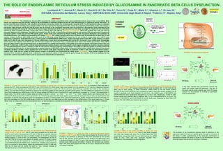
FIGURE 3: Tun and GlcN cause ER stress and a downregulation of specific beta cell genes in isolated pancreatic mouse islets. To evaluate if GlcN was able to
activate the UPR, firstly we measured BiP/GRP78 and CHOP/GADD153 mRNA levels. Islets were isolated from the pancreas of C57 mice by collagenase digestion
and groups of about 20 islets were treated with Tun (0.5 micrograms/ml) or GlcN (10 mM) for 24 hours. Total RNA was extracted from islets by the acid phenol
method, and real-time PCR was performed to analyze the expression pattern of both beta-cell and UPR specific genes. The 18S mRNA was measured as a control.
Upon GlcN treatment the mRNA levels of the ER stress markers BiP/GRP78 and Chop/GADD153 were significantly increased by 3,5 and 2 fold respectively, in
mouse islets. The mRNA levels in treated islets are relative to those in control. *, p < 0.05; **, p < 0.01; ***, p < 0.001. Similar results were obtained with Tun, a
classical ER stress inducer that inhibits glycosilation (A). Furthermore, given that ER stress causes a transcriptional reprogramming of cells, we hypothesized that
expression of specific beta cell genes could be susceptible to variation in stressed islets. Real- Time PCR experiments, indeed, showed that treatment with both Tun
and GlcN decreased by 70% the mRNA levels of both GLUT2 and GK, and determined an 80% decrease in the mRNA levels of the insulin gene (Ins-1). These
effects were very likely exerted at the transcriptional level, as demonstrated by a parallel downregulation of the transcription factors Pdx1 and NeuroD1 mRNA (B).
mRNAlevels(REU)
***
**
0
1
2
3
4
5
C Tun GlcN
A
**
CHOP/GADD153
***
C Tun GlcN
BiP/GRP78
Glut2
Tun -
GlcN
-
0
0.2
0.4
0.6
0.8
1.0
mRNAlevels(REU)
+
- +
GK
-+
- +
**
**
*
B
-
Ins-1
-+
- +
Pdx1
-+
- +
+
**
**
**
***
***
**
NeuroD1
-
- +
***
Tun
GlcN
-
0
1,0
1,5
2,0
2,5
3,0
Glucose-stimulated
InsulinSecretion(FoldControl)
+
--
-
-
+
-
+
-
-
+
-
+
PBA - - - - + - -
Gluc 2mM
Gluc 20mM
+ +
--
-
+
-
+
-
+
+
-
-
+
-
+
+
-
+
3,5
*** ***
*
***
*
FIGURE 6: Effect of Tun, GlcN and PBA on glucose-stimulated insulin
secretion in INS-1E cells. INS-1E cells were incubated for 24 hours with Tun
or GlcN in the presence or absence of PBA, then stimulated for 60 minutes
with 20 mM glucose. Insulin secreted into the medium was determined using
a radioimmunoassay kit. Both Tun and GlcN reduced capability of INS-1E to
secrete insulin after glucose stimulation. However, the secretion was restored,
when cells were preincubated with PBA for 24 hours. Results are the means
of 3 assay in triplicate.
FIGURE 7: Effect of Tun, GlcN and PBA on Glut2, Ins1 and
Pdx1 mRNA levels in INS-1E cells. INS-1E cells were incubated
with PBA, then treated with either Tun or GlcN. Real-Time PCR
experiments, showed that both Tun and GlcN reduced the mRNA
levels of Ins1, Glut2 and their upstream regulator Pdx1.
Furthermore, PBA prevented this downregulation.
Tun
GlcN
-
0
0.2
0.4
0.6
0.8
1.0
mRNAlevels(REU)
+
--
PBA -
+
-
- +
Ins1
-
+
-
-
+
+
Glut2
- +
--
-
+
-
- +
-
+
-
-
+
+
*
***
*
**
*
**
**
Pdx1
- +
--
-
+
-
- +
-
+
-
-
+
+
**
**
**
*
***
CONCLUSION
ER Stress Beta-cell
disfunction
GlcN
The activation of the hexosamine pathway leads to alterations in the
expression pattern of beta-cell specific genes through the induction of
ER stress. A better understanding of the molecular mechanisms of
glucosamine-induced ER stress is pivotal for the identification of novel
pharmacological interventions to treat type 2 diabetes.
It has been reported that glucosamine induces ER
stress and causes beta-cell disfunction. The aim of
this work was to study whether and how ER stress
induced by Glucosamine is involved in pancreatic
beta-cell dysfunction.
A BiP/GRP78
mRNAlevels(REU)
0
1
2
3
4
Tun +
GlcN
- +
- -
+
-- ++
- -
+
-
PBA +
NAC
- -
- -
-
+- --
- +
+
-
***
**
5 ***
*
B CHOP/GADD153
mRNAlevels(REU)
0
2
3
4
Tun +
GlcN
- +
- -
+
-- ++
- -
+
-
PBA +
NAC
- -
- -
-
+- --
- +
+
-
1
**
*
***
*
**
***
FIGURE 5: Effect of PBA and NAC on BiP/GRP78 and CHOP/GADD153
mRNA levels in INS-1E cells. INS-1E cells were incubated for 24 hours with
both Tun and GlcN in the presence of 2.5 mM of PBA (a non-specific chemical
chaperone that reduces the load of unfolded proteins in the ER by improving
folding capacity) or 1mM of NAC (an antioxidant). The pretreatment of INS-1E
cells with PBA for 24 hours was capable of partially prevent ER stress induced
by Tun and GlcN, as demonstrated by a reduced induction of BiP/GRP78 (A)
and CHOP/GADD153 (B) mRNA levels. Furthermore, oxidative stress appeared
not to be involved in these effects, indeed the pretreatment of INS1-E cells with
NAC for 24 hours did not modify the GlcN or Tun induced increase of
BiP/GRP78 (A) and CHOP/GADD153 (B) mRNA levels.
**
***
Translational
attenuation
(PERK,eIF2α)
Unfolded
Protein
Response RE
Degradation
misfolded proteins
(ERAD)
BIP/GRP78
NucleoERSE
XBP1 ATF6
Upregulation of genes
encoding ER Chaperones
(BiP/GRP78, GRP94 )
Cell death
FIGURE 1: The Unfolded Protein Response (UPR) FIGURE2: The Hexosamine pathway
**
**
*
*
***
GlcN
2,5mM
0
20
40
60
80
100
Relativecellsurvival%
CTRL
120
GlcN
5mM
GlcN
7,5mM
GlcN
10mM
GlcN
20mM
FIGURE 4: MTT assay and effect of Tun or GlcN on BiP/GRP78 and CHOP/GADD153 mRNA
levels in INS-1E cells: MTT assays showed that 24 hours incubation with 10 mM GlcN did not
affect INS-1E cells viability. In these conditions, mRNA levels of the ER stress markers
BiP/GRP78 and CHOP/GADD153 were increased yet after 6 hours and remain constant until 48
hours; in particular upon GlcN treatment for 24 hours the mRNA levels of BiP/GRP78 and
CHOP/GADD153 were significantly increased by 2,5 and 2 fold, respectively, in INS-1E cells.
Similar results were obtained after a treatment with Tun (0.5 micrograms/ml) for 24 hours. The
amount of BiP/GRP78 and CHOP/GADD153 mRNA levels were measured by Real Time PCR
analysis. The 18S mRNA was measured as a control.
BiP/GRP78
GlcN 10mM (hours)
*
**
**
CHOP/GADD153
GlcN 10mM (hours)
* **
AIM
ER Stress Beta-cell
disfunction
GlcN
?
? ?
Protein:
- Synthesis
- Folding
- Modification
- Transport