
Recommended
More Related Content
What's hot
What's hot (20)
Similar to Can diabetes make me blind AJAY DUDANI
Similar to Can diabetes make me blind AJAY DUDANI (20)
More from AjayDudani1
More from AjayDudani1 (20)
Recently uploaded
Recently uploaded (20)
Can diabetes make me blind AJAY DUDANI
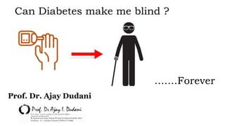
- 1. Can Diabetes make me blind ? .......Forever Prof. Dr. Ajay Dudani
- 2. 1. The Burden of Diabetes 2. DME Patient Journey 3. Diabetic Retinopathy and DME 4. Diagnosis 5. Management Agenda
- 3. Global estimates of diabetes: 7th edition of the IDF world atlas IDF Diabetes Atlas. 7th Edition, 2015: http://www.diabetesatlas.org/component/attachments/?task=download&id=90 [Accessed December 2015].
- 4. The cost of diabetes … In the US, 1 in 5 health care dollars is spent on the management of diabetes In the UK, £1 for every £5.50 is spent on diabetes … IDF Diabetes Atlas. 7th Edition, 2015: http://www.diabetesatlas.org/component/attachments/?task=download&id=90 [Accessed December 2015].
- 5. Loss of vision is the most feared complication of diabetes Feelings about complications at diagnosis: Complications patients were most concerned about: Strain WD, et al. Diabetes Res Clin Pract 2014;105:302-12. 7% 9% 10% 11% 21% 50% Other Problems with feet/legs Circulation problems Kidney/renal problems Heart/cardiac disease Problems with vision/loss of sight/retinopathy 63% knew these health problems might affect them in the future, but risk seemed remote 25%were devastated they might develop complications 9%were not really concerned 3%None of these
- 6. DME is the most common cause of visual impairment in patients with diabetes Patients with DR General population Patients with diabetes Patients with DME with visual impairment Patients with DME 35% of patients with diabetes have DR,2 of whom 7% have DME2,3 40% of patients with DME have associated visual impairment3 DR is one of the most common microvascular complications of diabetes, accounting for >10,000 new cases of blindness per year in the US alone4 DME, diabetic macular edema; DR, diabetic retinopathy 1. IDF Diabetes Atlas. 6th Edition, 2013: http://www.idf.org/sites/default/files/EN_6E_Atlas_Full_0.pdf [Accessed October 2015]; 2. Yau JW, et al. Diabetes Care 2012;35:556-64; 3. Minassian D, et al. Br J Ophthalmol 2012;96:345-9; 4. Fong DS, et al. Diabetes Care 2004;27:2540-53. 8.3% of people have diabetes1
- 7. The principle fears of the person with diabetes Self treated hypoglycaemia Kidney problems Blindness Not worried Very worried Male Female Male Female Male Female Visual analogue scale showing patients’ worries about hypoglycemic events and complications of diabetes, ranging from not worried to very worried Pramming S, et al. Diabet Med 1991;8:217-22.
- 8. The prevalence of visual impairment increases with type and management of diabetes 0 2 4 6 8 10 12 14 Type 2 diabetes not taking insulin Type 2 diabetes taking insulin DME prevalence differs according to type of diabetes and treatment1 Prevalence of CSME (%) The 25-year cumulative incidences of DME and CSME were 29% and 17%, respectively, in patients with Type 1 diabetes2 11.5 9.1 4.1 CSME, clinically significant diabetic macular edema; defined as the presence of retinal thickening at or within 500 µm of the center of the macula or hard exudates at or within 500 µm of the center of the macula if associated with thickening of the adjacent retina or zones of retinal thickening 1 disc area in size, at least part of which was within 1 disc diameter of the center 1. Ling R, et al. Eye 2002;16:140-5; 2. Klein R, et al. Ophthalmology 2009;116:497-503 Type 1 diabetes
- 9. Patients with diabetes are at increased risk of several comorbid and chronic conditions Diabetes is a leading cause of death worldwide1,5 Diabetes caused 5.1 million deaths worldwide in 20135 Every 6 seconds somebody dies from diabetes5 The prevalence of hypertension in patients with diabetes is 70%4 Diabetes doubles the risk of acute coronary syndrome and doubles the level of clinical risk once an event has occurred3 Stroke1,2 Retinopathy1,2 Neuropathy1,2 Nephropathy1,2 Coronary artery disease1,2 Myocardial infarction2 Congestive heart failure1,2 Dyslipidemia2 1. CDC. National Diabetes Fact Sheet, 2011: http://www.cdc.gov/diabetes/pubs/pdf/ndfs_2011.pdf [Accessed August 2015]; 2. Long AN, Dagogo-Jack S. J Clin Hypertens 2011;13:244-51; 3. Kapur A, De Palma R. Heart 2007;93:1504-06; 4. Klein R, et al. Arch Intern Med 1996;156:622-7; 5. IDF Diabetes Atlas. 6th Edition, 2013: http://www.idf.org/sites/default/files/EN_6E_Atlas_Full_0.pdf [Accessed October 2015].
- 10. The risk of comorbidities is increased further in patients with diabetes Diabetic nephropathy1 Dyslipidemia1 Cardiovascular disease3 Diabetes1 Cerebrovascular accident1-3 Diabetic retinopathy1 Diabetic neuropathy1 Compared with patients with diabetes without ocular complications, patients with DME and/or DR have a greater risk of stroke2,3 and myocardial infarction3 Compared with a healthy control cohort, a larger proportion of patients with DME are overweight or obese (~31%), have hypertension (66%), and have cardiovascular disease (25%)1 1. Petrella RJ, et al. J Ophthalmol 2012;159167; 2. Wong TY, et al. JAMA 2002;288:67-74; 3. Nguyen-Khoa BA, et al. BMC Ophthalmol 2012;12:11.
- 11. Cardiovascular risk is increased in patients with diabetes 0 50 100 150 200 250 300 <120 120-139 140-159 160-179 180-199 >200 Male patients with diabetes Male patients without diabetes Age-adjusted CVD death rate per 10,000 person-years3 Systolic blood pressure, mmHg 50% of people with diabetes go on to die of CVD1 Additional risk factors from: cholesterol, hypertension, and smoking Compared with healthy individuals, patients with diabetes are around 2-6 times more likely to develop CVD1 and have a life expectancy up to 8 years lower2 CVD, cardiovascular disease 1. IDF Diabetes Atlas. 6th Edition, 2013: http://www.idf.org/sites/default/files/EN_6E_Atlas_Full_0.pdf [Accessed October 2015]; 2. Gu K, et al. Diabetes Care 1998;21:1138-45; 3. Stamler J, et al. Diabetes Care 1993;16:434-44. CVD death rate per 10,000 person- years
- 12. Risk of stroke and cardiovascular disease increased Presence of DME is a predictor of cardiovascular morbidity and mortality 6.9 19.7 0 5 10 15 20 25 Patients with diabetes with no DR* Patients with DME Rate per 1,000 person-years 5.4 13.8 0 4 8 12 16 Patients with diabetes with no DR* Patients with DME Rate per 1,000 person-years Acute myocardial infarction Cerebrovascular accident *Age- and gender-matched patients with diabetes without ophthalmic manifestations, retinal disorders, or vitreous hemorrhage Nguyen-Khoa BA, et al. BMC Ophthalmol 2012;12:11.
- 13. Risk of stroke by presence of microvascular complication 1.4 4.0 4.7 20.0 0 2 4 6 8 10 12 14 16 18 20 Neither Cerebral WMLs Retinopathy Both Risk of stroke (%) WMLs, white matter lesions Wong TY, et al. JAMA 2002;288:67-74. WMLs are thought to be associated with cerebral microvascular disease Patients with WMLs and retinopathy had a significantly higher increased risk of stroke than patients without WMLs and/or retinopathy
- 14. The effects of hyperglycemia on the vascular system are the major source of morbidity and mortality in diabetes Diabetic nephropathy Diabetic neuropathy Diabetic retinopathy Coronary artery disease Peripheral arterial disease Stroke The incidence of microvascular and macrovascular complications is strongly correlated with extent and duration of hyperglycemia1,2 Macrovascular complications HbA1c, glycated hemoglobin 1. Fowler MJ. Clin Diabetes 2008;26:77-82; 2. Stratton IM, et al. BMJ 2000;321:405-12. Each 1% reduction in mean HbA1c reduces the risk of diabetes- related death by 21%, myocardial infarction by 14%, and microvascular complications by 37% (all p < 0.0001)2 Microvascular complications Hyperglycemia1
- 15. Damage to the microvasculature has potential serious systemic effects Microcirculatory function is extremely important1 Diabetes can damage the systems that regulate microcirculation, including the autonomic nervous system (diabetic autonomic neuropathy), affecting small vessels and organs throughout the body2 Cardiovascular autonomic neuropathy is one of the most important forms of diabetic autonomic neuropathy3 Clinical manifestations include sudden cardiac death, silent myocardial ischemia, resting tachycardia, orthostasis, high cardiovascular mortality rate2,3 1. Johnson PC. Overview of the microcirculation. In: Tuma RF, et al., eds. Handbook of Physiology: Microcirculation. 2nd ed USA: Elsevier, 2008: x-xxiv; 2. Dokken BB. Diabetes Spectrum 2008;21:160-5; 3. Boulton AJ, et al. Diabetes Care 2005;28:956-62.
- 16. The Anatomy of the eye http://www.eyedefectsresearch.org/mac-degen.html https://www.ncbi.nlm.nih.gov/pubmedhealth/PMHT0022378/?figure=1 https://www.aapos.org/terms/conditions/22
- 17. The anatomy of the eye(Layers of Retina) 1) Image: Caspi RR. J Clin Invest. 2010;120:3073-3083
- 19. • Corneal sensitivity is commonly impaired in diabetes- predispose to bacterial corneal ulcers, neurotropic ulcers and difficulties with contact lenses • Decreased reflex tear secretion- dry eye • Intrinsic abnormalities of the epithelial basement membrane complexes , with impaired barrier function lead to: Superficial punctate keratitis Poor healing after trauma Prolonged recovery after intraocular surgery CORNEA
- 20. Diabetes is one of the frequent etiology of acquired palsy The 3rd, 4th and 6th are affected (3rd and 6th are frequently cited) Extra Ocular Muscles
- 21. 6th Nerve Palsy • Most common • Horizontal diplopia in primary gaze and in gaze towards same side
- 22. Patching either eye or binasal occlusion Fresnel Prism To treat diplopia and alleviate face turn. Can be tried for small eso deviations or postoperatively if needed Botolinum toxin A Prevent contracture of medial rectus ◦ Successful use of botulinum toxin A in the early treatment of diplopia caused by 6th nerve palsy in two type 2 diabetic patients. (Anna Broniarezyk-Loba, 2004) Eye muscle surgery Longstanding esotropia ~ 6 months and above Control blood pressure and blood sugar High sugar and blood pressure not only impact the eye but has increased risk of stroke Management of 6th Nerve Palsy
- 23. • Cataract is one of the major cause of vision impairment in people with diabetes • Diabetics are 60% more likely to be develop cataract • It occurs 10-20 years after the onset of insulin dependent diabetes • Control of the diabetes with restoration of normal blood glucose levels stops progression of the opacity • True diabetes cataract (snow-flake/snow- storm cataract) Pre-senile cataract Cataract Snow-flake Cataract
- 26. Anterior Ischemic Optic Neuropathy(AION) Ischemic optic neuropathy is due to acute ischemia of the optic nerve. Based on the pattern of blood supply of the optic nerve, it can be divided into two distinct regions: 1.The anterior part (optic nerve head) which is supplied primarily by the posterior ciliary artery circulation 2.The posterior part which is supplied by multiple sources other than posterior ciliary artery circulation Abbreviations: A = arachnoid; C= choroid; CRA= central retinal artery; Col. Br.= Collateral branches; CRV= central retinal vein; D= dura; LC= lamina cribrosa; NFL= surface nerve fiber layer of the disc; OD= optic disc; ON= optic nerve; P= pia; PCA= posterior ciliary artery; PR and PLR= prelaminar region; R= retina; RA= retinal arteriole; S= sclera; SAS= subarachnoid space. Hayreh 1978 [16]. B Modified From Hayreh, S.S. 1974 [8].
- 27. Treatment of A-AION is actually treatment of GCA. It is unlikely that the patient will regain vision with treatment. The goal of treatment is to prevent further vision loss, especially in the contralateral eye. To prevent bilateral blindness, two things are crucial: Early diagnosis Immediate and adequate steroid therapy The set of clinical criteria most strongly suggestive of GCA in order are: Jaw Claudication, CRP > 2.45, Neck pain, ESR > 47 A combination of the ESR and CRP is 97% specific (best indicator of all). If there is a reasonable index of suspicion for GCA, the patient must be treated ASAP. A biopsy can confirm the diagnosis later. Management of AION
- 28. Glaucoma Glaucoma refers to a group of diseases characterized by • Optic neuropathy • Specific pattern of visual field defect • Higher intraocular pressure • Damage to optic nerve is irreversible process • Normal IOP is 10-21mmHg
- 29. Symptoms • Photophobia • Lacrimation • Blepharospasm • Enlarged eyeball Signs • Corneal edema, corneal enlargement more than 13mm diameter. • Sclera become thin and appers blue • Iris may show iridodonesis and atrophic patches in late stage • Lens becomes flat or subluxated • Optic disc shows increased cup/disc ratio and atrophy specially after third year. • IOP is invariably high. Symptoms and signs
- 31. Diabetic Retinopathy • Progressive dysfunction of the retinal blood vessels caused by chronic hyperglycemia. • DR can be a complication of diabetes type 1 or diabetes type 2. • Initially, DR is asymptomatic, if not treated though it can cause low vision and blindness.
- 32. Cotton Wool Spots Infarction of the nerve fiber layer, resulting in fluffy, white patches Microaneurism Outpouchings of the vessel Hemorrhage Vascular tortuosity Hard Exudates lipid byproducts, appears as waxy, yellow deposits Eye with Diabetic Retinopathy https://webeye.ophth.uiowa.edu/eyeforum/tutorials/diabetic-retinopathy-med-students/Classification.htm
- 33. • Global prevalence of DR among patients with diabetes to be 35.4%. • Prevalence of any DR was higher in those with type 1 diabetes • A UK study showed prevalence of DR-33.6% and a study from USA showed 50.3% respectively. • The incidence and prevalence of DR in India appears to be lower than that noted in western literature. Epidemiology of Diabetic Retinopathy
- 34. Healthy Retina Diabetic Retinopathy Diabetic Retinopathy and healthy Eye https://webeye.ophth.uiowa.edu/eyeforum/tutorials/diabetic-retinopathy-med-students/Classification.htm
- 35. Epidemiology of Diabetic Retinopathy in INDIA 1) http://bmctoday.net/retinatoday/2013/04/article.asp?f=diabetic-macular-edema-the-indian-perspective CURES APEDS SN-DREAMS NORTHESTERN 18% 22.40% 18% 28.90% PREVALENCE OF DR IN INDIA CURES: Chennai Urban Rural Epidemiological study APEDS: Andhrapradesh Eye Disease Study SN-DREAMS:Sankara Nethralaya-Diabetic Retinopathy Epidemiological and Molecular Genetic Study
- 36. Types of DR 1) Mild nonproliferative DR Small areas of balloon-like swelling in the retina’s tiny blood vessels. Occur at this earliest stage of the disease. 2) Moderate nonproliferative retinopathy Swell and distort blood vessel Blood supply to eye reduced 3) Severe nonproliferative retinopathy. Many more blood vessels are blocked, depriving blood supply to areas of the retina. 4) Proliferative diabetic retinopathy (PDR) Growth factor (VEGF) released and forms new tiny,leaky blood vessel. Risk of permanent vision loss https://emedicine.medscape.com/article/1224138-overview 5.Watkins PJ. BMJ 2003;326:924-6; 6.Minassian DC, et al. Br J Ophthalmol 2012;96:345-9
- 37. • Macular edema in diabetes, defined as retinal thickening within 2 disc diameters of the center of the macula, results from retinal microvascular changes • These changes compromise the blood-retinal barrier, causing leakage of plasma constituents into the surrounding retina and, consequently, retinal edema. • DME is a consequence of diabetic retinopathy Diabetic Macular Edema
- 38. Clinically significant macular edema (CSME) https://emedicine.medscape.com/article/1224138-overview • Retinal thickening within 500 µm of the center of the fovea • At least 1 disc area of retinal thickening, any part of which is within 1 disc diameter of the center of the fovea • Hard, yellow exudates within 500 µm of the center of the fovea with adjacent retinal thickening CSME as defined by the Early Treatment Diabetic Retinopathy Study (ETDRS):
- 39. Symptoms of DR Asymptomatic Blurred central vision Distortion of objects Difficult in reading and Floaters ! ! ! ! ! ! https://emedicine.medscape.com/article/1224138-overview
- 40. Diabetes and DR/ DME Pathway
- 41. Risk Factor for DR https://www.asrs.org/content/documents/fact_sheet_22_diabetic_retinopathy_new.pdf Increased total serum cholesterol associated with increased risk of hard exudates High blood pressure Duration of Diabetes and Levels of HbA1C Teresa,MEDtube Science Mar, 2017; Vol. V (1) Genetic factors Pregnant women with diabetes
- 43. Choice of therapy • Anti-VEGF • Laser • Steroids Modifia ble risk factors1 • Blood sugar control • Blood pressure • Cholesterol Disease stage • Non-proliferative DR • Proliferative DR DME, diabetic macular edema; DR, diabetic retinopathy; VEGF, vascular endothelial growth factor Ding J, Wong TY. Curr Diab Rep 2012;12:346-54 Factors to consider for treatment
- 44. https://webeye.ophth.uiowa.edu/eyeforum/tutorials/diabetic-retinopathy-med-students/TreatmentOpts.htm Treatment For NPDR: • Managed by optimizing the patient’s general health. • Patients should maintain a HbA1c ≤7%. • patients should be counseled to stop smoking. • if the patient has clinically significant macular edema (CSME) with NPDR: treat with laser therapy. If the leakage is more diffuse a grid of light laser burns can slow the edema. • Finally off-label medical options are available steroids and Anti-VEGF
- 45. 45 PDR Treatment PRP: • Portions of retina are destroyed using thousands of laser burns while sparing the macula. • But risk of vision loss. It has been found to be extremely effective, reducing the risk of severe vision loss by 50%. • Advised for patients with vitreous hemorrhage and neovascularization. https://nei.nih.gov/health/diabetic/retinopathy https://webeye.ophth.uiowa.edu/eyeforum/tutorials/diabetic-retinopathy-med-students/TreatmentOpts.htm
- 46. Vitrectomy: • Surgical removal of the vitreous gel in the center of the eye. • Procedure is used to treat severe bleeding into the vitreous. • The procedure is also indicated for certain tractional retinal detachments.
- 48. Prior to Anti- VEGF injections , patients lost vision, But with the advent of Anti- VEGF injection vision was restored and regained
- 49. One of the landmark trials shows the gain in vision by using Anti- VEGF as compared to Laser and Corticosteroids 49 0 1 2 3 4 5 6 7 8 9 10 11 0 4 8 12 16 20 24 28 32 36 40 44 48 52 56 60 64 68 72 76 80 84 88 92 96100 104 Sham+prom pt laser Ranibizumab +prompt laser Ranibizumab +deferred laser Triamcinolo ne+prompt laser * Values that were ±30 letters were assigned a value of 30 P-values for difference in mean change in visual acuity from sham+prompt laser at the 52-week visit: ranibizumab+prompt laser <0.001; ranibizumab+deferred laser <0.001; and triamcinolone+prompt laser=0.31. http://publicfiles.jaeb.org/drcrnet/presentation/protocol_I_Results_slides_4_26_10.ppt VA (no. of letters on ETDRS chart) Time (Weeks )
- 50. 50 DME management Anti-VEGF • Specifically designed for intraocular use • Monoclonal antibody fragment (Fab), maximizing biologic activity while minimizing systemic exposure • Inhibits the action of VEGF-A in the retina to decrease vascular permeability and edema • Very favorable safety profile. Approved for DME. Ranibizumab 1. Bayer. Eylea SmPC. 2013; 2. Roche. Avastin SmPC. 2014; 3. Avery R, et al. Data presented at ASRS, August 25, 2013, Toronto, Canada; 4. Avery R, et al. Br J Ophthalmol 2014; Epub ahead of print; 5. Matsuyama K, et al. Br J Ophthalmol 2010;94:1215-8
- 51. 51 Anti-VEGF • Anti-VEGF-A / PlGF / VEGF-B recombinant fusion protein containing the FC portion of IgG • Initially developed for systemic administration in oncology • Licensed for metastatic colorectal cancer in the EU and US Aflibercept 1. Bayer. Eylea SmPC. 2013; 2. Roche. Avastin SmPC. 2014; 3. Avery R, et al. Data presented at ASRS, August 25, 2013, Toronto, Canada; 4. Avery R, et al. Br J Ophthalmol 2014; Epub ahead of print; 5. Matsuyama K, et al. Br J Ophthalmol 2010;94:1215-8 • Anti-VEGF-A full-length monoclonal antibody containing the Fc portion of IgG • Licensed for multiple oncology indications in the EU and US • Not licensed for ophthalmology indications or compounding • Not manufactured for ophthalmic administrations Bevacizumab (Off-Label)
- 52. 52 Corticosteroids for DME • The dexamethasone intravitreal implant 0.7 mg is a long-acting sustained-release, biodegradable corticosteroid. • PLACID trial gain in mean BCVA was more in the group receiving Dex compared to that receiving laser therapy. • The Dex implant releases the corticosteroid into the vitreous over a period of ≤6 months. • Risk of IOP rise and cataract formation Dexamethasone Implant 1Rajendran A, Badole P. DME Management – Current Perspective and Therapeutic Strategies. Journal of Ophthalmology and Related Sciences. 2018;2(1):7–14REVIEW. Alimera Sciences. Iluvien SmPC. 2014; 2. Campochiaro PA, et al. Ophthalmology 2011;118:626-35 Messenger WB, et al. Drug Des Devel Ther 2013;7:425-34; 2. Al Dhibi HA, Arevalo JF. World J Diabetes 2013;4:295-302; 3. Campochiaro PA, et al. Ophthalmology 2012;119:2125-32; • Cylindrical polyamide tube containing 190 ug of fluocinolone acetate • Corticosteroids inhibit inflammation and reduce leakage from blood vessels in the retina • Risk of IOP rise and cataract formation Fluocinolone Acetonide
- 53. 53 Corticosteroids for DME • Show short-term efficacy, and effects on visual acuity are transient • Associated with a high incidence of drug-related cataracts and glaucoma • Increased incidence of sterile endophthalmitis may be associated with preserved triamcinolone acetonide • Not approved for ophthalmology indications Triamcinolone acetonide IV DRCR.net. Ophthalmology 2008;115:1447-59; 4. Jonisch J, et al. Br J Ophthalmol 2008;92:1051-54
- 54. 54
- 55. 55 Prevention of DR Take your medicines as prescribed by doctor T Reach and maintain a healthy weight R Add physical activity to your day A Control your ABCs—A1C, B.P, and cholesterol C Kick the smoking habit K https://www.asrs.org/patients/retinal-diseases/3/diabetic-retinopathy Do you know? DCCT trail shown that controlling diabetes slows the onset and worsening of DR DCCT: Diabetes control and complication trail
- 56. Conclusions Loss of vision is the most feared complication of diabetes1 DME is the most common cause of visual impairment in patients with diabetes2-4 DME often affects people of working age and therefore requires long-term management5,6 Patients with diabetes are at increased risk of several comorbid and chronic conditions,7-9 which is increased further in patients with DME5,10 1. Strain WD, et al. Diabetes Res Clin Pract 2014;105:302-12; 2. Yau JW, et al. Diabetes Care 2012;35:556-64; 3. Minassian DC, et al. Br J Ophthalmol 2012;96:345-9; 4. Fong DS, et al. Diabetes Care 2004;27:2540-53; 5. Petrella RJ, et al. J Ophthalmol 2012;159167; 6. Zheng Y, He M, Congdon N. Indian J Ophthalmol 2012;60:428-31; 7. IDF Diabetes Atlas. 6th Edition, 2013: http://www.idf.org/sites/default/files/EN_6E_Atlas_Full_0.pdf [Accessed October 2015]; 8. Kapur A, De Palma R. Heart 2007;93:1504-06; 9. Long AN, Dagogo-Jack S. J Clin Hypertens 2011;13:244-51; 10. Nguyen-Khoa B, et al. BMC Ophthalmol 2012;12:11.