
Recommended
More Related Content
What's hot
What's hot (20)
Similar to How to perform pericardiocentesis step-by-step
Similar to How to perform pericardiocentesis step-by-step (20)
More from AhmedElBorae1
More from AhmedElBorae1 (20)
Recently uploaded
Recently uploaded (20)
How to perform pericardiocentesis step-by-step
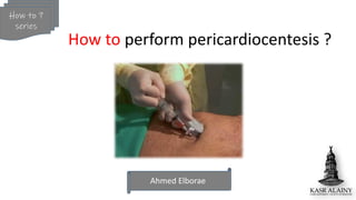
- 1. Ahmed Elborae How to ? series How to perform pericardiocentesis ?
- 3. Franz Schuh 1840, Vienna First percutaneous pericardiocentesis in human 24-year-old lady with mediastinal tumor Done via “Trocar” introduced in the left 3rd intercostal space
- 4. Antoine Marfan 1911, Paris First percutaneous sub-xiphoid approach
- 5. Indications?
- 7. Two layers : Visceral and outer fibrous parietal Normally: Volume: 25 ml Pressure: < 5 mmHg
- 8. Signs of tamponade Beck’s triad Electrical alternans Pulsus paradoxus > 10 mmHg drop Volume: Variable Pressure: “15-20 mmHg”
- 9. Rate dependent not volume dependent Cardiac tamponade as little as 50 cc can cause tamponade May exceed 1000 cc without tamponade
- 10. Measure at end diastole Grading effusion severity: -Mild (< 1 cm) -Moderate (1-2 cm) -Severe (> 2 cm)
- 11. Late diastole Inspiration Early diastole Inspiration A4 view A4 view+ PWD Hemodynamic significance is what really matter
- 13. All are relative because tamponade itself is life threatening • -Bleeding diathesis: - Platelets< 50,000 - High INR (Modifiable) - Anticoagulation • -Surgical candidates: - Rupture LV free wall - Ruptured aortic aneurysm - Posterior effusion - Loculated effusion
- 14. How?
- 15. Ideally Prepared kit 4- Tubes for cytology, chemistry, C/S 2- Puncture needle, dilator/sheath, 0.035 J-wire, Pigtail catheter 3- I.V line, 3-way,Urine bag, 20 cc syringes 1- Sterile drape, local anesthesia
- 16. Six Possible drainage site • Xiphoid: Sub-xiphoid (1), Left para- xiphoid (2), Right para-xiphoid (3) • Apical (4) • Parasternal: left parasternal(5) , right parasternal (6)
- 17. Step by step 1- Informed consent, check platelets count and coagulation status 2-Patient is positioned supine at (30º- 45º), monitored 3- Sterilization and local anesthesia, sterile probe cover Gravity ! Skin Liver * (30º- 45º) Head Leg
- 18. Step by step (sub-xiphoid approach) 4- Echo: Distance from probe to fluid & orientation * Skin Liver X * Either: Echo assisted: Memorise position and go Echo guided: Continuous monitoring
- 19. Step by step (sub-xiphoid approach) 5- Advance needle slowly at 30º angle to pass beneath the costal margin and directed towards tip of left shoulder (on negative pressure, feel characteristic pop once pass through the fibrous parietal pericardium ,stop once get fluid)
- 20. Step by step (sub-xiphoid approach) 6- Check position: -Echo method: Inject (5 cc ) of agitated saline and check in echo (Should seen in pericardial sac not RV) The most critical step
- 21. Step by step (sub-xiphoid approach) 6- Check position: -Pressure method : Just connect it to transducer using three-way Pericardial pressure wave RV pressure wave X The most critical step
- 22. Step by step (sub-xiphoid approach) 6- Check position: -Dye method: inject few cc of dye and check shadow by fluoroscopy The most critical step
- 23. Step by step (sub-xiphoid approach) 6- Check position: -ECG method: connect to v electrode using sterile alligator Pericardium “Isoelectric” Touching RV “ST elevation or PVCS” X The most critical step
- 24. Step by step (sub-xiphoid approach) 7- Advance 0.035- inch soft J shaped wire under fluoroscopy , then remove needle -J-Wire course: around heart shadow, check in 2 views ( AP& lateral) Don't advance if any resistance
- 25. Step by step (sub-xiphoid approach) 8- Advance sheath, then pigtail over the J-wire (Monitor course by fluoroscopy) Alternative sheathless technique (Safer) : Direct pigtail on J wire after using 6F dilator only without sheath
- 26. Step by step (sub-xiphoid approach) 9- Get your samples for analysis first: 1- Cytology 2-Histopathology 3- Chemistry (LDH, Glucose, protein), ADA for TB 4- Gram stain, Z/N stain, bacterial culture and sensitivity, TB C/S & PCR (Usually three 20 cc sterile syringes)
- 27. This end = close, so when face I.V set =suck from pigtail, when face pigtail =evacuate to I.V set 10- Use the “3-way connection” , aspirate fluid through the pigtail and inject into urine bag via I.V set Aspirate Inject
- 28. Is it blood or hemorrhagic effusion ? More likely hemorrhagic effusion if : 1- Lower hematocrit value than blood 2-Does not clot easily
- 29. Post procedure • Reading of intra-pericardial pressure “Post-drainage” > Especially If effusive-constrictive suspected (Remains elevated) • Fix catheter by suture, and cover with sterile dressing • No consensus about prophylactic antibiotics “preferred anti-staph if drain left” • Do CXR post procedure to exclude lung complications • Remove drain if less than 30 ml / 24 hours & no residual by echo • Echo FU before and after removal of drainage • If after 72 hours and still high output > 100 ml, consider further steps to prevent recurrence
- 30. Complications? “If you didn’t have complications, then you did not perform the number needed to get one”
- 31. “Expect the unexpected, then you will be ready” The PCR-EAPCI Textbook – Percutaneous interventional cardiovascular medicine John D. Groarke, Andrew O. Maree, Igor F. Palacios Liver laceration “Left lobe” LAD injury RV/RA/LV perforation Pericardial decompression If large amount drained
- 33. Daniel A Jones.Journal of thoracic oncology.2011 Percutaneous balloon pericardiotomy Intra-pericardial instillation of sclerosing agent Or
- 34. Thank You