This study demonstrates that anti-third party CTLs can efficiently kill B cell lymphomas through a novel TCR-independent mechanism that requires engagement of MHC class I molecules on the lymphoma cells with CD8 molecules on the CTLs. Using mutant cell lines and blocking antibodies, the researchers showed that lymphoma cell killing begins with conjugate formation between CTLs and tumor cells mediated by ICAM-1 and LFA-1 binding, followed by slower MHC-I dependent induction of apoptosis upon binding of MHC-I on tumor cells to CD8 on CTLs. This novel finding suggests MHC-I molecules can signal tumor cell death independent of antigen presentation when engaged by CD8, representing a new role for MHC-I in
![The Journal of Immunology
TCR-Independent Killing of B Cell Malignancies by
Anti–Third-Party CTLs: The Critical Role of MHC–CD8
Engagement
Assaf Lask,*,1
Polina Goichberg,*,1
Adva Cohen,* Rinat Goren-Arbel,* Oren Milstein,*
Shraga Aviner,* Ilan Feine,* Eran Ophir,* Shlomit Reich-Zeliger,* David Hagin,*
Tirza Klein,†
Arnon Nagler,‡
Alain Berrebi,x
and Yair Reisner*
We previously demonstrated that anti–third-party CTLs (stimulated under IL-2 deprivation against cells with an MHC class I
[MHC-I] background different from that of the host and the donor) are depleted of graft-versus-host reactivity and can eradicate
B cell chronic lymphocytic leukemia cells in vitro or in an HU/SCID mouse model. We demonstrated in the current study that
human allogeneic or autologous anti–third-party CTLs can also efficiently eradicate primary non-Hodgkin B cell lymphoma by
inducing slow apoptosis of the pathological cells. Using MHC-I mutant cell line as target cells, which are unrecognizable by the
CTL TCR, we demonstrated directly that this killing is TCR independent. Strikingly, this unique TCR-independent killing is
induced through lymphoma MHC-I engagement. We further showed that this killing mechanism begins with durable conjugate
formation between the CTLs and the tumor cells, through rapid binding of tumor ICAM-1 to the CTL LFA-1 molecule. This
conjugation is followed by a slower second step of MHC-I–dependent apoptosis, requiring the binding of the MHC-I a2/3 C region
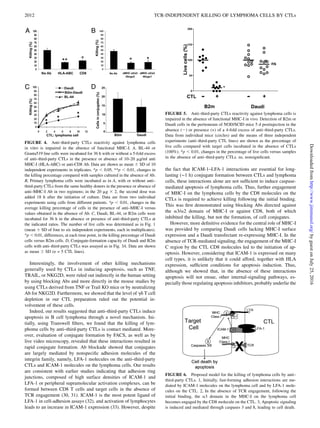
on tumor cells to the CTL CD8 molecule for killing to ensue. By comparing CTL-mediated killing of Daudi lymphoma cells
(lacking surface MHC-I expression) to Daudi cells with reconstituted surface MHC-I, we demonstrated directly for the first time
to our knowledge, in vitro and in vivo, a novel role for MHC-I in the induction of lymphoma cell apoptosis by CTLs. Additionally,
by using different knockout and transgenic strains, we further showed that mouse anti–third-party CTLs also kill lymphoma cells
using similar unique TCR-independence mechanism as human CTLs, while sparing normal naive B cells. The Journal of
Immunology, 2011, 187: 2006–2014.
D
evelopment of strategies to generate ex vivo immune
cells selectively endowed with graft-versus-leukemia
(GVL), while depleted of graft-versus-host (GVH) re-
activity, potentially represents a major challenge in bone marrow
(BM) transplantation and in cancer immunotherapy. Our previous
studies showed that ex vivo stimulation of mouse CD8 T cells
against third-party stimulators (carrying an MHC class I [MHC-I]
background different from that of the host and the donor, hence,
termed “third party”) under IL-2 deprivation led to the selective
growth of third-party–restricted CTL clones concomitantly with
a loss of other clones, including anti-host clones that are unable to
produce their own IL-2 (1–3). Such anti–third-party CTLs were
found to be markedly depleted of GVH reactivity upon trans-
plantation into lethally irradiated mice (2) and exhibited strong
veto activity in vitro (4) and in vivo (2). Thus, their potential role
in tolerance induction, especially in the context of BM allografting
under reduced-intensity conditioning (RIC), was demonstrated in
mouse models (2). This ability of donor anti–third-party CTLs to
kill cognate alloreactive host CTL precursors, when the donor
anti–third-party CTLs are recognized by the naive host CTL
precursors, is initiated upon immune-synapse formation through
TCR recognition of the donor cells by the host naive T cells.
Perhaps somewhat confusing is that the anti–third-party CTLs
serving as target are capable of killing the recognizing naive
T cells. Early studies using Fas and Fas ligand (FasL) knockout
(KO) T cells in long-term MLR cultures indicated a role for Fas–
FasL killing (4), whereas a more recent study using microscopy
imaging indicated that short-term killing through a perforin-
mediated mechanism can also occur (5). In both instances, the
interaction of CD8 on the veto anti–third-party CTL with a3
domain of MHC on the recognizing host CTL precursor is critical
for induction of the killing process.
Based on the insights from the mouse model, a new procedure for
the generation of human anti–third-party CTLs was developed (3).
When studied in a human–mouse chimeric model, these newly
generated human anti–third-party CTLs did not negatively affect
the engraftment of fully allogeneic normal B and T cells (6).
Surprisingly, we found that such allogeneic or autologous anti–
third-party CTLs exhibited potent killing of B cell chronic lym-
phocytic leukemia (B-CLL) (6) cells. Thus, these host nonreactive
anti–third-party CTLs could potentially be used to facilitate en-
graftment of allogeneic hematopoietic stem cells, as well as pro-
vide GVL reactivity.
*Department of Immunology, Weizmann Institute of Science, Rehovot 76100, Israel;
†
Tissue Typing, Rabin Medical Center, Petach Tikva 49100, Israel; ‡
Division of
Hematology, Sheba Medical Center, Tel-Hashomer 52621, Israel; and x
Hematology
Institute, Kaplan Medical Center, Rehovot 76100, Israel
1
A.L. and P.G. contributed equally to this study.
Received for publication January 24, 2011. Accepted for publication June 6, 2011.
This work was supported by the Gabriella Rich Center for Transplantation Biology
Research, Mrs. E. Drake, and by a research grant from Roberto and Renata Ruhman.
Address correspondence and reprint requests to Prof. Yair Reisner, Department of
Immunology, Weizmann Institute of Science, Rehovot 76100, Israel. E-mail address:
yair.reisner@weizmann.ac.il
The online version of this article contains supplemental material.
Abbreviations used in this article: B-CLL, B cell chronic lymphocytic leukemia;
B2m, b2-microglobulin–reconstituted Daudi; BM, bone marrow; B-NHL, B cell
non-Hodgkin’s lymphoma; DHE, dihydroethidium; FasL, Fas ligand; GVH, graft-
versus-host; GVL, graft-versus-leukemia; KO, knockout; b2m, b2-microglobulin;
MHC-I, MHC class I; RIC, reduced-intensity conditioning; wt, wild-type.
Copyright Ó 2011 by The American Association of Immunologists, Inc. 0022-1767/11/$16.00
www.jimmunol.org/cgi/doi/10.4049/jimmunol.1100095
byguestonJuly25,2016http://www.jimmunol.org/Downloadedfrom](https://image.slidesharecdn.com/6d7af24c-dbb7-494b-9f7a-43ec29cdf739-160725211322/85/J-Immunol-2011-Lask-2006-14-1-320.jpg)