Rho Kinase Promotes Alloimmune Responses by Regulating the Proliferation and ...
Characterization of novel T. gondii microtubule protein TgTLAP3
1. TgMorn1
Characterization of a novel microtubule-associated protein
in Toxoplasma gondii, a human parasite.
Phoebe (Yudou) He, Jun Liu, John Murray, Jacqueline Leung, Ke Hu Department of Biology, Indiana University, Bloomington, Indiana, 47405
Introduction
Toxoplasma gondii is a widespread protozoan parasite that can infect nearly
all warm-blooded animals. It causes toxoplasmosis in humans. Infections in
immunocompromised individuals and the unprotected fetus have
devastating consequences, including the development of lethal Toxoplasma
encephalitis. T. gondii is also a model for its relatives in the phylum
Apicomplexa, which includes the malarial parasites. These parasites have a
highly organized cytoskeleton that is essential to their replication and
infectiivity. Furthermore, the cytoskeleton of the parasite has many
properties distinct from that of the host cell and therefore an ideal target for
anti-parasitic therapy. One prominent component of the T. gondii
cytoskeleton is its set of 22 cortical microtubules. Compared with
microtubules found in mammalian cells, the cortical microtubules are
arranged in a reproducible, ordered array and are exceptionally stable. It is
believed that these special properties of the cortical microtubules are due to
novel microtubule associated proteins, because the major tubulin subunits in
T. gondii are essentially identical with those in mammalian cells. In our effort
to search for the proteins that determine the properties of the cortical
microtubules, we identified a group of proteins that coat the microtubules,
including TgTLAP3 (for TgTrxL associated protein 3). TgTLAP3 has no
close homolog in the sequenced genomes of mammals, thus is a potential
drug target. We have constructed a transgenic line of T. gondii through
homologous recombination. In this line, fluorescently tagged TgTLAP3 is
expressed from its endogenous locus. Interestingly, we found that the
localization of TgTLAP3 is restricted to an apical region close to the
presumed organizing center of the cortical microtubules. This may suggest
an organization role of TgTLAP3 in the initial construction of the cortical
microtubules. Consistent with this hypothesis, TgTLAP3 is found close to
the apex of developing daughter cells. To further test this possibility, we
generated a TgTLAP3 knockout parasite using Cre-LoxP mediated excision
to probe it’s function.
P140
B246
Figure 2 (A) A low magnification view of human foreskin fibroblast cells
infected with transgenic T. gondii (green) expressing YFP-α-tubulin.
(B) Drawings of T. gondii cytoskeleton (left panel) and apical complex
(right panel) (modified from Nichols and Chiappino 1987). T. gondii has
three membranes: a plasma membrane (blue) and two additional
membranes (the inner membrane complex, red), formed from a
patchwork of flattened vesicles. Cytoskeletal elements, including 22
subpellicular microtubules (green) are closely associated with the inner
membrane complex. The parasite also possesses an intricate apical
structure (right panel) consisting of the conoid (green), three polar rings
(brown) and two intra-conoid microtubules (green). The conoid is formed
of 14 spiraling 420nm long conoid fibers, a novel tubulin polymer (Hu et
al. 2002).
BA
C
A
B
Figure 3. (A) Scheme for generating loxP_mNeonGreenFP_TLAP3_knockin
parasites using a double homologous recombination mechanism. The
genomic copy of TgTLAP3 is replaced with the coding sequence of
TgTLAP3 fused to GFP and a drug resistance gene. (B) Scheme for
generating ∆TgTLAP3 parasites using an inducible Cre-loxP recombination
mechanism.
Parental loxP_mNeonGreenFP_TLAP3 parasites were co-transfected with a
plasmid transiently expressing Cre recombinase to excise the genome
fragment between the two loxP sites. (C) PCR confirmation of the knock-in
and knockout clones; location of primers are shown in the scheme.
C
3’UTR PCR
5’UTR PCR
Intron PCR
RHΔHXΔku80
Knock in
clones
2.6 kb
2 kb
2.5 kb
2 kb
400 bp
300 bp
RHΔHXΔku80
Plasmid PCR
Knock out PCR
Knock out
clones Knock-in
clone
1.3 kb
1 kb
1.2 kb
1 kb
Generation of TgTLAP3 knockin and knockout parasites
Over-expressed GFP_TgTLAP3 labels the entire cytoskeleton
Figure 4 Structured-illumination image of GFP_TLAP3 expressed under the strong tubulin
promoter( A.) Projection of two transfected parasites shows the cortical microtubules. (B)
Two mother cells each developing daughter cells. (C) One mother cell with more developed
daughter cells. Individual structures are labeled in the image.
C
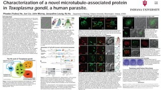
Figure 5. (A) TgTLAP3 knock-in parasites expressing mCherry_TgCentrin2, a Ca2+ binding protein
that has multiple localizations as shown in the drawing (Hu et al. 2006). In this image a clear gap is
seen between TgCentrin2 and TgTLAP3, revealing an unknown structural compartment in T.gondii.
(B-D)TgTLAP3 knock-in parasites expressing mCherryFP-TgMORN1, a cytoskeletal protein
recruited to the basal complex at the very beginning of cell division, also located at the spindle pole
and centrioles (Hu et al. 2006). The drawings show the localization of TgMORN1 during daughter
cell development. (B) Projection of four parasites at the initial stage of daughter cell development.
TgMORN1 is located at the spindle pole. TgTLAP3 is already seen at this stage and does not
co-localize with TgMORN1. (C) Projection of four parasites slightly later during daughter cell
development. TgMORN1 is located at the spindle pole, centrioles and the basal complex ring.
TgTLAP3 is at the apex of the developing daughter cells, and in many small dots in mother cells.
(D) Projection of two mother cells at the late stage of daughter cell development. The two pairs of
daughters are highlighted in the merged image. TgTLAP3 labels a truncated conical structure at the
apical end, as well as brighter dots in the mother cells, symmetrically placed relative to the
TgMORN1 ring.
Summary and Future Directions
A novel microtubule-associated protein, TgTLAP3, localizes to the very apical
end of the cortical microtubules, distal to the TgCentrin2 annuli. TgTLAP3 is
present very early in daughter cell generation and is expressed as multiple
symmetrically arranged dots in the cytoplasm of the mother-daughter complex,
while interphase parasites are usually found to have only one cytoplasmic dot.
TgTLAP3 expression in Vero cells suggests that it does not bind directly to
microtubules. In preliminary characterization, no obvious phenotype is found
in TgTLAP3 knockout parasites. Our future interest focuses on the further
characterization of TgTLAP3 knockout parasites, particularly time-lapse
imaging of daughter cell development.
References
Hu, K., Roos, D. S. and Murray, J. M. (2002). A novel polymer of tubulin forms the conoid of Toxoplasma gondii. J Cell Biol 156:,
1039-50.
Hu, K., Johnson J Florens L, Frauholz M, Suravajjala S, DiLullo C, Yates J, Roos DS and Murray JM. PLOS pathog. 2006, 2(2): e13
Worth A.R , Lymbery A. J, and Thompson R.C.(2013 ) Adaptive host manipulation by Toxoplasma gondii: fact or fiction? Trends in
Parasitology April 2013, Vol. 29, No. 4
This work was supported by grants from NIH(5R01A1098686-02)and the March of Dimes(6-FY12-258) to KH.
B
A
TgTLAP3 does not bind to microtubules in Vero cells
Figure 6 In Vero cells
TgTLAP3 does not directly
bind to microtubules. (A)
TgTLAP3 is seen as
vesicles inside of the cell.
(B) TgTLAP3 vesicles are
absent in the nuclear region.
B
TgTLAP3 knockout parasites appear normal
Figure 7 (A) Immunofluorescence staining of
TgTLAP3 knock-in parasites with
anti-TgTLAP3 antibody. TgTLAP3 is present
at the apical end and also as one dotted
structure in the body, which is different from
the images in Figure 5. (B). Staining of
TLAP3 knockout parasites with anti-TgTLAP3
antibody.
RHΔHXΔku80 ΔTgTLAP3TLAP3_Knockin
Figure 8 Plaque essay for
comparison of growth rates among
RH∆HX∆ku80 parental parasites,
TgTLAP3_knockin parasites and
∆TLAP3 parasites.
TgTLAP3 knockout parasites grow normally
The life cycle of Toxoplasma gondii
Figure 1 The flow chart shows the definitive host (Felidae) for T. gondii,
a variety of intermediate hosts, and three routes of transmission to
them: ingestion of oocysts from cat faeces, ingestion of tissue cysts via
predation/scavenging, and vertical transmission from mother to
offspring.(Worth, 2013)
Vertical transmissionIngestion of oocysts
shed in cat faeces
Predation by
definitive host
Definitive Host
Intermediate
Hosts
Trends in Parasitology
Predation/scavenging
by non-definitive host
Co-localization of TgTLAP3, TgCentrin2, and TgMORN1
TgMorn1
1μTLAP3 Merge
DIC
MORN1
B
Centrin2
1μTLAP3 Merge DICCentrin2
A
TgMorn1
1μ
C
MORN1 TLAP3 Merge DIC
MORN1 TLAP3 Merge DIC
D
1μ
A
B