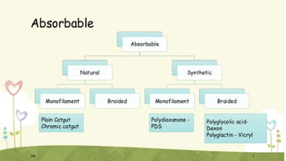
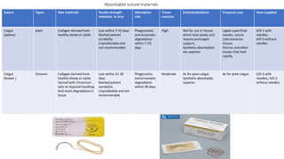
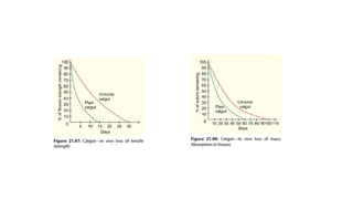
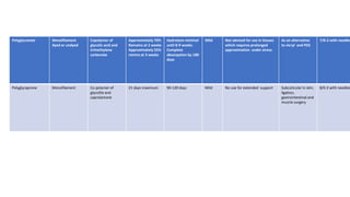
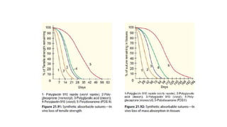
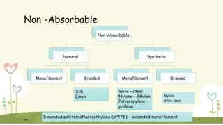
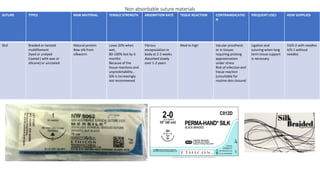
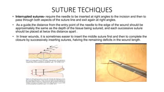
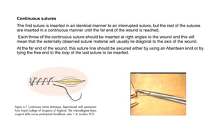
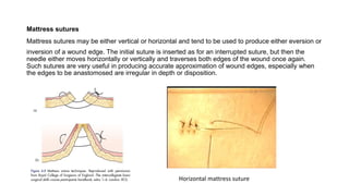
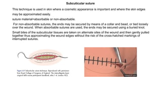
SUTURE MATERIALS
PRESENTED BY – INTERN KRITI GURUNG
For department of Obstetrics and Gynecology
Moderator- DR Jibanath Dhamala
Contents
• Introduction
• Absorbable and non absorbable sutures
• Suture techinques
• Needles
• Knots
• Healing time
A strand of material used to approximate tissues or ligate vessles
during the wound healing period
Suture characteristics
1.Physical structure- monofilament and multifilament
2. Strength- The tensile strength =the force required to break suture when pulling the two ends apart
3. Tensile behavior- Suture materials behave differently depending upon their deformability and
flexibility
Some may be ‘elastic’ in which the material will return to its original length once any tension is
released
Others may be ‘plastic’ in which case this phenomenon does not occur.
synthetic materials demonstrate ‘memory’ in which they keep curling up in the shape that they
adopted within the packaging. the more memory a suture material has, the less is the knot security
4. Absorbability-Sutures for use in the biliary or urinary tract need to be absorbable in order to
minimise the risk of stone production.
a vascular anastomosis requires a non-absorbable material and it is wise to avoid braided material as
platelet adherence may predispose to distal embolisation.
Non absorbable suture is preferred where persistent strength is required
5. Biological behavior-
Biological or natural sutures= are proteolyzed but this involves a process that is not entirely
predictable and can cause local irritation.
Man-made synthetic polymers are hydrolysed and their disappearance in the tissues is more
predictable.
the presence of pus, urine or faeces influences the final result and renders the outcome more
unpredictable.
There is also some evidence that in the gut, cancer cells may accumulate at sites where sutures
persist, possibly giving rise to local recurrence.
United States Pharmacopeia (U.S.P.)
Packaging
Depending on the number of strands in the suture materials, sutures may be:
Monofilament sutures:
• Consisting of a single strand of fiber
• smooth and strong.
• Chance of bacterial contamination is less.
• knot tied may become loose.
• Polypropylene, Polyamide, Catgut, Monocryl, Polydioxanone, Polyglactin finer sizes 6/0-9/0.
Polyfilament sutures:
• multiple strands braided together
• easier to handle and the knot tied does not slip.
• bacteria may lodge in the crevices of the sutures so these sutures are not suitable in presence of
infection, e.g. silk, linen, polyglycolic acid,polyglactin 910, braided polyamide and braided polyester
• What are the criteria of an ideal suture material?
• Should have adequate tensile strength
• Should incite minimal tissue reaction
• Should have easy handling property
• Should have good knotting quality
• Should be nonallergenic and noncarcinogenic
• Should be easily available and cheap
Absorbable suture materials
Suture Types Raw materials Tensile strength
retention in vivo
Absorption
role
Tissue
reaction
Contraindications Frequent uses How supplied
Catgut
(yellow)
plain Collagen derived from
healthy sheep or cattle
Lost within 7-10 days
Marked patient
variability
Unpredictable and
not recommended
Phagocytosis
and enzymatic
degradation
within 7-10
days
High Not for use in tissues
which heal slowly and
require prolonged
support,
Synthetic absorbables
are superior
Ligate superficial
vessles, suture
subcutaneous
tissues
Stomas and other
tissues that heal
rapidly
6/0-1 with
needles,
4/0-3 without
needles
Catgut
(brown )
Chromic Collagen derived from
healthy sheep or cattle
Tanned with chromium
salts to improve handling
And resist degradation in
tissue
Lost within 21-28
days
Marked patient
variability
Unpreditable and not
recommended
Phagocytisis
and enzymatic
degradation
within 90 days
Moderate As for plain catgut
Synthetic absorbable
superior
As for plain catgut 6/0-3 with
needles, 5/0-3
without needles
In presence of infection the catgut gets absorbed earlier
• Catgut is sterilized by gamma irradiation and is supplied
in a sterilized pack containing isopropyl alcohol.
Chromic catgut uses:
• Used to suture muscles, bowel anastomosis, closure of
peritoneum.
• Used during appendicectomy. The mesoappendix ,The base of
the appendix and The stump of the appendix is tied.
• The peritoneum, muscles and the external oblique aponeurosis
is apposed
• small gut resection anastomosis
• during gastrojejunostomy
• Episiotomy repair (1-0)
• Fallopian tube tie (2-0)
Synthetic absorbable- sterilized by ethylene oxide
Suture Types Raw materials Tensile strength
retention in
vivo
Absorption role Tissue
reaction
Contraindications Frequent uses How supplied
Polyglactin 910
(vicryl)
Braided
multifilament
Copolymer of
lacitide and
glycolide in a
ratio of 90:10 ,
coated with
polyglactin and
calcium stearate
Approximately
60% remains at 2
weeks
Approximately
30 % remains at
3 weeks
Hydrolysis
minimal until 5-6
weeks.
Complete
absorption by
days 60-90 days
Mild Not advised for use
in tissues which
require prolonged
approximation under
stress
General surgical
use e,g- gut
anastomoses,
vascular
ligatures. Has
become the
“workhorse”
suture.
Opthalmic
surgery
8/0-2 with
needles, 5/0-2
without needles
vicryl 1 used to close uterine incision during caesarean section
• Uses of polyglycolic acid and polyglactin sutures
• for closure of subcostal, paramedian, Pfannenstial or McBurney's incision
• biliary enteric anastomosis— choledochoduodenostomy, choledocho-jejunostomy,
hepaticodochojejunostomy
• pancreaticojejunal anastomosis
949
• In small gut resection anastomosis—seromuscular (anterior and posterior) and through
• (posterior and anterior) layers may be sutured with 2-0 polyglactin or polyglycollic acid
• sutures
• Single layered anastomosis in large gut may be done with 2-0 polyglactin or polyglycolic
• acid suture.
Polyglactin Rapide (Vicryl Rapide) Suture
• variety of polyglactin 910 suture
• The rapid absorption characteristics of vicryl rapide is achieved by exposure of coated vicryl to
gamma irradiation=low molecular weight
• This is undyed.
• Maintains tensile strength for 10–12 days and gets absorbed in tissues in 42 days
Uses
• for subcuticular sutures
• for skin or mucosal closure.
• Gets spontaneously absorbed.
• used for circumcision for approximation of cut margins of the prepuce.
• Perineal repair (episiotomy)
Suture Types Raw materials Tensile
strength
retention in
vivo
Absorption
role
Tissue reaction Contraindicatio
ns
Frequent uses How supplied
Polyglycolic acid
(dexon)
Braided
multifilament
Dyed or undyed
Coated or
uncoated
Polymer of
polyglycolic acid .
Available with
coating of inert
absorbable
surfactant
Poloxamer 188
to enchance
surface
smoothness
87%
Excreted in urine
within 3 days
Approximately
40%
Remains at 1
week
Approximately
20% remains at 3
weeks
Hydrolysis
minimal at 2
weeks,
significant at 4
weeks.
Complete
absorption 60-90
days
Minimal Not advised for
use in tissues
which require
prolonged
approximation
under stress
Uses as for other
absorbable
sutures, in
particular where
slightly longer
wound support is
required
9/0-2 with
needles, 9/0-2
without needles
undyed natural beige color and also
green dyed, violet dyed, or bi-color
Suture Types Raw materials Tensile strength
retention in vivo
Absorption role Tissue reaction Contraindications Frequent uses How supplied
Polydioxanone
(PDS)
Monofilament dyed
(violet) or undyed
Polyester polymer Approximately 70
% remains at 2
weeks
Approximately 50%
remain at 4 weeks
Approximately 14%
remain at 8 weeks
Hydrolysis minimal
at 90 days
Complete
absorption at 180
days
Mild Not for use in
association with
heart valves or
synthetic graft or
in situation s in
which prolonged
tissue
approximation
under stress is
required
Uses as other
absorbable sutures
, in particular
where slightly
longer wound
support is required
Polydiaxone suture
(PDS) 10/0-2 with
needles
Polyglyconate Monofilament
dyed or undyed
Copolymer of
glycolic acid and
trimethylene
carbonate
Approximately 70%
Remains at 2 weeks
Approximately 55%
ramins at 3 weeks
Hydrolysis minimal
until 8-9 weeks.
Complete
absorpytion by 180
days
Mild Not advised for use in tissues
which requires prolonged
approximation under stress
As an alternative
to vicryl and PDS
7/0-2 with needles
Polyglycaprone Monofilament Co-polymer of
glycolite and
caprolactone
21 days maximum 90-120 days Mild No use for extended support Subcuticular in skin,
ligation,
gastrointestinal and
muscle surgery
8/0-2 with needles
Nylon
Wire steel
Non absorbable suture materials
SUTURE TYPES RAW MATERIAL TENSILE STRENGTH ABSORPTION RATE TISSUE REACTION CONTRAINDICATIO
N
FREQUENT USES HOW SUPPLIED
SILK Braided or twisted
multifilament
Dyed or undyed
Coated ( with wax or
silicone) or uncoated
Natural protein
Raw silk from
silkworm
Loses 20% when
wet,
80-100% lost by 6
months
Because of the
tissue reactions and
unpredictability ,
Silk is increasingly
not recommened
Fibrous
encapsulation in
body at 2-3 weeks
Absorbed slowly
over 1-2 years
Mod to high Vacular prosthesis
or in tissues
requiring prolong
approximation
under stress
Risk of infection and
tissue reaction
(unsuitable for
routine skin closure)
Ligation and
suturing when long
term tissue support
is necessary
10/0-2 with needles
4/0-1 without
needles
• Uses
• No. 1 or 1-0 silk sutures are used as ligature:
• during cholecystectomy to ligate the cystic duct and cystic artery
• during small and large gut resection to ligate the mesenteric vessels
• ligate the pedicles during nephrectomy and splenectomy
• during truncal vagotomy to ligate the anterior and posterior vagus nerve before their division.
• for skin closure either with interrupted or continuous suture.
Uses of Mersilk:
• 2-0 and 3-0 mersilk is used for anterior and posterior seromuscular sutures in small gut
• anastomosis and in gastrojejunostomy
• May be used to repair the posterior wall of inguinal canal in herniorrhaphy
• 3-0 mersilk may be used for pancreaticojejunal anastomosis
• 4-0 mersilk may be used for nerve suture.
Polyproylene
(prolene)
monofilament
Dyed or undyed
Polymer of
propylene
Infinite (> 1 year) Non absorbable:
remains
encapsulated in
body tissues
Low None Cardiovascualr
surgery, plastic
surgery, ophthalmic
surgery , general
surgical subcuticular
skin closure
10/0-1 with
needles
SUTURE TYPES RAW MATERIAL TENSILE STRENGTH ABSORPTION RATE TISSUE REACTION CONTRAINDICATIO
N
FREQUENT USES HOW SUPPLIED
• It has low coefficient of friction and slides through the tissues readily
• The suture may extend up to 30% before breaking, hence is useful in situations where postoperatively
some give is required on the part of the suture to accommodate postoperative swelling ( prevent tissue
Strangulation)
• Uses
• Herniorrhaphy for repair of the posterior wall of inguinal canal
• Closure of midline abdominal incision
• Used for repair of incisional hernia
• Repair of tendon injuries
• vascular anastomosis and for repair of nerve injury.
SUTURE TYPES RAW MATERIAL TENSILE STRENGTH ABSORPTION RATE TISSUE
REACTION
CONTRAINDICATIO
N
FREQUENT USES HOW SUPPLIED
Nylon
(black)
Monofilament
Or braided
Multifilament
Dyed or undyed
Polyamide
Polymer
Loses 15-20% per
year
Degrades at
approximately 15-
20% per year
low None General surgical use,
eg- skin closure,
abdominal mass
closure, hernia repair,
plastic surgery,
neurosurgery,
microsurgery,
ophthalmic surgery
Monofiment: 11/0-
2 with needles,
multifilament
(including loops in
some sizes), 4/0-2
without needles,
Multifilament 6/0-2
with needles
3/0-1 without
needles
Very low coefficient of friction and readily passes through the tissues
This is an inert suture and incites minimal tissue reaction
Monofilament polyamide suture has a memory and knot security is poor so 4-5 throws are
required for proper knotting
Uses
• For closure of skin incision
• For closure of abdominal wall incision
• For herniorrhaphy
• Monofilament polyamide sutures are also available as finer sutures 3–0, 4–0, 5–0 up to 10–0 The
finer sutures are used in vascular surgery.
LINEN Twisted Long staple flax
Fibres
Stronger when wet
Loses 50% at 6
months, 30%
remains at 2 years
Infinite (>1 year)
Non absorbable
Remains
encapsulated in
body tissues
Moderate Not advised for
use with vascular
prostheses
Ligation and
suturing in
gastrointestinal
surgery
No longer common
in most centres
3/0-1 with needles,
3/0-1 without
Surgical steel Monofilament or
multifilament
An alloy of iron,
nickel and
chromium
Infinite >1 year Non absorbable
remains
encapsulated in
body tissues
Minimal Should not be
used in
conjucntion with
Prosthesis of
different metal
Closure of
sternotomy wounds
Previously found
devour for tendon
and hernia repair
Monofilament 5/0-
5 with needles
Multifilament: 5/0-
3/0 with needles
Monofilament
11/0-2 with
needles (including
loops in some
sizes), 4/0-2
without needles.
SUTURE TYPES RAW MATERIAL TENSILE STRENGTH ABSORPTION RATE TISSUE REACTION CONTRAINDICAT
ION
FREQUENT USES HOW SUPPLIED
SUTURE TYPES RAW MATERIAL TENSILE STRENGTH ABSORPTION RATE TISSUE
REACTION
CONTRAINDICA
TION
FREQUENT USES HOW SUPPLIED
Polyester Monofilament
Dyed or undyed
Coated
(polybutylene or silicone) or
Uncoated
Polyester(polyethyl
ene terephthalate)
Infinite (>1 year ) Non absorbable-
remains encapsulated
in body tissues
low None Cardiovascular, ophthalmic,
Plastic and general surgery
Monofilament:
(ophthalmic)
11/10; 10/0 with
needles,
multifilament: 5/0-
1 with needles
Polybutester Monofilament:
Dyed or undyed
Polybutylene
terephthalate and
polyteteramythlene
ether glycol
Infinite(> 1 year ) Non absorbable
remains encapsulated
in body tissues
Low None Exhibits a degree of
elasticity. Particularly
favored for use in pkastic
surgery
7/0-1 with needles
• Numbering of Suture Material
• 2-Thick. For pedicle ligation.
• 1-
• 0-zero.
• 1-zero.
• 2-zero. For bowel suturing.
• 3-zero.
• 4-zero.
• 5-zero. For vascular anastomosis.
• 6-zero.
• 7-zero.
• 8-zero.
• 9-zero. For ophthalmic surgery. Requires operating microscope
SUTURE TECHIQUES
• Interrupted sutures- require the needle to be inserted at right angles to the incision and then to
pass through both aspects of the suture line and exit again at right angles.
• As a guide,the distance from the entry point of the needle to the edge of the wound should be
approximately the same as the depth of the tissue being sutured, and each successive suture
should be placed at twice this distance apart .
• In linear wounds, it is sometimes easier to insert the middle suture first and then to complete the
closure by successively inserting sutures, halving the remaining deficits in the wound length.
Continuous sutures
The first suture is inserted in an identical manner to an interrupted suture, but the rest of the sutures
are inserted in a continuous manner until the far end of the wound is reached.
Each throw of the continuous suture should be inserted at right angles to the wound and this will
mean that the externally observed suture material will usually lie diagonal to the axis of the wound.
At the far end of the wound, this suture line should be secured either by using an Aberdeen knot or by
tying the free end to the loop of the last suture to be inserted.
Mattress sutures
Mattress sutures may be either vertical or horizontal and tend to be used to produce either eversion or
inversion of a wound edge. The initial suture is inserted as for an interrupted suture, but then the
needle either moves horizontally or vertically and traverses both edges of the wound once again.
Such sutures are very useful in producing accurate approximation of wound edges, especially when
the edges to be anastomosed are irregular in depth or disposition.
Horizontal mattress suture
This technique is used in skin where a cosmetic appearance is important and where the skin edges
may be approximated easily.
suture material=absorbable or non-absorbable.
For non-absorbable sutures, the ends may be secured by means of a collar and bead, or tied loosely
over the wound. When absorbable sutures are used, the ends may be secured using a buried knot.
Small bites of the subcuticular tissues are taken on alternate sites of the wound and then gently pulled
together thus approximating the wound edges without the risk of the cross-hatched markings of
interrupted sutures.
Subcuticular suture
Purse string suture
Continous stich paralleling to the edges of circular
wound
-used to close circular wound. Eg- hernia or
appendiceal stump
NEEDLES
made of stainless steel
Either straight or curved
Types-
• Round bodied
• Cutting
• Reverse cutting
• Taper cut
• Blunt
In the past, needles had eyes in them and suture material had to be loaded into them= time
consuming,
the needle holes in tissues were considerably larger than the suture material being used.
Currently, needles are eyeless or ‘atraumatic’ with the suture material embedded within the shank of
the needle. The needle has three main parts
1 Shank
2 Body
3 Point.
• Needle point or sharp apex of the needle
• Body:straight or curved.
• Eye: For threading a suture. In atraumatic
needles there are no eyes.
• Needle length: circumferential length of the needle.
• Needle chord length: The linear distance
between the pointed tip and the end of the needle.
Parts of needle
Round bodied needle
• The needle is uniformly round on cross section with a tapering tip.
• Designed to separate tissue fibres rather than cut them.
• Suitable for suturing soft tissues where easy splitting of tissue fibres are possible, e.g. muscles,
intestines, vessels.
Cutting needle
• Required for penetration of tough structures like fascia, aponeurosis, linea alba and skin.
• Has a triangular cross section with the apex on
the inside of the needle curvature.
• The reverse cutting needle- triangular in cross section having the apex of the cutting
edge on the outer surface of the needle curvature.
This improves the strength of the needle and increases the resistance to bending
Taper cut needle
• This needle combines the initial penetration of a reverse cutting needle with the minimized trauma
of a round bodied needle.
• The cutting tip is limited to the point of the needle, which then tapers out to merge smoothly into a
round cross section.
• Used mostly in vascular surgery
Blunt pointed needles
• Needles with a blunt tip is designed for suturing friable vascular tissues like, liver, spleen and
kidneys
• Also used in patients with blood born viruses such as Hepatitis B
The more confined the operative space, the more curved the needle.
Half circle needles are commonly utilised in the gastrointestinal tract
J-shaped needles(rectus sheath after laparoscopy),quarter circle needles and compound curvature
needles are used in special situations such as the vagina, eye and oral cavity, respectively.
The size of the needle tends to correspond with the gauge of the suture material, although it is
possible to get similar
Sterilization of needles
• Needles are sterilized by keeping them dipped in concentrated lysol for 1 hour or in dilute lysol
for 24 hours. Boiling or autoclaving damages the sharpness of the needle.
• 17. SKIN
Alternatives to sutures
Skin adhesive strips
used where there is no tension and not too much moisture, such as after a wide excision of a breast
lump.
• They may also be used to minimize ‘spreading’ of a scar.
Tissue glue
Tissue glue is also available based upon a solution of n-butyl-2-cyanoacrylate monomer.
When it is applied to a wound, it polymerises to form a firm adhesive bond
the wound does need to be clean, dry, with near perfect haemostasis and under no tension.
• This process has good adhesive properties and has been used for haemostasis in the liver and
spleen, for dural tears, in ear, nose and throat (ENT) and ophthalmic surgery, to attach skin grafts
and also to prevent haemoserous collections under flaps.
• Fibrin glues have also been used to control gastrointestinal haemorrhage endoscopically, but do not
work when the bleeding is brisk
Staples
linear, side-to-side and end-to-end stapling devices
Clips
Skin clips produce a very neat scar with good wound eversion.
placed faster than suture insertion
have a lower predisposition to infection as they do not penetrate entirely through the wound and do
not produce a complete track from one wound edge to the other.
uncomfortable for the patient and they require a special instrument to remove them.
Stapling devices
Types of knots
Reef knot or square knot
Granny’s knot
Surgeon’s knot
1.Square knot-
• formed by wrapping the suture around the needle holder once in opposite direction between ties
• 3 ties are recommended
• Easiest and most reliable
2. Granny knot
• Involve tie in 1 direction followed by tie in same
direction and third tie in opposite direction to
square the knot and hold it permanently.
• Tendency to slip with increasing pressure
Surgeon’s knot-
Formed by two throws of suture around needle holder on the first tie
and one throw in opposite direction on 2nd tie.
• The general principles behind knot tying include:
• The knot must be tied firmly, but without strangulating the tissues.
• The knot must be unable to slip or unravel.
• The knot must be as small as possible to minimise the amount of foreign material.
• The knot must be tightened without exerting any tension or pressure on the tissues being ligated.
• During tying, the suture material must not be ‘sawed’ as this weakens the thread.
• The suture material must be laid square during tying, otherwise tension during tightening may cause
breakage or fracture of the thread.
• When tying an instrument knot, the thread should only be grasped at the free end, as gripping the
thread with artery forceps or needle holders can damage the material and again result in breakage or
fracture
Healing Time
• As a general rule, the greater the tension is across a wound, the
longer the stitches need to remain. The following is a guideline
for the recommended removal times based on the location of
the sutures:
• Face: 3-5 days
• Neck: 7 days
• Scalp: 5-7 days
• Chest, Abdomen :7-10days
• Back or upper extremities: 12-14 days
• Lower extremities: 14-21 days
Thank You!!
Refernce
• Bailey and Love’s short practice of surgery 26th edition
• SRB’s manual of surgery 5th edition

SUTURE.pptx

  • 1.
    SUTURE MATERIALS PRESENTED BY– INTERN KRITI GURUNG For department of Obstetrics and Gynecology Moderator- DR Jibanath Dhamala
  • 2.
    Contents • Introduction • Absorbableand non absorbable sutures • Suture techinques • Needles • Knots • Healing time
  • 3.
    A strand ofmaterial used to approximate tissues or ligate vessles during the wound healing period
  • 4.
    Suture characteristics 1.Physical structure-monofilament and multifilament 2. Strength- The tensile strength =the force required to break suture when pulling the two ends apart 3. Tensile behavior- Suture materials behave differently depending upon their deformability and flexibility Some may be ‘elastic’ in which the material will return to its original length once any tension is released Others may be ‘plastic’ in which case this phenomenon does not occur. synthetic materials demonstrate ‘memory’ in which they keep curling up in the shape that they adopted within the packaging. the more memory a suture material has, the less is the knot security 4. Absorbability-Sutures for use in the biliary or urinary tract need to be absorbable in order to minimise the risk of stone production. a vascular anastomosis requires a non-absorbable material and it is wise to avoid braided material as platelet adherence may predispose to distal embolisation. Non absorbable suture is preferred where persistent strength is required
  • 5.
    5. Biological behavior- Biologicalor natural sutures= are proteolyzed but this involves a process that is not entirely predictable and can cause local irritation. Man-made synthetic polymers are hydrolysed and their disappearance in the tissues is more predictable. the presence of pus, urine or faeces influences the final result and renders the outcome more unpredictable. There is also some evidence that in the gut, cancer cells may accumulate at sites where sutures persist, possibly giving rise to local recurrence.
  • 6.
  • 7.
  • 8.
    Depending on thenumber of strands in the suture materials, sutures may be: Monofilament sutures: • Consisting of a single strand of fiber • smooth and strong. • Chance of bacterial contamination is less. • knot tied may become loose. • Polypropylene, Polyamide, Catgut, Monocryl, Polydioxanone, Polyglactin finer sizes 6/0-9/0. Polyfilament sutures: • multiple strands braided together • easier to handle and the knot tied does not slip. • bacteria may lodge in the crevices of the sutures so these sutures are not suitable in presence of infection, e.g. silk, linen, polyglycolic acid,polyglactin 910, braided polyamide and braided polyester
  • 9.
    • What arethe criteria of an ideal suture material? • Should have adequate tensile strength • Should incite minimal tissue reaction • Should have easy handling property • Should have good knotting quality • Should be nonallergenic and noncarcinogenic • Should be easily available and cheap
  • 11.
    Absorbable suture materials SutureTypes Raw materials Tensile strength retention in vivo Absorption role Tissue reaction Contraindications Frequent uses How supplied Catgut (yellow) plain Collagen derived from healthy sheep or cattle Lost within 7-10 days Marked patient variability Unpredictable and not recommended Phagocytosis and enzymatic degradation within 7-10 days High Not for use in tissues which heal slowly and require prolonged support, Synthetic absorbables are superior Ligate superficial vessles, suture subcutaneous tissues Stomas and other tissues that heal rapidly 6/0-1 with needles, 4/0-3 without needles Catgut (brown ) Chromic Collagen derived from healthy sheep or cattle Tanned with chromium salts to improve handling And resist degradation in tissue Lost within 21-28 days Marked patient variability Unpreditable and not recommended Phagocytisis and enzymatic degradation within 90 days Moderate As for plain catgut Synthetic absorbable superior As for plain catgut 6/0-3 with needles, 5/0-3 without needles
  • 13.
    In presence ofinfection the catgut gets absorbed earlier • Catgut is sterilized by gamma irradiation and is supplied in a sterilized pack containing isopropyl alcohol.
  • 14.
    Chromic catgut uses: •Used to suture muscles, bowel anastomosis, closure of peritoneum. • Used during appendicectomy. The mesoappendix ,The base of the appendix and The stump of the appendix is tied. • The peritoneum, muscles and the external oblique aponeurosis is apposed • small gut resection anastomosis • during gastrojejunostomy • during gastrojejunostomy • Episiotomy repair (1-0) • Fallopian tube tie (2-0)
  • 15.
  • 16.
    Suture Types Rawmaterials Tensile strength retention in vivo Absorption role Tissue reaction Contraindications Frequent uses How supplied Polyglactin 910 (vicryl) Braided multifilament Copolymer of lacitide and glycolide in a ratio of 90:10 , coated with polyglactin and calcium stearate Approximately 60% remains at 2 weeks Approximately 30 % remains at 3 weeks Hydrolysis minimal until 5-6 weeks. Complete absorption by days 60-90 days Mild Not advised for use in tissues which require prolonged approximation under stress General surgical use e,g- gut anastomoses, vascular ligatures. Has become the “workhorse” suture. Opthalmic surgery 8/0-2 with needles, 5/0-2 without needles vicryl 1 used to close uterine incision during caesarean section
  • 17.
    • Uses ofpolyglycolic acid and polyglactin sutures • for closure of subcostal, paramedian, Pfannenstial or McBurney's incision • biliary enteric anastomosis— choledochoduodenostomy, choledocho-jejunostomy, hepaticodochojejunostomy • pancreaticojejunal anastomosis 949 • In small gut resection anastomosis—seromuscular (anterior and posterior) and through • (posterior and anterior) layers may be sutured with 2-0 polyglactin or polyglycollic acid • sutures • Single layered anastomosis in large gut may be done with 2-0 polyglactin or polyglycolic • acid suture.
  • 18.
    Polyglactin Rapide (VicrylRapide) Suture • variety of polyglactin 910 suture • The rapid absorption characteristics of vicryl rapide is achieved by exposure of coated vicryl to gamma irradiation=low molecular weight • This is undyed. • Maintains tensile strength for 10–12 days and gets absorbed in tissues in 42 days Uses • for subcuticular sutures • for skin or mucosal closure. • Gets spontaneously absorbed. • used for circumcision for approximation of cut margins of the prepuce. • Perineal repair (episiotomy)
  • 19.
    Suture Types Rawmaterials Tensile strength retention in vivo Absorption role Tissue reaction Contraindicatio ns Frequent uses How supplied Polyglycolic acid (dexon) Braided multifilament Dyed or undyed Coated or uncoated Polymer of polyglycolic acid . Available with coating of inert absorbable surfactant Poloxamer 188 to enchance surface smoothness 87% Excreted in urine within 3 days Approximately 40% Remains at 1 week Approximately 20% remains at 3 weeks Hydrolysis minimal at 2 weeks, significant at 4 weeks. Complete absorption 60-90 days Minimal Not advised for use in tissues which require prolonged approximation under stress Uses as for other absorbable sutures, in particular where slightly longer wound support is required 9/0-2 with needles, 9/0-2 without needles undyed natural beige color and also green dyed, violet dyed, or bi-color
  • 20.
    Suture Types Rawmaterials Tensile strength retention in vivo Absorption role Tissue reaction Contraindications Frequent uses How supplied Polydioxanone (PDS) Monofilament dyed (violet) or undyed Polyester polymer Approximately 70 % remains at 2 weeks Approximately 50% remain at 4 weeks Approximately 14% remain at 8 weeks Hydrolysis minimal at 90 days Complete absorption at 180 days Mild Not for use in association with heart valves or synthetic graft or in situation s in which prolonged tissue approximation under stress is required Uses as other absorbable sutures , in particular where slightly longer wound support is required Polydiaxone suture (PDS) 10/0-2 with needles
  • 21.
    Polyglyconate Monofilament dyed orundyed Copolymer of glycolic acid and trimethylene carbonate Approximately 70% Remains at 2 weeks Approximately 55% ramins at 3 weeks Hydrolysis minimal until 8-9 weeks. Complete absorpytion by 180 days Mild Not advised for use in tissues which requires prolonged approximation under stress As an alternative to vicryl and PDS 7/0-2 with needles Polyglycaprone Monofilament Co-polymer of glycolite and caprolactone 21 days maximum 90-120 days Mild No use for extended support Subcuticular in skin, ligation, gastrointestinal and muscle surgery 8/0-2 with needles
  • 23.
  • 24.
    Non absorbable suturematerials SUTURE TYPES RAW MATERIAL TENSILE STRENGTH ABSORPTION RATE TISSUE REACTION CONTRAINDICATIO N FREQUENT USES HOW SUPPLIED SILK Braided or twisted multifilament Dyed or undyed Coated ( with wax or silicone) or uncoated Natural protein Raw silk from silkworm Loses 20% when wet, 80-100% lost by 6 months Because of the tissue reactions and unpredictability , Silk is increasingly not recommened Fibrous encapsulation in body at 2-3 weeks Absorbed slowly over 1-2 years Mod to high Vacular prosthesis or in tissues requiring prolong approximation under stress Risk of infection and tissue reaction (unsuitable for routine skin closure) Ligation and suturing when long term tissue support is necessary 10/0-2 with needles 4/0-1 without needles
  • 25.
    • Uses • No.1 or 1-0 silk sutures are used as ligature: • during cholecystectomy to ligate the cystic duct and cystic artery • during small and large gut resection to ligate the mesenteric vessels • ligate the pedicles during nephrectomy and splenectomy • during truncal vagotomy to ligate the anterior and posterior vagus nerve before their division. • for skin closure either with interrupted or continuous suture. Uses of Mersilk: • 2-0 and 3-0 mersilk is used for anterior and posterior seromuscular sutures in small gut • anastomosis and in gastrojejunostomy • May be used to repair the posterior wall of inguinal canal in herniorrhaphy • 3-0 mersilk may be used for pancreaticojejunal anastomosis • 4-0 mersilk may be used for nerve suture.
  • 26.
    Polyproylene (prolene) monofilament Dyed or undyed Polymerof propylene Infinite (> 1 year) Non absorbable: remains encapsulated in body tissues Low None Cardiovascualr surgery, plastic surgery, ophthalmic surgery , general surgical subcuticular skin closure 10/0-1 with needles SUTURE TYPES RAW MATERIAL TENSILE STRENGTH ABSORPTION RATE TISSUE REACTION CONTRAINDICATIO N FREQUENT USES HOW SUPPLIED
  • 27.
    • It haslow coefficient of friction and slides through the tissues readily • The suture may extend up to 30% before breaking, hence is useful in situations where postoperatively some give is required on the part of the suture to accommodate postoperative swelling ( prevent tissue Strangulation) • Uses • Herniorrhaphy for repair of the posterior wall of inguinal canal • Closure of midline abdominal incision • Used for repair of incisional hernia • Repair of tendon injuries • vascular anastomosis and for repair of nerve injury.
  • 28.
    SUTURE TYPES RAWMATERIAL TENSILE STRENGTH ABSORPTION RATE TISSUE REACTION CONTRAINDICATIO N FREQUENT USES HOW SUPPLIED Nylon (black) Monofilament Or braided Multifilament Dyed or undyed Polyamide Polymer Loses 15-20% per year Degrades at approximately 15- 20% per year low None General surgical use, eg- skin closure, abdominal mass closure, hernia repair, plastic surgery, neurosurgery, microsurgery, ophthalmic surgery Monofiment: 11/0- 2 with needles, multifilament (including loops in some sizes), 4/0-2 without needles, Multifilament 6/0-2 with needles 3/0-1 without needles
  • 29.
    Very low coefficientof friction and readily passes through the tissues This is an inert suture and incites minimal tissue reaction Monofilament polyamide suture has a memory and knot security is poor so 4-5 throws are required for proper knotting Uses • For closure of skin incision • For closure of abdominal wall incision • For herniorrhaphy • Monofilament polyamide sutures are also available as finer sutures 3–0, 4–0, 5–0 up to 10–0 The finer sutures are used in vascular surgery.
  • 30.
    LINEN Twisted Longstaple flax Fibres Stronger when wet Loses 50% at 6 months, 30% remains at 2 years Infinite (>1 year) Non absorbable Remains encapsulated in body tissues Moderate Not advised for use with vascular prostheses Ligation and suturing in gastrointestinal surgery No longer common in most centres 3/0-1 with needles, 3/0-1 without Surgical steel Monofilament or multifilament An alloy of iron, nickel and chromium Infinite >1 year Non absorbable remains encapsulated in body tissues Minimal Should not be used in conjucntion with Prosthesis of different metal Closure of sternotomy wounds Previously found devour for tendon and hernia repair Monofilament 5/0- 5 with needles Multifilament: 5/0- 3/0 with needles Monofilament 11/0-2 with needles (including loops in some sizes), 4/0-2 without needles. SUTURE TYPES RAW MATERIAL TENSILE STRENGTH ABSORPTION RATE TISSUE REACTION CONTRAINDICAT ION FREQUENT USES HOW SUPPLIED
  • 31.
    SUTURE TYPES RAWMATERIAL TENSILE STRENGTH ABSORPTION RATE TISSUE REACTION CONTRAINDICA TION FREQUENT USES HOW SUPPLIED Polyester Monofilament Dyed or undyed Coated (polybutylene or silicone) or Uncoated Polyester(polyethyl ene terephthalate) Infinite (>1 year ) Non absorbable- remains encapsulated in body tissues low None Cardiovascular, ophthalmic, Plastic and general surgery Monofilament: (ophthalmic) 11/10; 10/0 with needles, multifilament: 5/0- 1 with needles Polybutester Monofilament: Dyed or undyed Polybutylene terephthalate and polyteteramythlene ether glycol Infinite(> 1 year ) Non absorbable remains encapsulated in body tissues Low None Exhibits a degree of elasticity. Particularly favored for use in pkastic surgery 7/0-1 with needles
  • 33.
    • Numbering ofSuture Material • 2-Thick. For pedicle ligation. • 1- • 0-zero. • 1-zero. • 2-zero. For bowel suturing. • 3-zero. • 4-zero. • 5-zero. For vascular anastomosis. • 6-zero. • 7-zero. • 8-zero. • 9-zero. For ophthalmic surgery. Requires operating microscope
  • 34.
    SUTURE TECHIQUES • Interruptedsutures- require the needle to be inserted at right angles to the incision and then to pass through both aspects of the suture line and exit again at right angles. • As a guide,the distance from the entry point of the needle to the edge of the wound should be approximately the same as the depth of the tissue being sutured, and each successive suture should be placed at twice this distance apart . • In linear wounds, it is sometimes easier to insert the middle suture first and then to complete the closure by successively inserting sutures, halving the remaining deficits in the wound length.
  • 35.
    Continuous sutures The firstsuture is inserted in an identical manner to an interrupted suture, but the rest of the sutures are inserted in a continuous manner until the far end of the wound is reached. Each throw of the continuous suture should be inserted at right angles to the wound and this will mean that the externally observed suture material will usually lie diagonal to the axis of the wound. At the far end of the wound, this suture line should be secured either by using an Aberdeen knot or by tying the free end to the loop of the last suture to be inserted.
  • 36.
    Mattress sutures Mattress suturesmay be either vertical or horizontal and tend to be used to produce either eversion or inversion of a wound edge. The initial suture is inserted as for an interrupted suture, but then the needle either moves horizontally or vertically and traverses both edges of the wound once again. Such sutures are very useful in producing accurate approximation of wound edges, especially when the edges to be anastomosed are irregular in depth or disposition. Horizontal mattress suture
  • 37.
    This technique isused in skin where a cosmetic appearance is important and where the skin edges may be approximated easily. suture material=absorbable or non-absorbable. For non-absorbable sutures, the ends may be secured by means of a collar and bead, or tied loosely over the wound. When absorbable sutures are used, the ends may be secured using a buried knot. Small bites of the subcuticular tissues are taken on alternate sites of the wound and then gently pulled together thus approximating the wound edges without the risk of the cross-hatched markings of interrupted sutures. Subcuticular suture
  • 38.
    Purse string suture Continousstich paralleling to the edges of circular wound -used to close circular wound. Eg- hernia or appendiceal stump
  • 39.
    NEEDLES made of stainlesssteel Either straight or curved Types- • Round bodied • Cutting • Reverse cutting • Taper cut • Blunt
  • 40.
    In the past,needles had eyes in them and suture material had to be loaded into them= time consuming, the needle holes in tissues were considerably larger than the suture material being used. Currently, needles are eyeless or ‘atraumatic’ with the suture material embedded within the shank of the needle. The needle has three main parts 1 Shank 2 Body 3 Point.
  • 41.
    • Needle pointor sharp apex of the needle • Body:straight or curved. • Eye: For threading a suture. In atraumatic needles there are no eyes. • Needle length: circumferential length of the needle. • Needle chord length: The linear distance between the pointed tip and the end of the needle. Parts of needle
  • 44.
    Round bodied needle •The needle is uniformly round on cross section with a tapering tip. • Designed to separate tissue fibres rather than cut them. • Suitable for suturing soft tissues where easy splitting of tissue fibres are possible, e.g. muscles, intestines, vessels.
  • 45.
    Cutting needle • Requiredfor penetration of tough structures like fascia, aponeurosis, linea alba and skin. • Has a triangular cross section with the apex on the inside of the needle curvature. • The reverse cutting needle- triangular in cross section having the apex of the cutting edge on the outer surface of the needle curvature. This improves the strength of the needle and increases the resistance to bending
  • 46.
    Taper cut needle •This needle combines the initial penetration of a reverse cutting needle with the minimized trauma of a round bodied needle. • The cutting tip is limited to the point of the needle, which then tapers out to merge smoothly into a round cross section. • Used mostly in vascular surgery
  • 47.
    Blunt pointed needles •Needles with a blunt tip is designed for suturing friable vascular tissues like, liver, spleen and kidneys • Also used in patients with blood born viruses such as Hepatitis B
  • 48.
    The more confinedthe operative space, the more curved the needle. Half circle needles are commonly utilised in the gastrointestinal tract J-shaped needles(rectus sheath after laparoscopy),quarter circle needles and compound curvature needles are used in special situations such as the vagina, eye and oral cavity, respectively. The size of the needle tends to correspond with the gauge of the suture material, although it is possible to get similar
  • 49.
    Sterilization of needles •Needles are sterilized by keeping them dipped in concentrated lysol for 1 hour or in dilute lysol for 24 hours. Boiling or autoclaving damages the sharpness of the needle. • 17. SKIN
  • 50.
    Alternatives to sutures Skinadhesive strips used where there is no tension and not too much moisture, such as after a wide excision of a breast lump. • They may also be used to minimize ‘spreading’ of a scar. Tissue glue Tissue glue is also available based upon a solution of n-butyl-2-cyanoacrylate monomer. When it is applied to a wound, it polymerises to form a firm adhesive bond the wound does need to be clean, dry, with near perfect haemostasis and under no tension.
  • 51.
    • This processhas good adhesive properties and has been used for haemostasis in the liver and spleen, for dural tears, in ear, nose and throat (ENT) and ophthalmic surgery, to attach skin grafts and also to prevent haemoserous collections under flaps. • Fibrin glues have also been used to control gastrointestinal haemorrhage endoscopically, but do not work when the bleeding is brisk Staples linear, side-to-side and end-to-end stapling devices Clips Skin clips produce a very neat scar with good wound eversion. placed faster than suture insertion have a lower predisposition to infection as they do not penetrate entirely through the wound and do not produce a complete track from one wound edge to the other. uncomfortable for the patient and they require a special instrument to remove them.
  • 52.
  • 53.
    Types of knots Reefknot or square knot Granny’s knot Surgeon’s knot
  • 54.
    1.Square knot- • formedby wrapping the suture around the needle holder once in opposite direction between ties • 3 ties are recommended • Easiest and most reliable 2. Granny knot • Involve tie in 1 direction followed by tie in same direction and third tie in opposite direction to square the knot and hold it permanently. • Tendency to slip with increasing pressure
  • 55.
    Surgeon’s knot- Formed bytwo throws of suture around needle holder on the first tie and one throw in opposite direction on 2nd tie.
  • 56.
    • The generalprinciples behind knot tying include: • The knot must be tied firmly, but without strangulating the tissues. • The knot must be unable to slip or unravel. • The knot must be as small as possible to minimise the amount of foreign material. • The knot must be tightened without exerting any tension or pressure on the tissues being ligated. • During tying, the suture material must not be ‘sawed’ as this weakens the thread. • The suture material must be laid square during tying, otherwise tension during tightening may cause breakage or fracture of the thread. • When tying an instrument knot, the thread should only be grasped at the free end, as gripping the thread with artery forceps or needle holders can damage the material and again result in breakage or fracture
  • 57.
    Healing Time • Asa general rule, the greater the tension is across a wound, the longer the stitches need to remain. The following is a guideline for the recommended removal times based on the location of the sutures: • Face: 3-5 days • Neck: 7 days • Scalp: 5-7 days • Chest, Abdomen :7-10days • Back or upper extremities: 12-14 days • Lower extremities: 14-21 days
  • 59.
  • 60.
    Refernce • Bailey andLove’s short practice of surgery 26th edition • SRB’s manual of surgery 5th edition