SlideShare a Scribd company logo
1 of 31
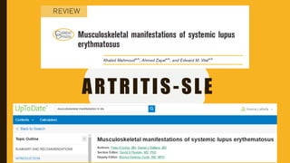
ARTRITIS-SLE
A LITTLE BIT OF HISTORY
Lupus is the Latin word for wolf. Erythematosus means red rashes. In 1851, Dr.
Cazenave discovered red rashes on a patient’s face that looked like wolf bites.
He named the rash Discoid Lupus Erythematosus (DLE).
In 1885, Sir William Osler recognized that many people with lupus had a disease
involving not only the skin but many other organs or systems. He named the
disease Systemic Lupus Erythematosus (SLE).
WHATCAUSES SLE?
SLE is an autoimmune disorder that develops when the body’s immune
system begins to attack its own tissues. Its cause is unknown, but it is likely
that a combination of genetic, environmental, and, possibly, hormonal
factors work together to cause SLE.
This occurs through the production of “auto-antibodies” that attack a person’s
own cells thus contributing to the inflammation of various parts of the
body, and may cause damage to organs and tissues.
The most common type of auto-antibody that develops in people with SLE
is called an antinuclear antibody (ANA) because it reacts with parts of the
cell’s nucleus (command centre).
WHATCAUSES SLE?
MUSCULOSKELETAL MANIFESTATION
IN SLE
• Musculoskeletal manifestations are among the most
common features of systemic lupus erythematosus (SLE)
both in initial diagnosis and in long-term management.
• They are crucial to overall patient outcome as well as the
development of new therapeutics.
• Of individual features of SLE disease activity, arthralgia
was the only one to be significantly associated with work
disability
• Arthralgia, arthritis, osteonecrosis (avascular necrosis
of bone), and myopathy are the principal
manifestations.
• Osteoporosis, often due to glucocorticoid therapy,
may increase the risk of fractures.
IMPACT OF MUSCULOSKELETAL MANIFESTATIONS OF
SYSTEMIC LUPUS ERYTHEMATOSUS
• Although other manifestations may be more important in causing organ
failure and early mortality, musculoskeletal manifestations are the key
determinant of impact of disease for a larger group of patients
• Apart from fatigue, the most frequent symptoms reported by 324 SLE
patients in answer to the question ‘What SLE-related symptoms have you
experienced as most difficult during your disease?’
– pain(50%)
– musculoskeletal (46%).
• Further, these symptoms were most strongly related to reduced health-
related quality of life.
• In systematic review, 47% of SLE patients were employed and 34% had
work disability
CLASSIFICATION OF MUSCULOSKELETAL
SYSTEMIC LUPUS ERYTHEMATOSUS
Many previous reviews, have focused on two clinically distinctive phenotypes of
lupus arthritis:
• Jaccoud’s arthropathy : a nonerosive arthropathy with reversible deformities,
quite uncommon
• Rhupus : an erosive arthritis with identical radiographic appearance to RA, and
and has also been associated with rheumatoid factor and anticitrullinated
peptide antibodies
• NDNE (nondeforming nonerosive) : the vast majority of patients with lupus
arthritis. these patients all have similar inflammatory features to other
inflammatory arthritis, such as symmetrical small joint distribution and
morning stiffness
ARTHRITIS AND ARTHRALGIAS
• Clinical characteristics — Arthritic symptoms in patients with SLE have the
following characteristics that are generally different from those in rheumatoid
arthritis (RA)
• The arthritis and arthralgias of SLE tend to be migratory; symptoms in a
particular joint may be gone within 24 hours.
• Involvement is usually symmetrical and polyarticular with a predilection for
the knees, carpal joints, and joints of the fingers, especially the proximal
interphalangeal (PIP) joint. The ankles, elbows, shoulders, and hips are less
frequently involved. Involvement of the sacroiliac joints and cervical spine
occur but is rare. Monoarticular arthritis is unusual and suggests an
cause such as infection.
• Morning stiffness is usually measured in minutes and is not prolonged as in
RA.
• The degree of pain often exceeds objective physical findings, and tenderness
tenderness may be difficult to assess because of increased pain sensitivity in
some patients, which can be associated with coexisting fibromyalgia.
ARTHRITIS AND ARTHRALGIAS
• Although the arthritis of SLE is generally considered to be nondeforming, flexion deformities,
ulnar deviation, soft tissue laxity, and swan neck deformities, as seen in RA, have been noted in
15 to 50 percent of patients with SLE
• However, unlike RA, erosions in the hand are rarely noted on plain radiographs of the hands in
SLE. Similar to magnetic resonance imaging, ultrasound can also reveal erosive changes and
abnormalities of the soft tissues, including capsular swelling, tenosynovitis, and synovial
proliferation. The presence of antibodies to citrullinated peptides/proteins in SLE patients is
strongly associated with erosive arthritis
• The hand deformity tends to occur in patients who have been receiving glucocorticoids, who
have anti-Ro and/or anti-La antibodies, or who have longstanding disease. In contrast to the
typical findings in RA, these deformities are usually easily reducible; they are thought to be due
to lax joint capsules, tendons, and ligaments that cause joint instability. Thus, the hand
deformities in patients with SLE resemble Jaccoud’s arthritis, a nonerosive chronic deforming
arthritis that may follow acute rheumatic fever.
• Tendons may also be involved in SLE. Tenosynovitis has been noted in 10 to 44 percent of
patients, including epicondylitis, rotator cuff tendinitis, Achilles tendinitis, tibialis posterior
tendinitis, and plantar fasciitis
ARTHRITIS AND ARTHRALGIAS
• Synovial involvement — Synovial effusions are infrequent in patients with SLE. When
they occur, they are usually small, and the fluid is clear or slightly cloudy.
• In contrast to the highly inflammatory exudates of RA, the synovial fluid is only mildly
inflammatory, with low protein levels and white blood cell counts (similar to a
transudate).
• Antinuclear antibodies (ANA) and lupus erythematosus (LE) cells have been observed
in synovial fluids and, if present, have been thought to be useful diagnostically.
However, they add little to a positive ANA in the serum.
• Synovitis in SLE has a molecular synovial signature distinct from that seen in
osteoarthritis or rheumatoid arthritis in that interferon-inducible genes are
upregulated
ARTHRITIS AND ARTHRALGIAS
• An MRI study found erosions in 45% of carpal bones, again present in all types of
lupus arthritis. These rates of erosion also seem to exceed prevalence of Rhupus and
Jaccoud’s in their conventional definitions, so their clinical significance is less clear
than in RA.
• In RA, synovitis then bone oedema are the precursors of bone erosion. However, in SLE
many bones affected by erosion had either no synovitis, or synovitis at a level that
would not lead to erosion in RA
• Although these RA and SLE populations had similar frequencies of erosions, bone
oedema was significantly less frequent in SLE.
IMMUNOPATHOGENESIS
• At a molecular level, synovial gene expression studies in SLE patients demonstrate a
distinct appearance from both osteoarthritis and RA.
• SLE synovium has marked upregulation of type I interferon-stimulated genes and
downregulation of extra-cellular matrix homeostasis
• Interferon (IFN)-a, primarily produced by circulating plasmacytoid dendritic cells and
monocytes is generally associated with more severe disease in SLE
• Synoviocytes and fibroblasts produce interferon-b and this has been shown
experimentally to have regulatory roles, with downregulation tumour necrotizing
factor- a and upregulation of tumour growth factor- b, Interleukin (IL)-10, and IL-1ra
• Understanding the roles of type I interferons in SLE is of renewed interest as therapies that
target this pathway are now in phase III trials and have demonstrated efficacy for arthritis-
specific outcomes when targeting either (IFN)-a alone or the interferon receptor that is
shared by IFN-a and IFN-b
CLINICAL ASSESSMENT OF LUPUS ARTHRITIS
• SLEDAI : the inclusion of erythema or warmth to define synovitis, as well as just joint
swelling.
– 4 points are scored for two or more joints with these signs (SLEDAI-2K) or more than two
joints (SELENA–SLEDAI)
– no points for lesser degrees of inflammation.
• BILAG-2004 index is semiquantitative for each organ system assessed. For the
musculoskeletal domain,
– BILAG A (the highest score) : active synovitis > 2 joints with marked loss of functional
range of movements.
– BILAG B : tendonitis/tenosynovitis or active synovitis > 1 joint (observed or through
history) with some loss of functional range of movement (or improving BILAG A disease).
– BILAG C : inflammatory pain (e.g. with morning stiffness) without synovitis (or improving
BILAG B disease).
– BILAG D : Pain without inflammatory symptoms (e.g. pain that clinically appears to be
because of osteoarthritis) is and patients with no current symptoms.
ARTHRITIS AND ARTHRALGIAS
• Treatment — The treatment of arthritis and arthralgias associated with SLE can often be limited
to a background of antimalarial drugs and symptomatic treatment with analgesic
medications.
• Arthritis in patients with SLE usually responds to nonsteroidal antiinflammatory drugs
or hydroxychloroquine. Patients with arthralgia may benefit from acetaminophen.
Glucocorticoids, methotrexate, and other immunosuppressives may be required in some patients
• Low doses of glucocorticoids may be required to treat the arthritis in patients also having a
systemic disease flare.
• For patients with persistent arthritis, additional immunosuppressive agents or disease-
modifying antirheumatic drugs (DMARDs) may also be used in a manner largely based upon
the treatment of rheumatoid arthritis (RA).
• Pain from arthritis must be distinguished from fibromyalgia, which is more common among
patients with SLE. The arthritis in patients with SLE must also be distinguished from osteoarthritis
• Total joint arthroplasty is infrequently required in patients with SLE.
SUBCUTANEOUS NODULES
• Subcutaneous nodules that occur characteristically in patients with
rheumatoid arthritis (RA) have been noted in 5 to 7 percent of patients
with SLE.
• Nodules are generally seen in association with active disease in patients
with disease that resembles RA (eg, rheumatoid factor positivity)
• The pathology of these nodules is similar to that of rheumatoid nodules.
OSTEONECROSIS
• Osteonecrosis (also called avascular, aseptic, or ischemic necrosis) in patients with
systemic lupus erythematosus (SLE) is most common in the femoral head, although the
humeral head, tibial plateau, and scaphoid navicular can also be affected.
• Osteonecrosis is usually bilateral and is often asymptomatic. When symptoms occur,
femoral head involvement usually manifests as pain in the groin, especially with
weightbearing
• Mechanism and risk factors — Osteonecrosis begins by interruption of blood
to the bone. Subsequently, the adjacent area becomes hyperemic, resulting in
emineralization, trabecular thinning, and, if stressed, collapse.
• Patients with SLE who have taken glucocorticoids are at greatest risk
• Osteonecrosis often develops a relatively short time after the onset of
glucocorticoid therapy, within a month in some patients who receive high doses
OSTEONECROSIS
• Other risk factors for osteonecrosis have also been suggested.
• The Raynaud phenomenon and hyperlipidemia were associated in one retrospective
series
• In the latter study, the presence of arthritis, use of glucocorticoids, and/or cytotoxic
medication were associated with a significantly increased risk of osteonecrosis.
Cytotoxic treatment was also identified in a prospective study of 571 patients with SLE
OSTEONECROSIS
• Treatment — The management of osteonecrosis is problematic. The best initial
approach is prevention by avoiding chronic therapy with high-dose glucocorticoids.
OSTEOPOROSIS
• Loss of trabecular bone density is a significant problem in patients with systemic lupus
erythematosus (SLE). Trabecular bones (eg, ribs, vertebrae) are more likely to be
involved than long cortical bones.
• There are no symptoms unless fractures occur.
• A study of 516 women with SLE seen between 1995 and 2000 found that 205 had a
determination of bone mineral density (BMD).
• Those who had a BMD measurement tended to have more traditional risk factors for
osteoporosis (eg, age, postmenopausal status), to have higher lupus disease activity,
to more frequently have renal involvement, to have evidence of increased end-
organ damage, and to have higher use of glucocorticoids and other
immunosuppressives.
• Among the women whose BMDs were measured, 18 percent had osteoporosis, and 49
percent had osteopenia
• Other risk factors for osteoporosis include smoking, physical inactivity, chronic
systemic inflammation, and premature gonadal failure
• Prevention and treatment — Certain general principles should be followed in all
patients to minimize bone loss, particularly in patients receiving therapy with
glucocorticoids.
Briefly, important elements in management include:
• Modification of lifestyle factors (ie, elimination of cigarette smoking, limitation of
alcohol consumption, and maintenance of a weight-bearing exercise regimen).
• Limitation of glucocorticoid therapy to the lowest possible dose and duration
• Administration of calcium and vitamin D.
• Measurement of bone mineral density and administration of pharmacologic therapy.
most postmenopausal patients, the therapy of choice is a bisphosphonate. Other
interventions may be appropriate in selected patients, such as hormone therapy and
use of teriparatide(parathyroid hormone).
FRACTURES
• Osteopenia and osteoporosis may account for much of the increased risk of fracture
seen in patients with systemic lupus erythematosus (SLE)
• The magnitude of the fracture risk is illustrated by a retrospective, cohort study of
approximately 700 women with SLE and an equivalent number of age-matched
controls; a fivefold increase in fracture among those with SLE was noted.
• Variables that were significantly associated with the time from diagnosis to that of
fracture included older age at the time of diagnosis of SLE, longer disease
duration, longer duration of glucocorticoid use, less use of oral contraceptives,
menopause status.
• The presence of antiphospholipid antibodies (aPL) may be associated with an increased
risk of nontraumatic fractures. This was illustrated in one study that linked aPL to an
increased risk of metatarsal (stress) fractures in patients with SLE.
• Vertebral fractures, found in up to 20 percent of patients with SLE, have been
associated with intravenous use of methylprednisolone at any time and with male sex
MUSCLE DISEASE
• Myalgias, muscle tenderness, or muscle weakness occurs in up to 70
percent of patients with systemic lupus erythematosus (SLE) and may be
the reason that the patient initially seeks medical attention.
• However, severe muscle weakness, atrophy, or myositis is relatively
uncommon (7 to 15 percent).
• Pathologic examination reveals perivascular and perifascicular
mononuclear cell infiltrates in 25 percent of patients. Other histologic
findings include muscle atrophy, microtubular inclusions, mononuclear
infiltrate, fiber necrosis, and, occasionally, vacuolated muscle fibers
• In addition to SLE itself, glucocorticoids and antimalarial drugs can cause muscle
weakness. Medication-induced myopathy is generally easily differentiated from SLE-
induced myopathy or myositis:
• Serum levels of creatine kinase (CK) and/or aldolase are usually normal in patients with
glucocorticoid-induced myopathy, although lactate dehydrogenase (LDH) values may
be elevated in some cases. Muscle biopsy reveals an increase in the number of
sarcolemmal nuclei, rowing and centralization of the nuclei, vacuolization and loss of
fiber cross-striations, and phagocytosis but does not reveal the inflammation
characteristic of SLE.
• Muscle enzymes are also typically normal in patients with antimalarial-induced
myopathy. Muscle biopsy may reveal vacuolar changes but not inflammation.
• Establishing the correct diagnosis has important implications for therapy. Lupus
myositis responds to treatment with glucocorticoids (using a regimen similar to that
in polymyositis), while glucocorticoid-induced myopathy responds to a reduction in
or withdrawal of glucocorticoid therapy. Antimalarial myopathy responds to stopping
the drug, but it may take months for the myopathy to improve, partly due to the long
half-life of hydroxychloroquine (months).
FIBROMYALGIA
• Reported prevalence rates of fibromyalgia in patients with
systemic lupus erythematosus (SLE) vary widely (from 5 to 25
percent), but in general are much higher than in the general
population.
• The symptoms are detrimental to the patient’s quality of life
and may increase disability
• It is important to remember that treatment with
glucocorticoids can cause myalgia, as can glucocorticoid
withdrawal, and that symptoms and tenderness suggestive of
fibromyalgia in patients with SLE usually do not represent
active lupus
• The treatment of the musculoskeletal symptoms of SLE can be
similar to those of other arthropathies.
• A formal physical therapy program with modalities in
conjunction with appropriate pain medication tailored to not
affect other involved organ systems is the key.
THANK YOU

More Related Content

Similar to Artritis-sle tifa.pptx

Rheumatoid arthritis by dr shaiesh gupta
Rheumatoid arthritis by dr shaiesh guptaRheumatoid arthritis by dr shaiesh gupta
Rheumatoid arthritis by dr shaiesh guptaSHAILESH GUPTA
 
Rheumatoid arthritis
Rheumatoid arthritis Rheumatoid arthritis
Rheumatoid arthritis Zahirulkhan1
 
rheumatic_diseases (2).ppt
rheumatic_diseases (2).pptrheumatic_diseases (2).ppt
rheumatic_diseases (2).pptEetaJain1
 
Rheumatoid arthritis
Rheumatoid arthritisRheumatoid arthritis
Rheumatoid arthritisJulie Jane
 
Approach towards a case of musculoskeletal disorder.#
Approach towards a case of musculoskeletal disorder.#Approach towards a case of musculoskeletal disorder.#
Approach towards a case of musculoskeletal disorder.#sirmohit
 
Rheumatic diseases
Rheumatic diseasesRheumatic diseases
Rheumatic diseasesAmer
 
Rheumatic disorders summary
Rheumatic disorders summaryRheumatic disorders summary
Rheumatic disorders summaryRasha Dabbagh
 
Juvenile arthritis an overview
Juvenile arthritis an overviewJuvenile arthritis an overview
Juvenile arthritis an overviewNilesh Jadhav
 
Rheumatoid arthritis nov 2020
Rheumatoid arthritis nov 2020Rheumatoid arthritis nov 2020
Rheumatoid arthritis nov 2020Mukiza1
 
Osteoarthritis and Rheumatoid Arthritis
Osteoarthritis and Rheumatoid Arthritis Osteoarthritis and Rheumatoid Arthritis
Osteoarthritis and Rheumatoid Arthritis Rifhan Kamaruddin
 
Systemic lupus erythematosus (sle)
Systemic lupus erythematosus (sle)Systemic lupus erythematosus (sle)
Systemic lupus erythematosus (sle)Qais Ali Asad
 
Rheumatoid arthritis by dr hari sharan aryal
Rheumatoid arthritis by dr hari sharan aryalRheumatoid arthritis by dr hari sharan aryal
Rheumatoid arthritis by dr hari sharan aryalHari Aryal
 
rheumatoid arthritis.
rheumatoid arthritis.rheumatoid arthritis.
rheumatoid arthritis.kajal sansoya
 
Approach to low back ache
Approach to low back acheApproach to low back ache
Approach to low back acheAlankar Tiwari
 
Approach to a patient with arthritis by Dr Imtiaz.pptx
Approach to a patient with arthritis by Dr Imtiaz.pptxApproach to a patient with arthritis by Dr Imtiaz.pptx
Approach to a patient with arthritis by Dr Imtiaz.pptxDRIMTIAZ3
 
Ankylosing spondylitis by dr ashutosh
Ankylosing spondylitis by dr ashutoshAnkylosing spondylitis by dr ashutosh
Ankylosing spondylitis by dr ashutoshAshutosh Kumar
 

Similar to Artritis-sle tifa.pptx (20)

Rheumatoid arthritis by dr shaiesh gupta
Rheumatoid arthritis by dr shaiesh guptaRheumatoid arthritis by dr shaiesh gupta
Rheumatoid arthritis by dr shaiesh gupta
 
Rheumatoid arthritis
Rheumatoid arthritis Rheumatoid arthritis
Rheumatoid arthritis
 
rheumatic_diseases (2).ppt
rheumatic_diseases (2).pptrheumatic_diseases (2).ppt
rheumatic_diseases (2).ppt
 
Rheumatoid arthritis
Rheumatoid arthritisRheumatoid arthritis
Rheumatoid arthritis
 
Approach towards a case of musculoskeletal disorder.#
Approach towards a case of musculoskeletal disorder.#Approach towards a case of musculoskeletal disorder.#
Approach towards a case of musculoskeletal disorder.#
 
Rheumatic diseases
Rheumatic diseasesRheumatic diseases
Rheumatic diseases
 
Rheumatic disorders summary
Rheumatic disorders summaryRheumatic disorders summary
Rheumatic disorders summary
 
Ankylos ing spondylitis
Ankylos ing spondylitisAnkylos ing spondylitis
Ankylos ing spondylitis
 
SLE.pdf
SLE.pdfSLE.pdf
SLE.pdf
 
Juvenile arthritis an overview
Juvenile arthritis an overviewJuvenile arthritis an overview
Juvenile arthritis an overview
 
Pearls and myths in sle
Pearls and myths in slePearls and myths in sle
Pearls and myths in sle
 
Rheumatoid arthritis nov 2020
Rheumatoid arthritis nov 2020Rheumatoid arthritis nov 2020
Rheumatoid arthritis nov 2020
 
Osteoarthritis and Rheumatoid Arthritis
Osteoarthritis and Rheumatoid Arthritis Osteoarthritis and Rheumatoid Arthritis
Osteoarthritis and Rheumatoid Arthritis
 
Systemic lupus erythematosus (sle)
Systemic lupus erythematosus (sle)Systemic lupus erythematosus (sle)
Systemic lupus erythematosus (sle)
 
Rheumatoid arthritis by dr hari sharan aryal
Rheumatoid arthritis by dr hari sharan aryalRheumatoid arthritis by dr hari sharan aryal
Rheumatoid arthritis by dr hari sharan aryal
 
rheumatoid arthritis.
rheumatoid arthritis.rheumatoid arthritis.
rheumatoid arthritis.
 
Approach to low back ache
Approach to low back acheApproach to low back ache
Approach to low back ache
 
Approach to a patient with arthritis by Dr Imtiaz.pptx
Approach to a patient with arthritis by Dr Imtiaz.pptxApproach to a patient with arthritis by Dr Imtiaz.pptx
Approach to a patient with arthritis by Dr Imtiaz.pptx
 
ANKYLOSING SPONDYLITIS.pptx
ANKYLOSING SPONDYLITIS.pptxANKYLOSING SPONDYLITIS.pptx
ANKYLOSING SPONDYLITIS.pptx
 
Ankylosing spondylitis by dr ashutosh
Ankylosing spondylitis by dr ashutoshAnkylosing spondylitis by dr ashutosh
Ankylosing spondylitis by dr ashutosh
 

Recently uploaded

VIP Call Girls Pune Vani 9907093804 Short 1500 Night 6000 Best call girls Ser...
VIP Call Girls Pune Vani 9907093804 Short 1500 Night 6000 Best call girls Ser...VIP Call Girls Pune Vani 9907093804 Short 1500 Night 6000 Best call girls Ser...
VIP Call Girls Pune Vani 9907093804 Short 1500 Night 6000 Best call girls Ser...Miss joya
 
Russian Call Girls in Pune Riya 9907093804 Short 1500 Night 6000 Best call gi...
Russian Call Girls in Pune Riya 9907093804 Short 1500 Night 6000 Best call gi...Russian Call Girls in Pune Riya 9907093804 Short 1500 Night 6000 Best call gi...
Russian Call Girls in Pune Riya 9907093804 Short 1500 Night 6000 Best call gi...Miss joya
 
Call Girls Service Chennai Jiya 7001305949 Independent Escort Service Chennai
Call Girls Service Chennai Jiya 7001305949 Independent Escort Service ChennaiCall Girls Service Chennai Jiya 7001305949 Independent Escort Service Chennai
Call Girls Service Chennai Jiya 7001305949 Independent Escort Service ChennaiNehru place Escorts
 
Call Girls Horamavu WhatsApp Number 7001035870 Meeting With Bangalore Escorts
Call Girls Horamavu WhatsApp Number 7001035870 Meeting With Bangalore EscortsCall Girls Horamavu WhatsApp Number 7001035870 Meeting With Bangalore Escorts
Call Girls Horamavu WhatsApp Number 7001035870 Meeting With Bangalore Escortsvidya singh
 
Kesar Bagh Call Girl Price 9548273370 , Lucknow Call Girls Service
Kesar Bagh Call Girl Price 9548273370 , Lucknow Call Girls ServiceKesar Bagh Call Girl Price 9548273370 , Lucknow Call Girls Service
Kesar Bagh Call Girl Price 9548273370 , Lucknow Call Girls Servicemakika9823
 
VIP Mumbai Call Girls Hiranandani Gardens Just Call 9920874524 with A/C Room ...
VIP Mumbai Call Girls Hiranandani Gardens Just Call 9920874524 with A/C Room ...VIP Mumbai Call Girls Hiranandani Gardens Just Call 9920874524 with A/C Room ...
VIP Mumbai Call Girls Hiranandani Gardens Just Call 9920874524 with A/C Room ...Garima Khatri
 
Call Girls Chennai Megha 9907093804 Independent Call Girls Service Chennai
Call Girls Chennai Megha 9907093804 Independent Call Girls Service ChennaiCall Girls Chennai Megha 9907093804 Independent Call Girls Service Chennai
Call Girls Chennai Megha 9907093804 Independent Call Girls Service ChennaiNehru place Escorts
 
Sonagachi Call Girls Services 9907093804 @24x7 High Class Babes Here Call Now
Sonagachi Call Girls Services 9907093804 @24x7 High Class Babes Here Call NowSonagachi Call Girls Services 9907093804 @24x7 High Class Babes Here Call Now
Sonagachi Call Girls Services 9907093804 @24x7 High Class Babes Here Call NowRiya Pathan
 
Call Girl Chennai Indira 9907093804 Independent Call Girls Service Chennai
Call Girl Chennai Indira 9907093804 Independent Call Girls Service ChennaiCall Girl Chennai Indira 9907093804 Independent Call Girls Service Chennai
Call Girl Chennai Indira 9907093804 Independent Call Girls Service ChennaiNehru place Escorts
 
Bangalore Call Girls Majestic 📞 9907093804 High Profile Service 100% Safe
Bangalore Call Girls Majestic 📞 9907093804 High Profile Service 100% SafeBangalore Call Girls Majestic 📞 9907093804 High Profile Service 100% Safe
Bangalore Call Girls Majestic 📞 9907093804 High Profile Service 100% Safenarwatsonia7
 
Call Girls Yelahanka Just Call 7001305949 Top Class Call Girl Service Available
Call Girls Yelahanka Just Call 7001305949 Top Class Call Girl Service AvailableCall Girls Yelahanka Just Call 7001305949 Top Class Call Girl Service Available
Call Girls Yelahanka Just Call 7001305949 Top Class Call Girl Service Availablenarwatsonia7
 
Aspirin presentation slides by Dr. Rewas Ali
Aspirin presentation slides by Dr. Rewas AliAspirin presentation slides by Dr. Rewas Ali
Aspirin presentation slides by Dr. Rewas AliRewAs ALI
 
CALL ON ➥9907093804 🔝 Call Girls Hadapsar ( Pune) Girls Service
CALL ON ➥9907093804 🔝 Call Girls Hadapsar ( Pune)  Girls ServiceCALL ON ➥9907093804 🔝 Call Girls Hadapsar ( Pune)  Girls Service
CALL ON ➥9907093804 🔝 Call Girls Hadapsar ( Pune) Girls ServiceMiss joya
 
Artifacts in Nuclear Medicine with Identifying and resolving artifacts.
Artifacts in Nuclear Medicine with Identifying and resolving artifacts.Artifacts in Nuclear Medicine with Identifying and resolving artifacts.
Artifacts in Nuclear Medicine with Identifying and resolving artifacts.MiadAlsulami
 
Call Girls Doddaballapur Road Just Call 7001305949 Top Class Call Girl Servic...
Call Girls Doddaballapur Road Just Call 7001305949 Top Class Call Girl Servic...Call Girls Doddaballapur Road Just Call 7001305949 Top Class Call Girl Servic...
Call Girls Doddaballapur Road Just Call 7001305949 Top Class Call Girl Servic...narwatsonia7
 
Vip Call Girls Anna Salai Chennai 👉 8250192130 ❣️💯 Top Class Girls Available
Vip Call Girls Anna Salai Chennai 👉 8250192130 ❣️💯 Top Class Girls AvailableVip Call Girls Anna Salai Chennai 👉 8250192130 ❣️💯 Top Class Girls Available
Vip Call Girls Anna Salai Chennai 👉 8250192130 ❣️💯 Top Class Girls AvailableNehru place Escorts
 
VIP Call Girls Indore Kirti 💚😋 9256729539 🚀 Indore Escorts
VIP Call Girls Indore Kirti 💚😋  9256729539 🚀 Indore EscortsVIP Call Girls Indore Kirti 💚😋  9256729539 🚀 Indore Escorts
VIP Call Girls Indore Kirti 💚😋 9256729539 🚀 Indore Escortsaditipandeya
 
Ahmedabad Call Girls CG Road 🔝9907093804 Short 1500 💋 Night 6000
Ahmedabad Call Girls CG Road 🔝9907093804  Short 1500  💋 Night 6000Ahmedabad Call Girls CG Road 🔝9907093804  Short 1500  💋 Night 6000
Ahmedabad Call Girls CG Road 🔝9907093804 Short 1500 💋 Night 6000aliya bhat
 

Recently uploaded (20)

sauth delhi call girls in Bhajanpura 🔝 9953056974 🔝 escort Service
sauth delhi call girls in Bhajanpura 🔝 9953056974 🔝 escort Servicesauth delhi call girls in Bhajanpura 🔝 9953056974 🔝 escort Service
sauth delhi call girls in Bhajanpura 🔝 9953056974 🔝 escort Service
 
VIP Call Girls Pune Vani 9907093804 Short 1500 Night 6000 Best call girls Ser...
VIP Call Girls Pune Vani 9907093804 Short 1500 Night 6000 Best call girls Ser...VIP Call Girls Pune Vani 9907093804 Short 1500 Night 6000 Best call girls Ser...
VIP Call Girls Pune Vani 9907093804 Short 1500 Night 6000 Best call girls Ser...
 
Russian Call Girls in Pune Riya 9907093804 Short 1500 Night 6000 Best call gi...
Russian Call Girls in Pune Riya 9907093804 Short 1500 Night 6000 Best call gi...Russian Call Girls in Pune Riya 9907093804 Short 1500 Night 6000 Best call gi...
Russian Call Girls in Pune Riya 9907093804 Short 1500 Night 6000 Best call gi...
 
Call Girls Service Chennai Jiya 7001305949 Independent Escort Service Chennai
Call Girls Service Chennai Jiya 7001305949 Independent Escort Service ChennaiCall Girls Service Chennai Jiya 7001305949 Independent Escort Service Chennai
Call Girls Service Chennai Jiya 7001305949 Independent Escort Service Chennai
 
Call Girls Horamavu WhatsApp Number 7001035870 Meeting With Bangalore Escorts
Call Girls Horamavu WhatsApp Number 7001035870 Meeting With Bangalore EscortsCall Girls Horamavu WhatsApp Number 7001035870 Meeting With Bangalore Escorts
Call Girls Horamavu WhatsApp Number 7001035870 Meeting With Bangalore Escorts
 
Kesar Bagh Call Girl Price 9548273370 , Lucknow Call Girls Service
Kesar Bagh Call Girl Price 9548273370 , Lucknow Call Girls ServiceKesar Bagh Call Girl Price 9548273370 , Lucknow Call Girls Service
Kesar Bagh Call Girl Price 9548273370 , Lucknow Call Girls Service
 
VIP Mumbai Call Girls Hiranandani Gardens Just Call 9920874524 with A/C Room ...
VIP Mumbai Call Girls Hiranandani Gardens Just Call 9920874524 with A/C Room ...VIP Mumbai Call Girls Hiranandani Gardens Just Call 9920874524 with A/C Room ...
VIP Mumbai Call Girls Hiranandani Gardens Just Call 9920874524 with A/C Room ...
 
Call Girls Chennai Megha 9907093804 Independent Call Girls Service Chennai
Call Girls Chennai Megha 9907093804 Independent Call Girls Service ChennaiCall Girls Chennai Megha 9907093804 Independent Call Girls Service Chennai
Call Girls Chennai Megha 9907093804 Independent Call Girls Service Chennai
 
Sonagachi Call Girls Services 9907093804 @24x7 High Class Babes Here Call Now
Sonagachi Call Girls Services 9907093804 @24x7 High Class Babes Here Call NowSonagachi Call Girls Services 9907093804 @24x7 High Class Babes Here Call Now
Sonagachi Call Girls Services 9907093804 @24x7 High Class Babes Here Call Now
 
Call Girl Chennai Indira 9907093804 Independent Call Girls Service Chennai
Call Girl Chennai Indira 9907093804 Independent Call Girls Service ChennaiCall Girl Chennai Indira 9907093804 Independent Call Girls Service Chennai
Call Girl Chennai Indira 9907093804 Independent Call Girls Service Chennai
 
Bangalore Call Girls Majestic 📞 9907093804 High Profile Service 100% Safe
Bangalore Call Girls Majestic 📞 9907093804 High Profile Service 100% SafeBangalore Call Girls Majestic 📞 9907093804 High Profile Service 100% Safe
Bangalore Call Girls Majestic 📞 9907093804 High Profile Service 100% Safe
 
Call Girls Yelahanka Just Call 7001305949 Top Class Call Girl Service Available
Call Girls Yelahanka Just Call 7001305949 Top Class Call Girl Service AvailableCall Girls Yelahanka Just Call 7001305949 Top Class Call Girl Service Available
Call Girls Yelahanka Just Call 7001305949 Top Class Call Girl Service Available
 
Aspirin presentation slides by Dr. Rewas Ali
Aspirin presentation slides by Dr. Rewas AliAspirin presentation slides by Dr. Rewas Ali
Aspirin presentation slides by Dr. Rewas Ali
 
CALL ON ➥9907093804 🔝 Call Girls Hadapsar ( Pune) Girls Service
CALL ON ➥9907093804 🔝 Call Girls Hadapsar ( Pune)  Girls ServiceCALL ON ➥9907093804 🔝 Call Girls Hadapsar ( Pune)  Girls Service
CALL ON ➥9907093804 🔝 Call Girls Hadapsar ( Pune) Girls Service
 
Artifacts in Nuclear Medicine with Identifying and resolving artifacts.
Artifacts in Nuclear Medicine with Identifying and resolving artifacts.Artifacts in Nuclear Medicine with Identifying and resolving artifacts.
Artifacts in Nuclear Medicine with Identifying and resolving artifacts.
 
Call Girls Doddaballapur Road Just Call 7001305949 Top Class Call Girl Servic...
Call Girls Doddaballapur Road Just Call 7001305949 Top Class Call Girl Servic...Call Girls Doddaballapur Road Just Call 7001305949 Top Class Call Girl Servic...
Call Girls Doddaballapur Road Just Call 7001305949 Top Class Call Girl Servic...
 
Escort Service Call Girls In Sarita Vihar,, 99530°56974 Delhi NCR
Escort Service Call Girls In Sarita Vihar,, 99530°56974 Delhi NCREscort Service Call Girls In Sarita Vihar,, 99530°56974 Delhi NCR
Escort Service Call Girls In Sarita Vihar,, 99530°56974 Delhi NCR
 
Vip Call Girls Anna Salai Chennai 👉 8250192130 ❣️💯 Top Class Girls Available
Vip Call Girls Anna Salai Chennai 👉 8250192130 ❣️💯 Top Class Girls AvailableVip Call Girls Anna Salai Chennai 👉 8250192130 ❣️💯 Top Class Girls Available
Vip Call Girls Anna Salai Chennai 👉 8250192130 ❣️💯 Top Class Girls Available
 
VIP Call Girls Indore Kirti 💚😋 9256729539 🚀 Indore Escorts
VIP Call Girls Indore Kirti 💚😋  9256729539 🚀 Indore EscortsVIP Call Girls Indore Kirti 💚😋  9256729539 🚀 Indore Escorts
VIP Call Girls Indore Kirti 💚😋 9256729539 🚀 Indore Escorts
 
Ahmedabad Call Girls CG Road 🔝9907093804 Short 1500 💋 Night 6000
Ahmedabad Call Girls CG Road 🔝9907093804  Short 1500  💋 Night 6000Ahmedabad Call Girls CG Road 🔝9907093804  Short 1500  💋 Night 6000
Ahmedabad Call Girls CG Road 🔝9907093804 Short 1500 💋 Night 6000
 

Artritis-sle tifa.pptx

  • 2. A LITTLE BIT OF HISTORY Lupus is the Latin word for wolf. Erythematosus means red rashes. In 1851, Dr. Cazenave discovered red rashes on a patient’s face that looked like wolf bites. He named the rash Discoid Lupus Erythematosus (DLE). In 1885, Sir William Osler recognized that many people with lupus had a disease involving not only the skin but many other organs or systems. He named the disease Systemic Lupus Erythematosus (SLE).
  • 3. WHATCAUSES SLE? SLE is an autoimmune disorder that develops when the body’s immune system begins to attack its own tissues. Its cause is unknown, but it is likely that a combination of genetic, environmental, and, possibly, hormonal factors work together to cause SLE. This occurs through the production of “auto-antibodies” that attack a person’s own cells thus contributing to the inflammation of various parts of the body, and may cause damage to organs and tissues. The most common type of auto-antibody that develops in people with SLE is called an antinuclear antibody (ANA) because it reacts with parts of the cell’s nucleus (command centre).
  • 5.
  • 6.
  • 7.
  • 8. MUSCULOSKELETAL MANIFESTATION IN SLE • Musculoskeletal manifestations are among the most common features of systemic lupus erythematosus (SLE) both in initial diagnosis and in long-term management. • They are crucial to overall patient outcome as well as the development of new therapeutics. • Of individual features of SLE disease activity, arthralgia was the only one to be significantly associated with work disability
  • 9. • Arthralgia, arthritis, osteonecrosis (avascular necrosis of bone), and myopathy are the principal manifestations. • Osteoporosis, often due to glucocorticoid therapy, may increase the risk of fractures.
  • 10. IMPACT OF MUSCULOSKELETAL MANIFESTATIONS OF SYSTEMIC LUPUS ERYTHEMATOSUS • Although other manifestations may be more important in causing organ failure and early mortality, musculoskeletal manifestations are the key determinant of impact of disease for a larger group of patients • Apart from fatigue, the most frequent symptoms reported by 324 SLE patients in answer to the question ‘What SLE-related symptoms have you experienced as most difficult during your disease?’ – pain(50%) – musculoskeletal (46%). • Further, these symptoms were most strongly related to reduced health- related quality of life. • In systematic review, 47% of SLE patients were employed and 34% had work disability
  • 11. CLASSIFICATION OF MUSCULOSKELETAL SYSTEMIC LUPUS ERYTHEMATOSUS Many previous reviews, have focused on two clinically distinctive phenotypes of lupus arthritis: • Jaccoud’s arthropathy : a nonerosive arthropathy with reversible deformities, quite uncommon • Rhupus : an erosive arthritis with identical radiographic appearance to RA, and and has also been associated with rheumatoid factor and anticitrullinated peptide antibodies • NDNE (nondeforming nonerosive) : the vast majority of patients with lupus arthritis. these patients all have similar inflammatory features to other inflammatory arthritis, such as symmetrical small joint distribution and morning stiffness
  • 12. ARTHRITIS AND ARTHRALGIAS • Clinical characteristics — Arthritic symptoms in patients with SLE have the following characteristics that are generally different from those in rheumatoid arthritis (RA) • The arthritis and arthralgias of SLE tend to be migratory; symptoms in a particular joint may be gone within 24 hours. • Involvement is usually symmetrical and polyarticular with a predilection for the knees, carpal joints, and joints of the fingers, especially the proximal interphalangeal (PIP) joint. The ankles, elbows, shoulders, and hips are less frequently involved. Involvement of the sacroiliac joints and cervical spine occur but is rare. Monoarticular arthritis is unusual and suggests an cause such as infection. • Morning stiffness is usually measured in minutes and is not prolonged as in RA. • The degree of pain often exceeds objective physical findings, and tenderness tenderness may be difficult to assess because of increased pain sensitivity in some patients, which can be associated with coexisting fibromyalgia.
  • 13. ARTHRITIS AND ARTHRALGIAS • Although the arthritis of SLE is generally considered to be nondeforming, flexion deformities, ulnar deviation, soft tissue laxity, and swan neck deformities, as seen in RA, have been noted in 15 to 50 percent of patients with SLE • However, unlike RA, erosions in the hand are rarely noted on plain radiographs of the hands in SLE. Similar to magnetic resonance imaging, ultrasound can also reveal erosive changes and abnormalities of the soft tissues, including capsular swelling, tenosynovitis, and synovial proliferation. The presence of antibodies to citrullinated peptides/proteins in SLE patients is strongly associated with erosive arthritis • The hand deformity tends to occur in patients who have been receiving glucocorticoids, who have anti-Ro and/or anti-La antibodies, or who have longstanding disease. In contrast to the typical findings in RA, these deformities are usually easily reducible; they are thought to be due to lax joint capsules, tendons, and ligaments that cause joint instability. Thus, the hand deformities in patients with SLE resemble Jaccoud’s arthritis, a nonerosive chronic deforming arthritis that may follow acute rheumatic fever. • Tendons may also be involved in SLE. Tenosynovitis has been noted in 10 to 44 percent of patients, including epicondylitis, rotator cuff tendinitis, Achilles tendinitis, tibialis posterior tendinitis, and plantar fasciitis
  • 14. ARTHRITIS AND ARTHRALGIAS • Synovial involvement — Synovial effusions are infrequent in patients with SLE. When they occur, they are usually small, and the fluid is clear or slightly cloudy. • In contrast to the highly inflammatory exudates of RA, the synovial fluid is only mildly inflammatory, with low protein levels and white blood cell counts (similar to a transudate). • Antinuclear antibodies (ANA) and lupus erythematosus (LE) cells have been observed in synovial fluids and, if present, have been thought to be useful diagnostically. However, they add little to a positive ANA in the serum. • Synovitis in SLE has a molecular synovial signature distinct from that seen in osteoarthritis or rheumatoid arthritis in that interferon-inducible genes are upregulated
  • 15. ARTHRITIS AND ARTHRALGIAS • An MRI study found erosions in 45% of carpal bones, again present in all types of lupus arthritis. These rates of erosion also seem to exceed prevalence of Rhupus and Jaccoud’s in their conventional definitions, so their clinical significance is less clear than in RA. • In RA, synovitis then bone oedema are the precursors of bone erosion. However, in SLE many bones affected by erosion had either no synovitis, or synovitis at a level that would not lead to erosion in RA • Although these RA and SLE populations had similar frequencies of erosions, bone oedema was significantly less frequent in SLE.
  • 16. IMMUNOPATHOGENESIS • At a molecular level, synovial gene expression studies in SLE patients demonstrate a distinct appearance from both osteoarthritis and RA. • SLE synovium has marked upregulation of type I interferon-stimulated genes and downregulation of extra-cellular matrix homeostasis • Interferon (IFN)-a, primarily produced by circulating plasmacytoid dendritic cells and monocytes is generally associated with more severe disease in SLE • Synoviocytes and fibroblasts produce interferon-b and this has been shown experimentally to have regulatory roles, with downregulation tumour necrotizing factor- a and upregulation of tumour growth factor- b, Interleukin (IL)-10, and IL-1ra • Understanding the roles of type I interferons in SLE is of renewed interest as therapies that target this pathway are now in phase III trials and have demonstrated efficacy for arthritis- specific outcomes when targeting either (IFN)-a alone or the interferon receptor that is shared by IFN-a and IFN-b
  • 17. CLINICAL ASSESSMENT OF LUPUS ARTHRITIS • SLEDAI : the inclusion of erythema or warmth to define synovitis, as well as just joint swelling. – 4 points are scored for two or more joints with these signs (SLEDAI-2K) or more than two joints (SELENA–SLEDAI) – no points for lesser degrees of inflammation. • BILAG-2004 index is semiquantitative for each organ system assessed. For the musculoskeletal domain, – BILAG A (the highest score) : active synovitis > 2 joints with marked loss of functional range of movements. – BILAG B : tendonitis/tenosynovitis or active synovitis > 1 joint (observed or through history) with some loss of functional range of movement (or improving BILAG A disease). – BILAG C : inflammatory pain (e.g. with morning stiffness) without synovitis (or improving BILAG B disease). – BILAG D : Pain without inflammatory symptoms (e.g. pain that clinically appears to be because of osteoarthritis) is and patients with no current symptoms.
  • 18.
  • 19. ARTHRITIS AND ARTHRALGIAS • Treatment — The treatment of arthritis and arthralgias associated with SLE can often be limited to a background of antimalarial drugs and symptomatic treatment with analgesic medications. • Arthritis in patients with SLE usually responds to nonsteroidal antiinflammatory drugs or hydroxychloroquine. Patients with arthralgia may benefit from acetaminophen. Glucocorticoids, methotrexate, and other immunosuppressives may be required in some patients • Low doses of glucocorticoids may be required to treat the arthritis in patients also having a systemic disease flare. • For patients with persistent arthritis, additional immunosuppressive agents or disease- modifying antirheumatic drugs (DMARDs) may also be used in a manner largely based upon the treatment of rheumatoid arthritis (RA). • Pain from arthritis must be distinguished from fibromyalgia, which is more common among patients with SLE. The arthritis in patients with SLE must also be distinguished from osteoarthritis • Total joint arthroplasty is infrequently required in patients with SLE.
  • 20. SUBCUTANEOUS NODULES • Subcutaneous nodules that occur characteristically in patients with rheumatoid arthritis (RA) have been noted in 5 to 7 percent of patients with SLE. • Nodules are generally seen in association with active disease in patients with disease that resembles RA (eg, rheumatoid factor positivity) • The pathology of these nodules is similar to that of rheumatoid nodules.
  • 21. OSTEONECROSIS • Osteonecrosis (also called avascular, aseptic, or ischemic necrosis) in patients with systemic lupus erythematosus (SLE) is most common in the femoral head, although the humeral head, tibial plateau, and scaphoid navicular can also be affected. • Osteonecrosis is usually bilateral and is often asymptomatic. When symptoms occur, femoral head involvement usually manifests as pain in the groin, especially with weightbearing • Mechanism and risk factors — Osteonecrosis begins by interruption of blood to the bone. Subsequently, the adjacent area becomes hyperemic, resulting in emineralization, trabecular thinning, and, if stressed, collapse. • Patients with SLE who have taken glucocorticoids are at greatest risk • Osteonecrosis often develops a relatively short time after the onset of glucocorticoid therapy, within a month in some patients who receive high doses
  • 22. OSTEONECROSIS • Other risk factors for osteonecrosis have also been suggested. • The Raynaud phenomenon and hyperlipidemia were associated in one retrospective series • In the latter study, the presence of arthritis, use of glucocorticoids, and/or cytotoxic medication were associated with a significantly increased risk of osteonecrosis. Cytotoxic treatment was also identified in a prospective study of 571 patients with SLE
  • 23. OSTEONECROSIS • Treatment — The management of osteonecrosis is problematic. The best initial approach is prevention by avoiding chronic therapy with high-dose glucocorticoids.
  • 24. OSTEOPOROSIS • Loss of trabecular bone density is a significant problem in patients with systemic lupus erythematosus (SLE). Trabecular bones (eg, ribs, vertebrae) are more likely to be involved than long cortical bones. • There are no symptoms unless fractures occur. • A study of 516 women with SLE seen between 1995 and 2000 found that 205 had a determination of bone mineral density (BMD). • Those who had a BMD measurement tended to have more traditional risk factors for osteoporosis (eg, age, postmenopausal status), to have higher lupus disease activity, to more frequently have renal involvement, to have evidence of increased end- organ damage, and to have higher use of glucocorticoids and other immunosuppressives. • Among the women whose BMDs were measured, 18 percent had osteoporosis, and 49 percent had osteopenia • Other risk factors for osteoporosis include smoking, physical inactivity, chronic systemic inflammation, and premature gonadal failure
  • 25. • Prevention and treatment — Certain general principles should be followed in all patients to minimize bone loss, particularly in patients receiving therapy with glucocorticoids. Briefly, important elements in management include: • Modification of lifestyle factors (ie, elimination of cigarette smoking, limitation of alcohol consumption, and maintenance of a weight-bearing exercise regimen). • Limitation of glucocorticoid therapy to the lowest possible dose and duration • Administration of calcium and vitamin D. • Measurement of bone mineral density and administration of pharmacologic therapy. most postmenopausal patients, the therapy of choice is a bisphosphonate. Other interventions may be appropriate in selected patients, such as hormone therapy and use of teriparatide(parathyroid hormone).
  • 26. FRACTURES • Osteopenia and osteoporosis may account for much of the increased risk of fracture seen in patients with systemic lupus erythematosus (SLE) • The magnitude of the fracture risk is illustrated by a retrospective, cohort study of approximately 700 women with SLE and an equivalent number of age-matched controls; a fivefold increase in fracture among those with SLE was noted. • Variables that were significantly associated with the time from diagnosis to that of fracture included older age at the time of diagnosis of SLE, longer disease duration, longer duration of glucocorticoid use, less use of oral contraceptives, menopause status. • The presence of antiphospholipid antibodies (aPL) may be associated with an increased risk of nontraumatic fractures. This was illustrated in one study that linked aPL to an increased risk of metatarsal (stress) fractures in patients with SLE. • Vertebral fractures, found in up to 20 percent of patients with SLE, have been associated with intravenous use of methylprednisolone at any time and with male sex
  • 27. MUSCLE DISEASE • Myalgias, muscle tenderness, or muscle weakness occurs in up to 70 percent of patients with systemic lupus erythematosus (SLE) and may be the reason that the patient initially seeks medical attention. • However, severe muscle weakness, atrophy, or myositis is relatively uncommon (7 to 15 percent). • Pathologic examination reveals perivascular and perifascicular mononuclear cell infiltrates in 25 percent of patients. Other histologic findings include muscle atrophy, microtubular inclusions, mononuclear infiltrate, fiber necrosis, and, occasionally, vacuolated muscle fibers
  • 28. • In addition to SLE itself, glucocorticoids and antimalarial drugs can cause muscle weakness. Medication-induced myopathy is generally easily differentiated from SLE- induced myopathy or myositis: • Serum levels of creatine kinase (CK) and/or aldolase are usually normal in patients with glucocorticoid-induced myopathy, although lactate dehydrogenase (LDH) values may be elevated in some cases. Muscle biopsy reveals an increase in the number of sarcolemmal nuclei, rowing and centralization of the nuclei, vacuolization and loss of fiber cross-striations, and phagocytosis but does not reveal the inflammation characteristic of SLE. • Muscle enzymes are also typically normal in patients with antimalarial-induced myopathy. Muscle biopsy may reveal vacuolar changes but not inflammation. • Establishing the correct diagnosis has important implications for therapy. Lupus myositis responds to treatment with glucocorticoids (using a regimen similar to that in polymyositis), while glucocorticoid-induced myopathy responds to a reduction in or withdrawal of glucocorticoid therapy. Antimalarial myopathy responds to stopping the drug, but it may take months for the myopathy to improve, partly due to the long half-life of hydroxychloroquine (months).
  • 29. FIBROMYALGIA • Reported prevalence rates of fibromyalgia in patients with systemic lupus erythematosus (SLE) vary widely (from 5 to 25 percent), but in general are much higher than in the general population. • The symptoms are detrimental to the patient’s quality of life and may increase disability • It is important to remember that treatment with glucocorticoids can cause myalgia, as can glucocorticoid withdrawal, and that symptoms and tenderness suggestive of fibromyalgia in patients with SLE usually do not represent active lupus
  • 30. • The treatment of the musculoskeletal symptoms of SLE can be similar to those of other arthropathies. • A formal physical therapy program with modalities in conjunction with appropriate pain medication tailored to not affect other involved organ systems is the key.