SlideShare a Scribd company logo
1 of 8
Download to read offline
CASE OF THE WEEK
                         PROFESSOR YASSER METWALLY
CLINICAL PICTURE

CLINICAL PICTURE:

A 33 years old male patient presented with gradual progressive hemiplegia with a dorsal sensory level at D 4.

RADIOLOGICAL FINDINGS

RADIOLOGICAL FINDINGS:


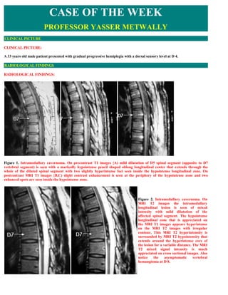
Figure 1. Intramedullary cavernoma. On precontrast T1 images {A) mild dilatation of D5 spinal segment (opposite to D7
vertebral segment) is seen with a markedly hypointense pencil shaped oblong longitudinal center that extends through the
whole of the dilated spinal segment with two slightly hyperintense foci seen inside the hypointense longitudinal zone. On
postcontrast MRI T1 images {B,C) slight contrast enhancement is seen at the periphery of the hypointense zone and two
enhanced spots are seen inside the hypointense zone.


                                                                                    Figure 2. Intramedullary cavernoma. On
                                                                                    MRI T2 images the intramedullary
                                                                                    longitudinal lesion is seen of mixed
                                                                                    intensity with mild dilatation of the
                                                                                    affected spinal segment. The hypointense
                                                                                    longitudinal zone that is appreciated on
                                                                                    the MRI T1 images appears hyperintense
                                                                                    on the MRI T2 images with irregular
                                                                                    contour, This MRI T2 hyperintensity is
                                                                                    surrounded by MRI T2 hypointensity that
                                                                                    extends around the hyperintense core of
                                                                                    the lesion for a variable distance. The MRI
                                                                                    T2 mixed signal intensity is much
                                                                                    appreciated on cross sectional images. Also
                                                                                    notice    the     asymptomatic      vertebral
                                                                                    hemangioma at D 8.
Figure 3. Intramedullary cavernoma.
                                                                                     On MRI T2 images (A,B) the
                                                                                     intramedullary longitudinal lesion is
                                                                                     seen of mixed intensity with mild
                                                                                     dilatation of the affected spinal
                                                                                     segment.       The        hypointense
                                                                                     longitudinal zone that is appreciated
                                                                                     on the MRI T1 (C,D) images appears
                                                                                     hyperintense on the MRI T2 images
                                                                                     with irregular contour, This MRI T2
                                                                                     hyperintensity is surrounded by MRI
                                                                                     T2 hypointensity that extends around
                                                                                     the hyperintense core of the lesion for
                                                                                     a variable distance. The MRI T2
                                                                                     mixed signal intensity is much
                                                                                     appreciated on cross sectional
                                                                                     images.


44


Figure 4. Intramedullary cavernoma. On MRI T2 images (B) the intramedullary longitudinal lesion is seen of mixed intensity
with mild dilatation of the affected spinal segment. The hypointense longitudinal zone that is appreciated on the MRI T1
images (A) appears hyperintense on the MRI T2 images with irregular contour, This MRI T2 hyperintensity is surrounded by
MRI T2 hypointensity that extends around the hyperintense core of the lesion for a variable distance. The MRI T2 mixed
signal intensity is much appreciated on cross sectional images. Also notice the asymptomatic vertebral hemangioma at D 8.
Figure 5. Intramedullary cavernoma. MRI T2 images (A,B) and precontrast MRI T1 images showing a mixed signal intensity
intramedullary lesion, The MRI mixed signal intensity of the precontrast MRI T1 images and the MRI T2 images is due to the
presence of altered blood of different ages.

      Conclusion

MRI picture of intramedullary cavernoma.

The MRI mixed signal intensity of the precontrast MRI T1 images and the MRI T2 images is due to the presence of altered
blood of different ages.

  1. The MRI T1 precontrast hyperintensity is due to methemoglobin

  2. The MRI T2 hypointensity is due to hemosiderin pigments.

Table 1. Evolution of MR imaging signal characteristics

                                Iron-containing           Iron oxidation     Red cell
    Phase           Time                                                                    T1            T2          T2*
                                   molecule                    state        membranes
Hyperacute     Hours         Oxyhemoglobin               Fe2+            Intact         Hypointense      Hyperintense
Acute          Hours to days Deoxyhemoglobin             Fe 2+           Intact            iso/
                                                                                                    Hypointense Hypointense
                                                                                        Hypointense
Early          Days to 1       Methemoglobin             Fe2+          Intact
                                                                                        Hyperintense       Hypointense
subacute       week
Late           1 week to       Methemoglobin             Fe2+          Degraded
                                                                                        Hyperintense       Hyperintense
subacute       months
Chronic        >months         Hemosiderin               Fe2+          Degraded            iso/
                                                                                                    Hypointense Hypointense
                                                                                        Hypointense
Abbreviations: Fe, iron; iso, isointense relative to normal brain.

DIAGNOSIS:

DIAGNOSIS: INTRAMEDULLARY CAVERNOMA

DISCUSSION

DISCUSSION:

Although they are not considered vascular lesions in the Anson and Spetzler grading system because they do not shunt blood
and have no arterial feeders and draining veins, spinal cavernous malformations (spinal cavernoma) (SCM) are another cause
of treatable myelopathy. 1 The frequency of spinal cavernomas is difficult to ascertain because of the low number of spinal
cord autopsies. In 1976, Voigt and Yasargil could find only 164 cases of cavernous malformations (cavernoma) in the literature.
13 In anther review, Ghogawala and Ogilvy reported only 67 intramedullary spinal cavernomas in the English-language
literature. 18 With the development of CT scanning and particularly MR imaging, these lesions are being reported more often.

It is estimated that cavernomas have an incidence of between 0.02% and 4% in the population 8 and that 3% to 5% of
cavernomas occur in the spinal cord. 15 Spinal cavernous malformations are considered to be congenital lesions that may
enlarge with time. 8 They are slow-flow lesions without arteriovenous shunting. They are found throughout the cord and are
often intramedullary Unlike their intracranial counterparts, which have no gender preponderance, 70% of spinal cavernoma
occur in women. 56 Sporadic forms and familial forms with autosomal dominant inheritance with variable penetrance have
been described. 4,6,7

Clinical features of spinal cavernomas vary from acute to progressive to asymptomatic. 10 Ogilvy et al described four types of
clinical presentations: (1) acute episodes with stepwise neurologic decline, (2) acute onset of a neurologic deficit with rapid
neurologic deterioration, (3) acute onset of a mild neurologic deficit with gradual neurologic deterioration, and (4) slowly
progressive neurologic decline. 16

Acute symptoms are caused by hemorrhage, which may cause enlargement of the spinal cavernoma or hematomyelia. Patients
often present with sudden back pain, often at the level of the lesion. Neurologic defects may follow many hours after the attack
of pain. This delayed neurologic deterioration may help differentiate spinal cavernoma hemorrhages from those of spinal
AVMS.

The progressive course of spinal cavernomas could be caused by small microhemorrhages, which may cause enlargement of the
spinal cavernoma. Small hemorrhages with organization, capillary budding, and propagation and repetitive hemorrhages are
two possible reasons for progression. 8 Other theories for progressive neurologic decline include a neurotoxic effect from red
blood cell degradation or an effect on the spinal cord microcirculation. 8

On gross inspection, cavernomas are well-defined, red-to-purple, lobulated lesions often described as looking like a mulberry.
Microscopically, cavernomas are made up of enlarged capillaries or sinusoidal vascular spaces with a simple endothelial lining,
thin adventitia, and no elastic or smooth muscle fibers in its walls. Evidence of prior hemorrhage is commonly seen. The
surrounding tissue usually has gliosis and hemosiderin. Calcification of spinal cavernoma is rare. 5 Spinal cavernomas are not
pathologically distinguishable from their intracranial counterparts. 9

Because so few spinal cavernomas have been studied, the natural history of these lesions is not completely known. Management
of spinal cavernoma depends on the patient's clinical symptoms and age and on the characteristics of the lesion. 5,8 For
asymptomatic lesions, no treatment is currently advised. 5,8 Symptomatic lesions are often surgically explored because of the
risk of future neurologic deterioration. The hemorrhage rate for cerebral cavernomas has been estimated to be between 0.5%
and 1.0% per year. 16,17 Whether this rate is valid for spinal cavernomas is unclear. In a review of 57 symptomatic patients
with spinal cavernomas, Canavero et al, 3 suggested a hemorrhage rate of 1.6% per year. 13 As with any spinal vascular
malformation, postoperative outcome depends largely on the level of preoperative neurologic function. 12 Residual tumor is
often difficult to distinguish from postoperative changes. Recurrence of spinal cavernoma after total removal and negative
postoperative MR imaging has been mentioned in only one review. 8,17

      Imaging of Spinal Cavernous Malformations

As with other vascular lesions of the spine, MR imaging should be the first modality used to look for spinal cavernomas.
Cavernous malformations are considered to be one of the angiographically occult lesions, and spinal angiography has no role in
its diagnosis or treatment. Although spinal cavernoma can enlarge the cord, this enlargement is often not detected with
myelographic studies. 12

MR imaging appearances of spinal cavernoma are quite similar to cerebral cavernomas. On MR imaging spinal cavernomas
are round or oblong lesions with mixed signal intensity from different stages of red blood cell breakdown. Cavernous
malformations are often encircled by a dark hemosiderin ring that shows characteristic blooming on gradient- recalled images.
Labauge et al, 7 found gradient-echo sequences to be the most sensitive for detecting small cavernomas. There is usually no or
minimal enhancement with the administration of contrast. Surrounding edema can often be seen on T2-weighted images.
Whereas the MR imaging appearance is characteristic, it is not pathognomonic. Other vascular malformations or hemorrhagic
neoplasms may have a similar appearance. Also, some spinal cavernomas may be hidden by hemorrhage and not visualized on
MR imaging.
Figure 6. Spinal cavernous malformation. A, Sagittal Tl -weighted MR image of the cervical spine shows an area of
heterogeneous signal intensity (arrow) at the level of C6 with some increased signal seen extending caudally (open arrow). B,
Sagittal T2-weighted MR image demonstrates heterogeneous signal (arrow) with some decreased signal seen extending
caudally (open arrow), representing subacute hemorrhage. C, Axial Tl -weighted MR image at the level of C6 shows
intramedullary mixed signal in the left aspect of the spinal cord (arrow). D, Axial T2*-weighted gradient-recalled MR image at
the level of C6 demonstrates low signal with a small area of high signal (arrow).


SUMMARY


SUMMARY

Table 2. Pathological characteristics of cavernomas that differentiate them from arteriovenous malformation

      Cavernous angiomas (cavernomas) consist of enlarged capillaries with the following characteristics:

             Sinusoidal

             Single layer of endothelium

             Thin collagenous wall
   Lack of smooth muscle fibers and elastic fibers

      Capillaries are immediately adjacent to each other, with no intervening neural tissue.

      They typically are not associated with enlarged feeding arteries or draining veins, and blood flow is low or even
       stagnated (cavernomas are angiographically occult).

      The surrounding parenchyma contains hemosiderin-laden macrophages and iron-laden glial cells.

      Grossly, they may range from soft to hard.

      Thrombosis, calcification, or ossification results in a harder lesion.

      Adjacent neural tissue may be affected, and small hemorrhages result in hemosiderin staining (iron salts may incite an
       epileptogenic focus). Tissues also may be gliotic.


Figure 7. Histopathology. (A) Microscopic section (trichrome stain) shows a mass of closely apposed vessels of varying size. The
vessel walls have a variable thickness, and there is no intervening brain. (B) Whole mount (trichrome stain) of a CM of the
third ventricle shows a plexus of vessels with some intervening brain at the periphery.


Figure 8. Example of cavernomas, notice absence of feeding arteries or draining veins
Figure 9. Pontine cavernoma, notice absence of feeding
                                                                       arteries or draining veins


Figure 10. For comparison, an arteriovenous malformation, notice the feeding arteries and the draining veins


      Addendum

            A new version of this PDF file (with a new case) is uploaded in my web site every week (every Saturday and remains
             available till Friday.)

            To download the current version follow the link quot;http://pdf.yassermetwally.com/case.pdfquot;.
            You can also download the current version from my web site at quot;http://yassermetwally.comquot;.
            To download the software version of the publication (crow.exe) follow the link:
             http://neurology.yassermetwally.com/crow.zip
            The case is also presented as a short case in PDF format, to download the short case follow the link:
             http://pdf.yassermetwally.com/short.pdf
            At the end of each year, all the publications are compiled on a single CD-ROM, please contact the author to know more
             details.
   Screen resolution is better set at 1024*768 pixel screen area for optimum display.
            For an archive of the previously reported cases go to www.yassermetwally.net, then under pages in the right panel, scroll
             down and click on the text entry quot;downloadable case records in PDF formatquot;


REFERENCES

References

  1.  Anson  JA,  Spetzler  RF:  Classification  of  spinal  arteriovenous  malformations  and  implications  for  treatment.  Barrow 
     Neurological Institute Quarterly 8:2-8, 1992

  2.  Awad IA: Patients with spinal cord cavernous malformations are at an increased risk for multiple neuraxis cavernous 
     malformations [comment]. Neurosurgery 45:33,1999

  3.  Canavero S, Pagni C, Kornfield M, et al: Spinal intramedullary cavernous angiomas: A literature meta- analysis. Surg
     Neurol 43:448-452,1995

  4.  Halbach VV, Higashida RT, Hiehima GB: Treatment of vertebral arteriovenous fistulas. AJR Am J Roentgenol 150:405-
     412,1988

  5.  Hurst  RW:  Spinal  vascular  disorders.  In  Atlas  SW  (ed):  Magnetic  Resonance  Imaging  of  the  Brain  and  Spine,  ed  2. 
     Philadelphia. Lippincott-Raven, 1996 pp 1387- 1412

  6.  Johnson  PC,  Wascher  TM,  Golfinos  J,  et  al:  Definition  and  pathologic  features.  In  Awad  IA,  Barrow  DL  (eds): 
     Cavernous Malformations. Park Ridge, IL, American Association of Neurological Surgeons, 1993, pp 1-11

  7.  Labauge  P,  Labauge  S,  Levy  C,  et  al:  Hereditary  cerebral  cavernous  angiomas:  Clinical  and  genetic  features  in  57 
     French families. Lancet 352:1892-1897,1998

  8.  McCormick WF, Nofzinger JD: quot;Cryptickquot; vascular malformations of the central nervous system. J Neurosurg 24:865-
     875, 1966

  9.  Mehmet KY, Wecht DA, Awad IA: Clinical spectrum and natural history of spinal vascular malformations. In Barrow 
     DL, Awad IA (eds): Spinal Vascular Malformations. Park Ridge, IL, American Association of Neurological Surgeons,
     1999, pp 45-55

 10.  Ogilvy  C,  Louis  D,  Ojemann  R:  Intramedullary  cavernous  angiomas  of  the  spinal  cord:  Clinical  presentation, 
     pathological features, and surgical management. Neurosurgery 31:219-230,1992

 11.  Oldfield EH: Spinal vascular malformations. In Wilkins RH, Rengachary SS (eds): Neurosurgery, ed 2, vol 2. New York, 
     McGraw-Hill, 1996, pp 2541-2558

 12.  Vishteh GA, Zabramski JM,  Spetzler RF: Patients with spinal cord cavernous  malformations  are  at an  increased  risk 
     for multiple neuraxis cavernous malformations. Neurosurgery 45:30-32, 1999

 13. Voigt K, Yasargil MG: Cerebral cavernous hemangiomas or cavernomas: Incidence, pathology, localization, diagnosis,
     clinical features and treatment: Review of the literature and report of an unusual case. Neurochirurgia 19:59,1976

 14. Wong JH, Awad IA: Spinal vascular malformations. In Jafar jj, Awad IA, Rosenwasser RH (eds): Vascular
     Malformations of the Central Nervous System. Philadelphia, Lippincott Williams & Wilkins, 1999, pp 155-160

 15.  Zimmerman R, Spetzler R, Lee K, et al: Cavernous malformations of the brainstem. J Neurosurg 75:32- 39,1991

 16. Ogilvy C, Louis D, Ojemann R: Intramedullary cavernous angiomas of the spinal cord: Clinical presentation,
     pathological features, and surgical management. Neurosurgery 31:219-230,1992

 17. Metwally, MYM: Textbook of neuroimaging, A CD-ROM publication, (Metwally, MYM editor) WEB-CD agency for
     electronic publication, version 9.2a April 2008

 18. Ghogawala Z, Ogilvy OS: Intramedullary cavernous malformations of the spinal cord. Neurosurg Clin North Am
     10:101-109,1999

More Related Content

Viewers also liked

Myasthenia Gravis..A quick guide to management
Myasthenia Gravis..A quick guide to managementMyasthenia Gravis..A quick guide to management
Myasthenia Gravis..A quick guide to managementProfessor Yasser Metwally
 
Issues in brain mapping...EEG patterns of encephalopathies and altered state ...
Issues in brain mapping...EEG patterns of encephalopathies and altered state ...Issues in brain mapping...EEG patterns of encephalopathies and altered state ...
Issues in brain mapping...EEG patterns of encephalopathies and altered state ...Professor Yasser Metwally
 
Topic of the month...Neuroimaging of embolic cerebral infarctions
Topic of the month...Neuroimaging of embolic cerebral infarctionsTopic of the month...Neuroimaging of embolic cerebral infarctions
Topic of the month...Neuroimaging of embolic cerebral infarctionsProfessor Yasser Metwally
 
Thesis section: The role of neuroimaging in muscle and peripheral nerve disor...
Thesis section: The role of neuroimaging in muscle and peripheral nerve disor...Thesis section: The role of neuroimaging in muscle and peripheral nerve disor...
Thesis section: The role of neuroimaging in muscle and peripheral nerve disor...Professor Yasser Metwally
 
Topic of the month.... Pathogenesis of pituitary adenomas
Topic of the month.... Pathogenesis of pituitary adenomasTopic of the month.... Pathogenesis of pituitary adenomas
Topic of the month.... Pathogenesis of pituitary adenomasProfessor Yasser Metwally
 
Thesis section...Management of hyperacute stroke
Thesis section...Management of hyperacute strokeThesis section...Management of hyperacute stroke
Thesis section...Management of hyperacute strokeProfessor Yasser Metwally
 
Thesis section...Management of hyperacute stroke
Thesis section...Management of hyperacute strokeThesis section...Management of hyperacute stroke
Thesis section...Management of hyperacute strokeProfessor Yasser Metwally
 
Short case...Subependymal primary CNS lymphoma
Short case...Subependymal primary CNS lymphomaShort case...Subependymal primary CNS lymphoma
Short case...Subependymal primary CNS lymphomaProfessor Yasser Metwally
 
Topic of the month...Neuroimaging of pituitary adenomas
Topic of the month...Neuroimaging of pituitary adenomasTopic of the month...Neuroimaging of pituitary adenomas
Topic of the month...Neuroimaging of pituitary adenomasProfessor Yasser Metwally
 
Issues in brainmapping...Nonspecific EEG patterns
Issues in brainmapping...Nonspecific EEG patternsIssues in brainmapping...Nonspecific EEG patterns
Issues in brainmapping...Nonspecific EEG patternsProfessor Yasser Metwally
 

Viewers also liked (20)

Short case...Congenital syringomyelia
Short case...Congenital syringomyeliaShort case...Congenital syringomyelia
Short case...Congenital syringomyelia
 
Myasthenia Gravis..A quick guide to management
Myasthenia Gravis..A quick guide to managementMyasthenia Gravis..A quick guide to management
Myasthenia Gravis..A quick guide to management
 
Issues in brain mapping...EEG patterns of encephalopathies and altered state ...
Issues in brain mapping...EEG patterns of encephalopathies and altered state ...Issues in brain mapping...EEG patterns of encephalopathies and altered state ...
Issues in brain mapping...EEG patterns of encephalopathies and altered state ...
 
Case record...Tuberous sclerosis
Case record...Tuberous sclerosisCase record...Tuberous sclerosis
Case record...Tuberous sclerosis
 
Topic of the month...Neuroimaging of embolic cerebral infarctions
Topic of the month...Neuroimaging of embolic cerebral infarctionsTopic of the month...Neuroimaging of embolic cerebral infarctions
Topic of the month...Neuroimaging of embolic cerebral infarctions
 
Case record...Subependymal CNS lymphoma
Case record...Subependymal CNS lymphomaCase record...Subependymal CNS lymphoma
Case record...Subependymal CNS lymphoma
 
Thesis section: The role of neuroimaging in muscle and peripheral nerve disor...
Thesis section: The role of neuroimaging in muscle and peripheral nerve disor...Thesis section: The role of neuroimaging in muscle and peripheral nerve disor...
Thesis section: The role of neuroimaging in muscle and peripheral nerve disor...
 
Topic of the month.... Pathogenesis of pituitary adenomas
Topic of the month.... Pathogenesis of pituitary adenomasTopic of the month.... Pathogenesis of pituitary adenomas
Topic of the month.... Pathogenesis of pituitary adenomas
 
Thesis section...Management of hyperacute stroke
Thesis section...Management of hyperacute strokeThesis section...Management of hyperacute stroke
Thesis section...Management of hyperacute stroke
 
Case record...Glomus jugulare tumor
Case record...Glomus jugulare tumorCase record...Glomus jugulare tumor
Case record...Glomus jugulare tumor
 
Case record...Spinal metastasis
Case record...Spinal metastasisCase record...Spinal metastasis
Case record...Spinal metastasis
 
Case record...Tectal plate glioma
Case record...Tectal plate gliomaCase record...Tectal plate glioma
Case record...Tectal plate glioma
 
Short case...Rasmussen's encephalitis
Short case...Rasmussen's encephalitisShort case...Rasmussen's encephalitis
Short case...Rasmussen's encephalitis
 
Thesis section...Management of hyperacute stroke
Thesis section...Management of hyperacute strokeThesis section...Management of hyperacute stroke
Thesis section...Management of hyperacute stroke
 
Short case...Subependymal primary CNS lymphoma
Short case...Subependymal primary CNS lymphomaShort case...Subependymal primary CNS lymphoma
Short case...Subependymal primary CNS lymphoma
 
Topic of the month...Neuroimaging of pituitary adenomas
Topic of the month...Neuroimaging of pituitary adenomasTopic of the month...Neuroimaging of pituitary adenomas
Topic of the month...Neuroimaging of pituitary adenomas
 
Issues in brainmapping...Nonspecific EEG patterns
Issues in brainmapping...Nonspecific EEG patternsIssues in brainmapping...Nonspecific EEG patterns
Issues in brainmapping...Nonspecific EEG patterns
 
Short case...Neurofibromatosis type 2
Short case...Neurofibromatosis type 2Short case...Neurofibromatosis type 2
Short case...Neurofibromatosis type 2
 
Neurological lectures...Vertigo
Neurological lectures...VertigoNeurological lectures...Vertigo
Neurological lectures...Vertigo
 
Case record...Lumbar spondylosis
Case record...Lumbar spondylosisCase record...Lumbar spondylosis
Case record...Lumbar spondylosis
 

More from Professor Yasser Metwally

The Snake, the Scorpion, the turtle in Egypt
The Snake, the Scorpion, the turtle in EgyptThe Snake, the Scorpion, the turtle in Egypt
The Snake, the Scorpion, the turtle in EgyptProfessor Yasser Metwally
 
Radiological pathology of epileptic disorders
Radiological pathology of epileptic disordersRadiological pathology of epileptic disorders
Radiological pathology of epileptic disordersProfessor Yasser Metwally
 
Radiological pathology of cerebrovascular disorders
Radiological pathology of cerebrovascular disordersRadiological pathology of cerebrovascular disorders
Radiological pathology of cerebrovascular disordersProfessor Yasser Metwally
 
Radiological pathology of spontaneous cerebral hemorrhage
Radiological pathology of spontaneous cerebral hemorrhageRadiological pathology of spontaneous cerebral hemorrhage
Radiological pathology of spontaneous cerebral hemorrhageProfessor Yasser Metwally
 
Radiological pathology of cerebral amyloid angiography
Radiological pathology of cerebral amyloid angiographyRadiological pathology of cerebral amyloid angiography
Radiological pathology of cerebral amyloid angiographyProfessor Yasser Metwally
 
Radiological pathology of cerebral microbleeds
Radiological pathology of cerebral microbleedsRadiological pathology of cerebral microbleeds
Radiological pathology of cerebral microbleedsProfessor Yasser Metwally
 
Issues in radiological pathology: Radiological pathology of watershed infarct...
Issues in radiological pathology: Radiological pathology of watershed infarct...Issues in radiological pathology: Radiological pathology of watershed infarct...
Issues in radiological pathology: Radiological pathology of watershed infarct...Professor Yasser Metwally
 
Radiological pathology of cortical laminar necrosis
Radiological pathology of cortical laminar necrosisRadiological pathology of cortical laminar necrosis
Radiological pathology of cortical laminar necrosisProfessor Yasser Metwally
 

More from Professor Yasser Metwally (20)

The Egyptian Zoo in Cairo 2015
The Egyptian Zoo in Cairo 2015The Egyptian Zoo in Cairo 2015
The Egyptian Zoo in Cairo 2015
 
End of the great nile river in Ras Elbar
End of the great nile river in Ras ElbarEnd of the great nile river in Ras Elbar
End of the great nile river in Ras Elbar
 
The Lion and The tiger in Egypt
The Lion and The tiger in EgyptThe Lion and The tiger in Egypt
The Lion and The tiger in Egypt
 
The monkeys in Egypt
The monkeys in EgyptThe monkeys in Egypt
The monkeys in Egypt
 
The Snake, the Scorpion, the turtle in Egypt
The Snake, the Scorpion, the turtle in EgyptThe Snake, the Scorpion, the turtle in Egypt
The Snake, the Scorpion, the turtle in Egypt
 
The Egyptian Parrot
The Egyptian ParrotThe Egyptian Parrot
The Egyptian Parrot
 
The Egyptian Deer
The Egyptian DeerThe Egyptian Deer
The Egyptian Deer
 
The Egyptian Pelican
The Egyptian PelicanThe Egyptian Pelican
The Egyptian Pelican
 
The Flamingo bird in Egypt
The Flamingo bird in EgyptThe Flamingo bird in Egypt
The Flamingo bird in Egypt
 
Egyptian Cats
Egyptian CatsEgyptian Cats
Egyptian Cats
 
Radiological pathology of epileptic disorders
Radiological pathology of epileptic disordersRadiological pathology of epileptic disorders
Radiological pathology of epileptic disorders
 
Radiological pathology of cerebrovascular disorders
Radiological pathology of cerebrovascular disordersRadiological pathology of cerebrovascular disorders
Radiological pathology of cerebrovascular disorders
 
Radiological pathology of spontaneous cerebral hemorrhage
Radiological pathology of spontaneous cerebral hemorrhageRadiological pathology of spontaneous cerebral hemorrhage
Radiological pathology of spontaneous cerebral hemorrhage
 
Radiological pathology of cerebral amyloid angiography
Radiological pathology of cerebral amyloid angiographyRadiological pathology of cerebral amyloid angiography
Radiological pathology of cerebral amyloid angiography
 
Radiological pathology of cerebral microbleeds
Radiological pathology of cerebral microbleedsRadiological pathology of cerebral microbleeds
Radiological pathology of cerebral microbleeds
 
The Egyptian Zoo in Cairo
The Egyptian Zoo in CairoThe Egyptian Zoo in Cairo
The Egyptian Zoo in Cairo
 
Progressive multifocal leukoencephalopathy
Progressive multifocal leukoencephalopathyProgressive multifocal leukoencephalopathy
Progressive multifocal leukoencephalopathy
 
Progressive multifocal leukoencephalopathy
Progressive multifocal leukoencephalopathyProgressive multifocal leukoencephalopathy
Progressive multifocal leukoencephalopathy
 
Issues in radiological pathology: Radiological pathology of watershed infarct...
Issues in radiological pathology: Radiological pathology of watershed infarct...Issues in radiological pathology: Radiological pathology of watershed infarct...
Issues in radiological pathology: Radiological pathology of watershed infarct...
 
Radiological pathology of cortical laminar necrosis
Radiological pathology of cortical laminar necrosisRadiological pathology of cortical laminar necrosis
Radiological pathology of cortical laminar necrosis
 

Recently uploaded

Earth Day Presentation wow hello nice great
Earth Day Presentation wow hello nice greatEarth Day Presentation wow hello nice great
Earth Day Presentation wow hello nice greatYousafMalik24
 
ECONOMIC CONTEXT - LONG FORM TV DRAMA - PPT
ECONOMIC CONTEXT - LONG FORM TV DRAMA - PPTECONOMIC CONTEXT - LONG FORM TV DRAMA - PPT
ECONOMIC CONTEXT - LONG FORM TV DRAMA - PPTiammrhaywood
 
Gas measurement O2,Co2,& ph) 04/2024.pptx
Gas measurement O2,Co2,& ph) 04/2024.pptxGas measurement O2,Co2,& ph) 04/2024.pptx
Gas measurement O2,Co2,& ph) 04/2024.pptxDr.Ibrahim Hassaan
 
Barangay Council for the Protection of Children (BCPC) Orientation.pptx
Barangay Council for the Protection of Children (BCPC) Orientation.pptxBarangay Council for the Protection of Children (BCPC) Orientation.pptx
Barangay Council for the Protection of Children (BCPC) Orientation.pptxCarlos105
 
Like-prefer-love -hate+verb+ing & silent letters & citizenship text.pdf
Like-prefer-love -hate+verb+ing & silent letters & citizenship text.pdfLike-prefer-love -hate+verb+ing & silent letters & citizenship text.pdf
Like-prefer-love -hate+verb+ing & silent letters & citizenship text.pdfMr Bounab Samir
 
Roles & Responsibilities in Pharmacovigilance
Roles & Responsibilities in PharmacovigilanceRoles & Responsibilities in Pharmacovigilance
Roles & Responsibilities in PharmacovigilanceSamikshaHamane
 
Field Attribute Index Feature in Odoo 17
Field Attribute Index Feature in Odoo 17Field Attribute Index Feature in Odoo 17
Field Attribute Index Feature in Odoo 17Celine George
 
ENGLISH 7_Q4_LESSON 2_ Employing a Variety of Strategies for Effective Interp...
ENGLISH 7_Q4_LESSON 2_ Employing a Variety of Strategies for Effective Interp...ENGLISH 7_Q4_LESSON 2_ Employing a Variety of Strategies for Effective Interp...
ENGLISH 7_Q4_LESSON 2_ Employing a Variety of Strategies for Effective Interp...JhezDiaz1
 
AMERICAN LANGUAGE HUB_Level2_Student'sBook_Answerkey.pdf
AMERICAN LANGUAGE HUB_Level2_Student'sBook_Answerkey.pdfAMERICAN LANGUAGE HUB_Level2_Student'sBook_Answerkey.pdf
AMERICAN LANGUAGE HUB_Level2_Student'sBook_Answerkey.pdfphamnguyenenglishnb
 
Judging the Relevance and worth of ideas part 2.pptx
Judging the Relevance  and worth of ideas part 2.pptxJudging the Relevance  and worth of ideas part 2.pptx
Judging the Relevance and worth of ideas part 2.pptxSherlyMaeNeri
 
Grade 9 Q4-MELC1-Active and Passive Voice.pptx
Grade 9 Q4-MELC1-Active and Passive Voice.pptxGrade 9 Q4-MELC1-Active and Passive Voice.pptx
Grade 9 Q4-MELC1-Active and Passive Voice.pptxChelloAnnAsuncion2
 
What is Model Inheritance in Odoo 17 ERP
What is Model Inheritance in Odoo 17 ERPWhat is Model Inheritance in Odoo 17 ERP
What is Model Inheritance in Odoo 17 ERPCeline George
 
Difference Between Search & Browse Methods in Odoo 17
Difference Between Search & Browse Methods in Odoo 17Difference Between Search & Browse Methods in Odoo 17
Difference Between Search & Browse Methods in Odoo 17Celine George
 
ECONOMIC CONTEXT - PAPER 1 Q3: NEWSPAPERS.pptx
ECONOMIC CONTEXT - PAPER 1 Q3: NEWSPAPERS.pptxECONOMIC CONTEXT - PAPER 1 Q3: NEWSPAPERS.pptx
ECONOMIC CONTEXT - PAPER 1 Q3: NEWSPAPERS.pptxiammrhaywood
 

Recently uploaded (20)

Earth Day Presentation wow hello nice great
Earth Day Presentation wow hello nice greatEarth Day Presentation wow hello nice great
Earth Day Presentation wow hello nice great
 
Model Call Girl in Tilak Nagar Delhi reach out to us at 🔝9953056974🔝
Model Call Girl in Tilak Nagar Delhi reach out to us at 🔝9953056974🔝Model Call Girl in Tilak Nagar Delhi reach out to us at 🔝9953056974🔝
Model Call Girl in Tilak Nagar Delhi reach out to us at 🔝9953056974🔝
 
ECONOMIC CONTEXT - LONG FORM TV DRAMA - PPT
ECONOMIC CONTEXT - LONG FORM TV DRAMA - PPTECONOMIC CONTEXT - LONG FORM TV DRAMA - PPT
ECONOMIC CONTEXT - LONG FORM TV DRAMA - PPT
 
Gas measurement O2,Co2,& ph) 04/2024.pptx
Gas measurement O2,Co2,& ph) 04/2024.pptxGas measurement O2,Co2,& ph) 04/2024.pptx
Gas measurement O2,Co2,& ph) 04/2024.pptx
 
Barangay Council for the Protection of Children (BCPC) Orientation.pptx
Barangay Council for the Protection of Children (BCPC) Orientation.pptxBarangay Council for the Protection of Children (BCPC) Orientation.pptx
Barangay Council for the Protection of Children (BCPC) Orientation.pptx
 
TataKelola dan KamSiber Kecerdasan Buatan v022.pdf
TataKelola dan KamSiber Kecerdasan Buatan v022.pdfTataKelola dan KamSiber Kecerdasan Buatan v022.pdf
TataKelola dan KamSiber Kecerdasan Buatan v022.pdf
 
YOUVE_GOT_EMAIL_PRELIMS_EL_DORADO_2024.pptx
YOUVE_GOT_EMAIL_PRELIMS_EL_DORADO_2024.pptxYOUVE_GOT_EMAIL_PRELIMS_EL_DORADO_2024.pptx
YOUVE_GOT_EMAIL_PRELIMS_EL_DORADO_2024.pptx
 
Like-prefer-love -hate+verb+ing & silent letters & citizenship text.pdf
Like-prefer-love -hate+verb+ing & silent letters & citizenship text.pdfLike-prefer-love -hate+verb+ing & silent letters & citizenship text.pdf
Like-prefer-love -hate+verb+ing & silent letters & citizenship text.pdf
 
OS-operating systems- ch04 (Threads) ...
OS-operating systems- ch04 (Threads) ...OS-operating systems- ch04 (Threads) ...
OS-operating systems- ch04 (Threads) ...
 
Roles & Responsibilities in Pharmacovigilance
Roles & Responsibilities in PharmacovigilanceRoles & Responsibilities in Pharmacovigilance
Roles & Responsibilities in Pharmacovigilance
 
Field Attribute Index Feature in Odoo 17
Field Attribute Index Feature in Odoo 17Field Attribute Index Feature in Odoo 17
Field Attribute Index Feature in Odoo 17
 
ENGLISH 7_Q4_LESSON 2_ Employing a Variety of Strategies for Effective Interp...
ENGLISH 7_Q4_LESSON 2_ Employing a Variety of Strategies for Effective Interp...ENGLISH 7_Q4_LESSON 2_ Employing a Variety of Strategies for Effective Interp...
ENGLISH 7_Q4_LESSON 2_ Employing a Variety of Strategies for Effective Interp...
 
AMERICAN LANGUAGE HUB_Level2_Student'sBook_Answerkey.pdf
AMERICAN LANGUAGE HUB_Level2_Student'sBook_Answerkey.pdfAMERICAN LANGUAGE HUB_Level2_Student'sBook_Answerkey.pdf
AMERICAN LANGUAGE HUB_Level2_Student'sBook_Answerkey.pdf
 
Judging the Relevance and worth of ideas part 2.pptx
Judging the Relevance  and worth of ideas part 2.pptxJudging the Relevance  and worth of ideas part 2.pptx
Judging the Relevance and worth of ideas part 2.pptx
 
YOUVE GOT EMAIL_FINALS_EL_DORADO_2024.pptx
YOUVE GOT EMAIL_FINALS_EL_DORADO_2024.pptxYOUVE GOT EMAIL_FINALS_EL_DORADO_2024.pptx
YOUVE GOT EMAIL_FINALS_EL_DORADO_2024.pptx
 
Grade 9 Q4-MELC1-Active and Passive Voice.pptx
Grade 9 Q4-MELC1-Active and Passive Voice.pptxGrade 9 Q4-MELC1-Active and Passive Voice.pptx
Grade 9 Q4-MELC1-Active and Passive Voice.pptx
 
LEFT_ON_C'N_ PRELIMS_EL_DORADO_2024.pptx
LEFT_ON_C'N_ PRELIMS_EL_DORADO_2024.pptxLEFT_ON_C'N_ PRELIMS_EL_DORADO_2024.pptx
LEFT_ON_C'N_ PRELIMS_EL_DORADO_2024.pptx
 
What is Model Inheritance in Odoo 17 ERP
What is Model Inheritance in Odoo 17 ERPWhat is Model Inheritance in Odoo 17 ERP
What is Model Inheritance in Odoo 17 ERP
 
Difference Between Search & Browse Methods in Odoo 17
Difference Between Search & Browse Methods in Odoo 17Difference Between Search & Browse Methods in Odoo 17
Difference Between Search & Browse Methods in Odoo 17
 
ECONOMIC CONTEXT - PAPER 1 Q3: NEWSPAPERS.pptx
ECONOMIC CONTEXT - PAPER 1 Q3: NEWSPAPERS.pptxECONOMIC CONTEXT - PAPER 1 Q3: NEWSPAPERS.pptx
ECONOMIC CONTEXT - PAPER 1 Q3: NEWSPAPERS.pptx
 

Case record...Intramedullary cavernoma

  • 1. CASE OF THE WEEK PROFESSOR YASSER METWALLY CLINICAL PICTURE CLINICAL PICTURE: A 33 years old male patient presented with gradual progressive hemiplegia with a dorsal sensory level at D 4. RADIOLOGICAL FINDINGS RADIOLOGICAL FINDINGS: Figure 1. Intramedullary cavernoma. On precontrast T1 images {A) mild dilatation of D5 spinal segment (opposite to D7 vertebral segment) is seen with a markedly hypointense pencil shaped oblong longitudinal center that extends through the whole of the dilated spinal segment with two slightly hyperintense foci seen inside the hypointense longitudinal zone. On postcontrast MRI T1 images {B,C) slight contrast enhancement is seen at the periphery of the hypointense zone and two enhanced spots are seen inside the hypointense zone. Figure 2. Intramedullary cavernoma. On MRI T2 images the intramedullary longitudinal lesion is seen of mixed intensity with mild dilatation of the affected spinal segment. The hypointense longitudinal zone that is appreciated on the MRI T1 images appears hyperintense on the MRI T2 images with irregular contour, This MRI T2 hyperintensity is surrounded by MRI T2 hypointensity that extends around the hyperintense core of the lesion for a variable distance. The MRI T2 mixed signal intensity is much appreciated on cross sectional images. Also notice the asymptomatic vertebral hemangioma at D 8.
  • 2. Figure 3. Intramedullary cavernoma. On MRI T2 images (A,B) the intramedullary longitudinal lesion is seen of mixed intensity with mild dilatation of the affected spinal segment. The hypointense longitudinal zone that is appreciated on the MRI T1 (C,D) images appears hyperintense on the MRI T2 images with irregular contour, This MRI T2 hyperintensity is surrounded by MRI T2 hypointensity that extends around the hyperintense core of the lesion for a variable distance. The MRI T2 mixed signal intensity is much appreciated on cross sectional images. 44 Figure 4. Intramedullary cavernoma. On MRI T2 images (B) the intramedullary longitudinal lesion is seen of mixed intensity with mild dilatation of the affected spinal segment. The hypointense longitudinal zone that is appreciated on the MRI T1 images (A) appears hyperintense on the MRI T2 images with irregular contour, This MRI T2 hyperintensity is surrounded by MRI T2 hypointensity that extends around the hyperintense core of the lesion for a variable distance. The MRI T2 mixed signal intensity is much appreciated on cross sectional images. Also notice the asymptomatic vertebral hemangioma at D 8.
  • 3. Figure 5. Intramedullary cavernoma. MRI T2 images (A,B) and precontrast MRI T1 images showing a mixed signal intensity intramedullary lesion, The MRI mixed signal intensity of the precontrast MRI T1 images and the MRI T2 images is due to the presence of altered blood of different ages.  Conclusion MRI picture of intramedullary cavernoma. The MRI mixed signal intensity of the precontrast MRI T1 images and the MRI T2 images is due to the presence of altered blood of different ages. 1. The MRI T1 precontrast hyperintensity is due to methemoglobin 2. The MRI T2 hypointensity is due to hemosiderin pigments. Table 1. Evolution of MR imaging signal characteristics Iron-containing Iron oxidation Red cell Phase Time T1 T2 T2* molecule state membranes Hyperacute Hours Oxyhemoglobin Fe2+ Intact Hypointense Hyperintense Acute Hours to days Deoxyhemoglobin Fe 2+ Intact iso/ Hypointense Hypointense Hypointense Early Days to 1 Methemoglobin Fe2+ Intact Hyperintense Hypointense subacute week Late 1 week to Methemoglobin Fe2+ Degraded Hyperintense Hyperintense subacute months Chronic >months Hemosiderin Fe2+ Degraded iso/ Hypointense Hypointense Hypointense Abbreviations: Fe, iron; iso, isointense relative to normal brain. DIAGNOSIS: DIAGNOSIS: INTRAMEDULLARY CAVERNOMA DISCUSSION DISCUSSION: Although they are not considered vascular lesions in the Anson and Spetzler grading system because they do not shunt blood and have no arterial feeders and draining veins, spinal cavernous malformations (spinal cavernoma) (SCM) are another cause of treatable myelopathy. 1 The frequency of spinal cavernomas is difficult to ascertain because of the low number of spinal cord autopsies. In 1976, Voigt and Yasargil could find only 164 cases of cavernous malformations (cavernoma) in the literature. 13 In anther review, Ghogawala and Ogilvy reported only 67 intramedullary spinal cavernomas in the English-language
  • 4. literature. 18 With the development of CT scanning and particularly MR imaging, these lesions are being reported more often. It is estimated that cavernomas have an incidence of between 0.02% and 4% in the population 8 and that 3% to 5% of cavernomas occur in the spinal cord. 15 Spinal cavernous malformations are considered to be congenital lesions that may enlarge with time. 8 They are slow-flow lesions without arteriovenous shunting. They are found throughout the cord and are often intramedullary Unlike their intracranial counterparts, which have no gender preponderance, 70% of spinal cavernoma occur in women. 56 Sporadic forms and familial forms with autosomal dominant inheritance with variable penetrance have been described. 4,6,7 Clinical features of spinal cavernomas vary from acute to progressive to asymptomatic. 10 Ogilvy et al described four types of clinical presentations: (1) acute episodes with stepwise neurologic decline, (2) acute onset of a neurologic deficit with rapid neurologic deterioration, (3) acute onset of a mild neurologic deficit with gradual neurologic deterioration, and (4) slowly progressive neurologic decline. 16 Acute symptoms are caused by hemorrhage, which may cause enlargement of the spinal cavernoma or hematomyelia. Patients often present with sudden back pain, often at the level of the lesion. Neurologic defects may follow many hours after the attack of pain. This delayed neurologic deterioration may help differentiate spinal cavernoma hemorrhages from those of spinal AVMS. The progressive course of spinal cavernomas could be caused by small microhemorrhages, which may cause enlargement of the spinal cavernoma. Small hemorrhages with organization, capillary budding, and propagation and repetitive hemorrhages are two possible reasons for progression. 8 Other theories for progressive neurologic decline include a neurotoxic effect from red blood cell degradation or an effect on the spinal cord microcirculation. 8 On gross inspection, cavernomas are well-defined, red-to-purple, lobulated lesions often described as looking like a mulberry. Microscopically, cavernomas are made up of enlarged capillaries or sinusoidal vascular spaces with a simple endothelial lining, thin adventitia, and no elastic or smooth muscle fibers in its walls. Evidence of prior hemorrhage is commonly seen. The surrounding tissue usually has gliosis and hemosiderin. Calcification of spinal cavernoma is rare. 5 Spinal cavernomas are not pathologically distinguishable from their intracranial counterparts. 9 Because so few spinal cavernomas have been studied, the natural history of these lesions is not completely known. Management of spinal cavernoma depends on the patient's clinical symptoms and age and on the characteristics of the lesion. 5,8 For asymptomatic lesions, no treatment is currently advised. 5,8 Symptomatic lesions are often surgically explored because of the risk of future neurologic deterioration. The hemorrhage rate for cerebral cavernomas has been estimated to be between 0.5% and 1.0% per year. 16,17 Whether this rate is valid for spinal cavernomas is unclear. In a review of 57 symptomatic patients with spinal cavernomas, Canavero et al, 3 suggested a hemorrhage rate of 1.6% per year. 13 As with any spinal vascular malformation, postoperative outcome depends largely on the level of preoperative neurologic function. 12 Residual tumor is often difficult to distinguish from postoperative changes. Recurrence of spinal cavernoma after total removal and negative postoperative MR imaging has been mentioned in only one review. 8,17  Imaging of Spinal Cavernous Malformations As with other vascular lesions of the spine, MR imaging should be the first modality used to look for spinal cavernomas. Cavernous malformations are considered to be one of the angiographically occult lesions, and spinal angiography has no role in its diagnosis or treatment. Although spinal cavernoma can enlarge the cord, this enlargement is often not detected with myelographic studies. 12 MR imaging appearances of spinal cavernoma are quite similar to cerebral cavernomas. On MR imaging spinal cavernomas are round or oblong lesions with mixed signal intensity from different stages of red blood cell breakdown. Cavernous malformations are often encircled by a dark hemosiderin ring that shows characteristic blooming on gradient- recalled images. Labauge et al, 7 found gradient-echo sequences to be the most sensitive for detecting small cavernomas. There is usually no or minimal enhancement with the administration of contrast. Surrounding edema can often be seen on T2-weighted images. Whereas the MR imaging appearance is characteristic, it is not pathognomonic. Other vascular malformations or hemorrhagic neoplasms may have a similar appearance. Also, some spinal cavernomas may be hidden by hemorrhage and not visualized on MR imaging.
  • 5. Figure 6. Spinal cavernous malformation. A, Sagittal Tl -weighted MR image of the cervical spine shows an area of heterogeneous signal intensity (arrow) at the level of C6 with some increased signal seen extending caudally (open arrow). B, Sagittal T2-weighted MR image demonstrates heterogeneous signal (arrow) with some decreased signal seen extending caudally (open arrow), representing subacute hemorrhage. C, Axial Tl -weighted MR image at the level of C6 shows intramedullary mixed signal in the left aspect of the spinal cord (arrow). D, Axial T2*-weighted gradient-recalled MR image at the level of C6 demonstrates low signal with a small area of high signal (arrow). SUMMARY SUMMARY Table 2. Pathological characteristics of cavernomas that differentiate them from arteriovenous malformation  Cavernous angiomas (cavernomas) consist of enlarged capillaries with the following characteristics:  Sinusoidal  Single layer of endothelium  Thin collagenous wall
  • 6. Lack of smooth muscle fibers and elastic fibers  Capillaries are immediately adjacent to each other, with no intervening neural tissue.  They typically are not associated with enlarged feeding arteries or draining veins, and blood flow is low or even stagnated (cavernomas are angiographically occult).  The surrounding parenchyma contains hemosiderin-laden macrophages and iron-laden glial cells.  Grossly, they may range from soft to hard.  Thrombosis, calcification, or ossification results in a harder lesion.  Adjacent neural tissue may be affected, and small hemorrhages result in hemosiderin staining (iron salts may incite an epileptogenic focus). Tissues also may be gliotic. Figure 7. Histopathology. (A) Microscopic section (trichrome stain) shows a mass of closely apposed vessels of varying size. The vessel walls have a variable thickness, and there is no intervening brain. (B) Whole mount (trichrome stain) of a CM of the third ventricle shows a plexus of vessels with some intervening brain at the periphery. Figure 8. Example of cavernomas, notice absence of feeding arteries or draining veins
  • 7. Figure 9. Pontine cavernoma, notice absence of feeding arteries or draining veins Figure 10. For comparison, an arteriovenous malformation, notice the feeding arteries and the draining veins  Addendum  A new version of this PDF file (with a new case) is uploaded in my web site every week (every Saturday and remains available till Friday.)  To download the current version follow the link quot;http://pdf.yassermetwally.com/case.pdfquot;.  You can also download the current version from my web site at quot;http://yassermetwally.comquot;.  To download the software version of the publication (crow.exe) follow the link: http://neurology.yassermetwally.com/crow.zip  The case is also presented as a short case in PDF format, to download the short case follow the link: http://pdf.yassermetwally.com/short.pdf  At the end of each year, all the publications are compiled on a single CD-ROM, please contact the author to know more details.
  • 8. Screen resolution is better set at 1024*768 pixel screen area for optimum display.  For an archive of the previously reported cases go to www.yassermetwally.net, then under pages in the right panel, scroll down and click on the text entry quot;downloadable case records in PDF formatquot; REFERENCES References 1.  Anson  JA,  Spetzler  RF:  Classification  of  spinal  arteriovenous  malformations  and  implications  for  treatment.  Barrow  Neurological Institute Quarterly 8:2-8, 1992 2.  Awad IA: Patients with spinal cord cavernous malformations are at an increased risk for multiple neuraxis cavernous  malformations [comment]. Neurosurgery 45:33,1999 3.  Canavero S, Pagni C, Kornfield M, et al: Spinal intramedullary cavernous angiomas: A literature meta- analysis. Surg Neurol 43:448-452,1995 4.  Halbach VV, Higashida RT, Hiehima GB: Treatment of vertebral arteriovenous fistulas. AJR Am J Roentgenol 150:405- 412,1988 5.  Hurst  RW:  Spinal  vascular  disorders.  In  Atlas  SW  (ed):  Magnetic  Resonance  Imaging  of  the  Brain  and  Spine,  ed  2.  Philadelphia. Lippincott-Raven, 1996 pp 1387- 1412 6.  Johnson  PC,  Wascher  TM,  Golfinos  J,  et  al:  Definition  and  pathologic  features.  In  Awad  IA,  Barrow  DL  (eds):  Cavernous Malformations. Park Ridge, IL, American Association of Neurological Surgeons, 1993, pp 1-11 7.  Labauge  P,  Labauge  S,  Levy  C,  et  al:  Hereditary  cerebral  cavernous  angiomas:  Clinical  and  genetic  features  in  57  French families. Lancet 352:1892-1897,1998 8.  McCormick WF, Nofzinger JD: quot;Cryptickquot; vascular malformations of the central nervous system. J Neurosurg 24:865- 875, 1966 9.  Mehmet KY, Wecht DA, Awad IA: Clinical spectrum and natural history of spinal vascular malformations. In Barrow  DL, Awad IA (eds): Spinal Vascular Malformations. Park Ridge, IL, American Association of Neurological Surgeons, 1999, pp 45-55 10.  Ogilvy  C,  Louis  D,  Ojemann  R:  Intramedullary  cavernous  angiomas  of  the  spinal  cord:  Clinical  presentation,  pathological features, and surgical management. Neurosurgery 31:219-230,1992 11.  Oldfield EH: Spinal vascular malformations. In Wilkins RH, Rengachary SS (eds): Neurosurgery, ed 2, vol 2. New York,  McGraw-Hill, 1996, pp 2541-2558 12.  Vishteh GA, Zabramski JM,  Spetzler RF: Patients with spinal cord cavernous  malformations  are  at an  increased  risk  for multiple neuraxis cavernous malformations. Neurosurgery 45:30-32, 1999 13. Voigt K, Yasargil MG: Cerebral cavernous hemangiomas or cavernomas: Incidence, pathology, localization, diagnosis, clinical features and treatment: Review of the literature and report of an unusual case. Neurochirurgia 19:59,1976 14. Wong JH, Awad IA: Spinal vascular malformations. In Jafar jj, Awad IA, Rosenwasser RH (eds): Vascular Malformations of the Central Nervous System. Philadelphia, Lippincott Williams & Wilkins, 1999, pp 155-160 15.  Zimmerman R, Spetzler R, Lee K, et al: Cavernous malformations of the brainstem. J Neurosurg 75:32- 39,1991 16. Ogilvy C, Louis D, Ojemann R: Intramedullary cavernous angiomas of the spinal cord: Clinical presentation, pathological features, and surgical management. Neurosurgery 31:219-230,1992 17. Metwally, MYM: Textbook of neuroimaging, A CD-ROM publication, (Metwally, MYM editor) WEB-CD agency for electronic publication, version 9.2a April 2008 18. Ghogawala Z, Ogilvy OS: Intramedullary cavernous malformations of the spinal cord. Neurosurg Clin North Am 10:101-109,1999