3. MUTATIONS
• PERMANENT change in DNA
–GENOME MUTATION: (whole
chromosome)
–CHROMOSOME MUTATION: (visible
chromosome change)
–GENE MUTATION: (may, and often, result
in a single base error)
4. GENE MUTATION
• DELETION OF A SINGLE BASE
• SUBSTITUTION OF A SINGLE BASE
6. GENE MUTATION
• POINT MUTATION within a coding sequence:
VAL-GLU
• MUTATIONS in NON-coding sequences
defective transcription
• DELETIONS/INSERTIONS frameshift
mutation, involvement is NOT a multiple of 3
• Tri-nucleotide REPEATS, e.g., CGG repeats
many times in fragile X syndrome
7. GENE MUTATIONS
• INTERFERE with protein synthesis
• SUPPRESS transcription, DNARNA
• PRODUCE abnormal mRNA
• DEFECTS carried over into TRANSLATION
• ABNORMAL proteins WITHOUT
impairing syntheses
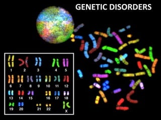
8. GENETIC DISORDERS
• SINGLE gene mutations, following
classical MENDELIAN inheritance
patterns
• MULTIFACTORIAL inheritance
• CHROMOSOMAL disorders
10. AUTOSOMAL DOMINANT
• Disease is in HETEROZYGOTES
• NEITHER parent may have the disease (NEW
mut.)
• REDUCED PENETRANCE (env?, other genes?)
• VARIABLE EXPRESSIVITY (env?, other
genes?)
• May have a DELAYED ONSET
• Usually result in a REDUCED PRODUCTION
or INACTIVE protein
13. AUTOSOMAL RECESSIVE
• Disease is in HOMOZYGOTES
• More UNIFORM expression than AD
• Often COMPLETE PENETRANCE
• Onset usually EARLY in life
• NEW mutations rarely detected clinically
• Proteins show LOSS of FUNCTION
• Include ALL inborn errors of metabolism
• MUCH more common that autosomal dominant
16. SEX (“X”) LINKED
• MALES ONLY
• HIS SONS are OK
• ALL his DAUGHTERS are CARRIERS
• The “Y” chromosome is NOT homologous to
the “X”, i.e., the concept of
dominant/recessive has no meaning here
• HETEROZYGOUS FEMALES have no
phenotypic expression (carriers)
17. SEX (“X”) LINKED
• DUCHENNE MUSCULAR DYSTROPHY
• HEMOPHILIA , A and B
• G6PD DEFICIENCY
• AGAMMAGLOBULINEMIA
• WISKOTT-ALDRICH SYNDROME
• DIABETES INSIPIDUS
• LESCH-NYHAN SYNDROME
• FRAGILE-X SYNDROME
19. SINGLE GENE DISORDERS
• ENZYME DEFECT (Most of them, e.g., PKU)
– Accumulation of substrate
– Lack of product
– Failure to inactivate a protein which causes damage
• RECEPTOR/TRANSPORT PROTEIN DEFECT (Familial
Hypercholesterolemia)
• STRUCTURAL PROTEIN DEFECT (Marfan, Ehl-Dan)
– Structure
– Function
– Quantity
• ENZYME DEFECT WHICH INCREASES DRUG
SUSCEPTIBILITY: G6PDPrimaquine
20. STRUCTURAL PROTEIN DEFECTS
• Marfan Syndrome
– Fibrillin-1 defect
– Tall, dislocated lens, aortic arch aneurysms, etc.
– Abraham Lincoln?, Osama bin-Laden
• Ehlers-Danlos Syndromes (AD, AR)
– Multiple (6?) different types
– Classical, Hypermob., Vasc., KyphoSc., ArthChal., Derm
– Various collagen defects
– Hyperelastic skin, hyperextensible joints
21. RECEPTOR PROTEIN DEFECTS
• FAMILIAL HYPERCHOLESTEROLEMIA
– LDL RECEPTOR defect
– Cholesterol TRANSPORT across liver cell impaired
– ergo, CHOLESTEROL BUILDUP IN BLOOD
• “Scavenger System” for CHOL kicks in, i.e.,
MACROPHAGES
• YOU KNOW THE REST OF THE STORY
• YOU KNOW WHY MACROPHAGES are “FOAMY”
22. ENZYME DEFICIENCIES
• BY FAR, THE LARGEST KNOWN
CATEGORY
– SUBSTRATE BUILDUP
– PRODUCT LACK
– SUBSTRATE could be HARMFUL
• LYSOSOMAL STORAGE DISEASES
comprise MOST of them
24. GLYCOGEN STORAGE DISEASES
• MANY TYPES (at least 10)
• Type 2 (Pompe), von Gierke, McArdle, most
studied and discussed, and referred to
• Storage sites: Liver, Muscle, Heart
25. SPHINGOLIPIDOSES
• MANY types, Tay-Sachs most often referred to
– GANGLIOSIDES are ACCUMULATED
– Ashkenazi Jews (1/30 are carriers)
– CNS neurons a site of accumulation
– CHERRY RED spot in Macula
26. SULFATIDOSES
• MANY types, but the metachromatic
leukodystrophies (CNS), Krabbe, Fabry,
Gaucher, and Niemann-Pick (A and B) are
most commonly referred to
• SULFATIDES, CEREBROSIDES,
SPHINGOMYELIN are the accumulations
27. NIEMANN-PICK
• TYPES A, B, C
• SPHINGOMYELIN
• MASSIVE SPLENOMEGALY
• ALSO in ASHKANAZI JEWS
• OFTEN FATAL in EARLY LIFE, CNS, ORGANOMEGALY
28. GAUCHER DISEASE
• GLUCOCEREBROSIDE BUILDUP
• 99% are type I, NO CNS involvement
• ALL MACROPHAGES, liv, spl, nodes, marrow
29. MUCOPOLYSACCHARIDOSES
• HURLER/HUNTER, for I and II, respectively
• DERMATAN sulfate, HEPARAN sulfate buildup
– coarse facial features
– clouding of the cornea
– joint stiffness
–mental retardation
– URINARY EXCRETION of SULFATES COMMON
31. ALCAPTONURIA
• NOT a LYSOSOMAL ENZYME DISEASE
• FIRST ONE TO BE DESCRIBED
• HOMOGENTISIC ACID
• HOMOGENTISIC ACID OXIDASE
–BLACK URINE
–BLACK NAILS (OCHRONOSIS), SKIN
–BLACK JOINT CARTILAGE (SEVERE ARTHRITIS)
36. MULTIFACTORIAL INHERITANCE
• Multi-”FACTORIAL”, not just multi-GENIC
• “SOIL” theory
• Common phenotypic expressions governed by
“multifactorial” inheritance
– Hair color
– Eye color
– Skin color
– Height
– Intelligence
– Diabetes, type II
37. FEATURES of
multifactorial inheritance
• Expression determined by NUMBER of genes
• Overall 5% chance of 1st degree relatives having it
• Identical twins >>>5%, but WAY less than 100%
• This 5% is increased if more children have it
• Expression of CONTINUOUS traits (e.g., height) vs.
DISCONTINUOUS traits (e.g., diabetes)
39. KARYOTYPING
• Defined as the study of CHROMOSOMES
• 46 = (22x2) + X = Y
• Conventional notation is “46,XY” or “46,XX”
• G(iemsa)-banding, 500 bands per haploid
recognizable
• Short (“p”-etit) arm = p, other (long) arm = q
40.
41. More KARYOTYPING info
• A,B,C,D,E,F,G depends on chromosome length
– A longest
– G shortest
• Groups within these letters depend on the p/q
ratio
• ARMREGIONBANDSub-BAND,
numbering from the centromere progressing
distad
42.
43. F.I.S.H.
greatly enhances G-banding
• Fluorescent In-
Situ
Hybridization
• Uses fluorescent
labelled DNA
fragments, ~10,000
base pairs, to bind (or
not bind) to its
complement
44. FISH
• SUBTLE MICRODELETIONS
• COMPLEX TRANSLOCATIONS
• AND TELOMERE ALTERATIONS
47. CYTOGENETIC DISORDERS
• DEFINITIONS:
– EUPLOID
– ANEUPLOID (NOT AN EXACT MULTIPLE OF 23)
–MONOSOMY, AUTOSOME OR SEX
– TRISOMY, AUTOSOME OR SEX
–DELETION
– BREAKAGE
51. TRISOMY-21
• Most trisomies (monosomies, aneuploidy) are
from maternal non-disjunction
• (non-disjunction or anaphase lag are BOTH
possible)
• #1 cause of mental retardation
• Maternal age related
• Congenital Heart Defects, risk for acute leukemias,
GI atresias
• Most LOVABLE of all God’s children
52.
53. Chromosome 22q11.2
Deletion Syndrome
• Because of a DELETION, this cannot be
detected by standard karyotyping and
needs FISH
• Cardiac defects, DiGeorge syndrome,
velocardiofacial
54.
55. SEX CHROMOSOME DISORDERS
• Problems related to sexual development and
fertility
• Discovered at time of puberty
• Retardation related to the number of X
chromosomes
• If you have at least ONE “Y” chromosome,
you are male
56. KLINEFELTER (XXY, XXXY, etc.)
• Hypogonadism found at puberty
• #1 cause of male infertility
• NO retardation unless more X’s
• 47, XXY 82% of the time
• L----O----N----G legs, atrophic testes,
small penis
57.
58. TURNER (XO)
• 45, X is the “proper” designation
• Mosaics common
• Often, the WHOLE chromosome is not
missing, but just part
• NECK “WEBBING”
• EDEMA of HAND DORSUM
• CONGENITAL HEART DEFECTS most
FEARED
59.
60. HERMAPHRODITES
• GENETIC SEX is determined by the PRESENCE or ABSENCE
of a “Y” chromosome, but there is also, GONADAL
(phenotypic), and DUCTAL sex
• TRUE HERMAPHRODITE: OVARIES AND TESTES, often on
opposite sides
• PSEUDO-HERMAPHRODITE:
– MALE: TESTES with female characteristics (Y-)
– FEMALE: OVARIES with male characteristics (XX)
61. SINGLE GENE, NON-Mendelian
• Triplet repeats
–Fragile X (CGG)
–Others: ataxias, myotonic dystrophy
• Mitochondrial Mutations: (maternal)
(LEBER HEREDITARY OPTIC NEUROPATHY)
• Genomic “IMPRINTING”: (Inactivation of
maternal or paternal allele)
• Gonadal “MOSAICISM”: (only gametes have
mutated cells)
62. MOLECULAR DX by DNA PROBES
• BIRTH DEFECTS, PRE- or POST- NATAL
• TUMOR CELLS
• CLASSIFICATIONS of TUMORS
• IDENTIFICATION of PATHOGENS
• DONOR COMPATIBILITY
• PATERNITY
• FORENSIC
Editor's Notes
Classical concept of a point mutation
The “official” notation for the normal male pattern is: “47, XY”
You do not have to see many trisomy-21 patients until you can recognize them very quickly and easily