CSF - Cerebrospinal fluid examination - from tapping to pathological diagnosis
•
34 likes•14,410 views

This is a series of notes on clinical pathology, useful for undergraduate and postgraduate students, as well as practising pathologists. Prepared from standard text books with data in tabular and easily readable format

Recommended
More Related Content
What's hot
What's hot (20)
Viewers also liked
Viewers also liked (20)
Similar to CSF - Cerebrospinal fluid examination - from tapping to pathological diagnosis
Similar to CSF - Cerebrospinal fluid examination - from tapping to pathological diagnosis (20)
More from Ashish Jawarkar
More from Ashish Jawarkar (20)
Recently uploaded
Recently uploaded (20)
CSF - Cerebrospinal fluid examination - from tapping to pathological diagnosis
- 1. PREVIEW ONLY 1 DOWNLOAD ENTIRE DOCUMENT FROM http://www.scribd.com/doc/231931268/Cerebrospinal-fluid-pathologic-examination Cerebrospinal Fluid Examination
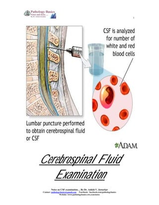
- 2. PREVIEW ONLY 2 DOWNLOAD ENTIRE DOCUMENT FROM http://www.scribd.com/doc/231931268/Cerebrospinal-fluid-pathologic-examination OVERVIEW 1. Physiology 2. Functions of CSF 3. Indications 4. Recommended laboratory tests 5. Specimen collection 6. Opening pressure 7. Gross examination Color Appearance (Clear/clot/cobweb/coagulum) Viscosity 8. Microscopic examination Total count Differential count i. Lymphocytes ii. Neutrophils iii. Plasma cells iv. Eosinophils v. Monocytes and macrophages vi. Tumor cells 9. Chemical examination Proteins i. Total protein ii. Albumin iii. IgG iv. Other CSF proteins Glucose Lactate F2 isoprostanes Enzymes i. Adenosine Deaminase (ADA) ii. Creatinine Kinase (CK) iii. Lactate Dehydrogenase (LDH) iv. Lysozyme Ammonia, amines and aminoacids 10. Microbiological examination Bacterial meningitis Spirochetal meningitis Viral meningitis Fungal meningitis Tuberculous meningitis Primary amebic meningoencephalitis 11. Reference values
- 3. PREVIEW ONLY 3 DOWNLOAD ENTIRE DOCUMENT FROM http://www.scribd.com/doc/231931268/Cerebrospinal-fluid-pathologic-examination * Physiology 1. CSF is derived from ultrafilteration and secretion through the choroid plexus. 2. CSF resorption occurs at arachnoidal villi predominantly along superior sagittal sinus.
- 4. PREVIEW ONLY 8 DOWNLOAD ENTIRE DOCUMENT FROM http://www.scribd.com/doc/231931268/Cerebrospinal-fluid-pathologic-examination * Opening pressure 1. Opening pressure can be measured by a manometer before collection of CSF 2. The pressure varies with postural changes, blood pressure, venous return and valsalva maneuver etc. 3. Pressure should be noted in lateral decubitus position with legs and neck in neutral position. manometer tube with graduation from -4 cm to +34 cm and attached to three way tap Normals CSF opening pressure Adult – 90-180 mm of water Children (upto 8 years) – 10-100 mm of water Abnormals If pressure is elevated more than 200 mm of water, no more than 2 ml should be withdrawn as it can lead to herniation Elevated pressure Decreased pressure 1. straining 2. congestive heart failure 3. meningitis 4. superior venacaval syndrome 5. thrombosis of venous sinuses 6. cerebral edema 7. mass lesions 8. hypoosmolality 9. Idiopathic intracranial hypertension (pseudotumor cerebri) 1. spinal-subarachnoid block 2. dehydration 3. circulatory collapse 4. CSF leakage – like from cribriform plate in case of head injury
- 5. PREVIEW ONLY 11 DOWNLOAD ENTIRE DOCUMENT FROM http://www.scribd.com/doc/231931268/Cerebrospinal-fluid-pathologic-examination B. Appearance Normal Appearance Clear Abnormals Turbid/cloudy Leucocyte count >200 cells/mm3 RBCs >400 cells/ mm3 Microorganisms (bacteria, fungi, amebas) Radiographic contrast material Aspirated epidural fat Protein level greater than 150mg/dl Bloody RBC counts >6000 cells/mm3 Clot Traumatic tap Complete spinal block (Froin’s syndrome) Suppurative or tuberculous meningitis *Not seen in patients with subarachnoid hemorrhage Cobweb Tuberculous meningitis Cobweb in tuberculous meningitis in CSF
- 6. PREVIEW ONLY 13 DOWNLOAD ENTIRE DOCUMENT FROM http://www.scribd.com/doc/231931268/Cerebrospinal-fluid-pathologic-examination * Microscopic examination (A) Total cell count Methods: 1. Manual count using Neubauer’s chamber or a Fuchs-Rosenthal type chamber (most commonly used) 2. Count with an automated cell counter (poor precision) 3. automated flow cytometry of CSF (rapid and reliable, but expensive) Counting using a neubauer’s chamber: 1. Sample in tube 3 is used 2. No dilution of CSF is usually required. A diluent (0.05ml CSF + 0.95 ml diluent, 1:20 dilution) is used only if CSF is cloudy and likely to contain increased number of leucocytes. Diluent mostly used is Turk solution (glacial acetic acid + methylene blue + distilled water) 3. Put coverslip on chamber. 4. Charge it from sides, take care that no fluid goes into the drain. 5. allow to stand for two minutes, cells will settle down. 6. Cells are counted in four corner WBC counting squares, marked ‘W’ in the figure. 7. Total count (per/mm3 )= No. of cells counted x 10 No. of squares counted Improved Neubauer’s chamber
- 7. PREVIEW ONLY 14 DOWNLOAD ENTIRE DOCUMENT FROM http://www.scribd.com/doc/231931268/Cerebrospinal-fluid-pathologic-examination Counting cells in WBC counting chamber Normals Total count Adults - 0-5 cells/mm3 Children – 0-30 cells/mm3 RBCs – Zero / hpf Abnormals Increased counts 1. Meningitis and other infections of CNS 2. Intracranial hemorrhage 3. Meningeal infiltration by malignancy 4. Repeated lumbar punctures 5. Injection of foreign substances (contrast media/drugs) in subarachnoid space. 6. Multiple sclerosis Correction for presence of blood in CSF Presence of blood either due to traumatic tap or subarachnoid hemorrhage artefactually raises the total count. This needs to be corrected by the following formula - Corrected WBC (/mm3 ) = WBC counted - WBC count in blood x RBC count in CSF RBC count in blood
- 8. PREVIEW ONLY 15 DOWNLOAD ENTIRE DOCUMENT FROM http://www.scribd.com/doc/231931268/Cerebrospinal-fluid-pathologic-examination (B) Differential cell count Methods: 1. counting chamber – poor precision, identification of different cell types difficult, not recommended 2. Direct smears of centrifuged CSF specimen – subjected to significant error from cellular distortion# and fragmentation, but most commonly performed 3. Using a cytocentrifuge – recommended method for all body fluids # cellular distortion can be minimized by adding 2 drops of 22% bovine albumin to the specimen Normals: Cell type Adults (%) Children (%) Lymphocytes # 62 +/- 34 20 +/- 18 Monocytes 36 +/- 20 72 +/- 22 Neutrophils 2 +/- 5 3 +/- 5 Histiocytes Rare 5 +/- 4 Ependymal cells Rare Rare Eosinophils Rare Rare #Blast like lymphocytes may be seen admixed with small and large lymphocytes in CSF of neonates Abnormals: 1. Increased neutrophils Meningitis 1. Bacterial meningitis # (PMN >60%) 2. Early viral meningoencephalitis (PMN <60%, changes to lymphocytic in 2-3 days) 3. Early tuberculous meningitis 4. Early mycotic meningitis 5. Amebic encephalomyelitis Other infections 1. Cerebral abscess 2. Subdural empyema 3. AIDS related CMV radiculopathy Following seizures Following CNS hemorrhage 1. subarachnoid 2. Intracerebral Following CNS infarct Reaction to repeated lumbar punctures Injection of foreign material in subarachnoid space (e.g. methotrexate, contrast media) Metastatic tumor in contact with CSF #A total neutrophil count of >1180 cells/mm3 has 99% predictive value for bacterial meningitis
- 9. PREVIEW ONLY 21 DOWNLOAD ENTIRE DOCUMENT FROM http://www.scribd.com/doc/231931268/Cerebrospinal-fluid-pathologic-examination (B) Albumin 1. Albumin is around 56-76% of total proteins in CSF. 2. Normal CSF albumin (in gm/dl) : serum albumin (in gm/dl) ratio is 1:230. 3. But this yields a very difficult decimal of 0.004 to deal with. 4. Hence the permeability of Blood brain barrier is assessed by CSF albumin : serum albumin index, where value of CSF albumin is taken in mg/dl. 5. A traumatic tap invalidates the calculation. CSF ALBUMIN / SERUM ALBUMIN ratio = CSF ALBUMIN (g/dl) Serum albumin (g/dl) CSF ALBUMIN / SERUM ALBUMIN INDEX = CSF ALBUMIN (mg/dl) Serum albumin (g/dl) Normals: CSF albumin: Serum albumin ratio 0.004 CSF albumin:Serum albumin index (mg/gm) <9 Slightly elevated in infants upto 6 months of age Reflects immaturity of blood brain barrier Index increases gradually after age 40 Abnormals: 9-14 Slight impairment 14-30 Moderate impairment >30 Severe impairment
- 10. PREVIEW ONLY 24 DOWNLOAD ENTIRE DOCUMENT FROM http://www.scribd.com/doc/231931268/Cerebrospinal-fluid-pathologic-examination Glucose 1. CSF glucose levels should be compared with plasma levels, ideally following a 4 hour fast, for adequate clinical interpretation. 2. CSF glucose levels normalize before protein levels and cell counts following recovery from meningitis, hence it is a useful parameter in assessing response to treatment. Normals: Fasting CSF glucose levels 60% of plasma level (50-80 mg/dl) Normal CSF glucose:Plasma glucose ratio 0.3-0.9 Abnormals: Decreased CSF fasting glucose (<40mg/dl or ratio <0.3) a.k.a. Hypoglycorrhachia Increased CSF fasting glucose values Due to: increased anaerobic glycolysis in brain tissue and leucocytes Due to: No clinical significance Seen in 1. Bacterial, tuberculous and fungal meningitis 2. meningeal involvement by malignant tumor, sarcoidosis, cysticercosis, trichinosis, ameba, syphilis 3. intrathecal administration of radioiodinated serum albumin 4. subarachnoid hemorrhage 5. symptomatic hypocglycemia 6. rheumatoid meningitis
- 11. PREVIEW ONLY 28 DOWNLOAD ENTIRE DOCUMENT FROM http://www.scribd.com/doc/231931268/Cerebrospinal-fluid-pathologic-examination (B) Creatine Kinase (CK) 1. CK-BB comprises of nearly 90% of brain CK activity, other 10 % being contributed by mitochondrial CK (CKmt) 2. CK-BB starts rising in CSF after about 6 hours of ischemic insult with peak levels in about 48 hours. 3. It is also raised following a subarachnoid hemorrhage and predicts chance of unfavourable outcome. Abnormals: FOLLOWING ISCHEMIC INSULT CK-BB <5 U/L Minimal neurologic damage CK-BB 5-20 U/L Mild to moderate CNS injury CK-BB 21-50 U/L Correlated with death CK-BB >50 U/L Death occurs in all patients FOLLOWING SUBARACHNOID HEMORRHAGE CK-BB >40 U/L Death
- 12. PREVIEW ONLY 37 DOWNLOAD ENTIRE DOCUMENT FROM http://www.scribd.com/doc/231931268/Cerebrospinal-fluid-pathologic-examination (D) Fungal Meningitis Cryptococcus is the most common fungus isolated from CSF Microbiological Methods: 1. India ink or nigrosin stains for capsule 2. Detection of cryptococcal antigen from CSF using latex agglutination 3. Culture Cryptococcus in CSF stained with India Ink Findings in CSF: Test Findings Opening pressure Variable Leucocyte count Variable Differential count Mainly lymphocytes Protein Increased Glucose Decreased CSF : serum glucose ratio Low Lactic acid Mild to moderate increased
- 13. PREVIEW ONLY 41 DOWNLOAD ENTIRE DOCUMENT FROM http://www.scribd.com/doc/231931268/Cerebrospinal-fluid-pathologic-examination Test Bacterial meningitis Viral Meningitis Fungal meningitis Tuberculous Meningitis Opening pressure Elevated Usually normal Variable Variable Leucocyte count >/= 1000/mm3 <100 / mm3 Variable Variable Differential count Mainly neutrophils Mainly lymphocytes Mainly lymphocytes Mainly lymphocytes Protein Mild-moderate increase Normal – mild increase Increased Increased Glucose Usually <40 mg/dL Normal Decreased Decreased (may be <45 mg/dL) CSF : serum glucose ratio Normal / decreased Usually normal Low Low Lactic acid Increased Normal – mild increase Mild to moderate increased Mild to moderate increased
- 14. PREVIEW ONLY 41 DOWNLOAD ENTIRE DOCUMENT FROM http://www.scribd.com/doc/231931268/Cerebrospinal-fluid-pathologic-examination Test Bacterial meningitis Viral Meningitis Fungal meningitis Tuberculous Meningitis Opening pressure Elevated Usually normal Variable Variable Leucocyte count >/= 1000/mm3 <100 / mm3 Variable Variable Differential count Mainly neutrophils Mainly lymphocytes Mainly lymphocytes Mainly lymphocytes Protein Mild-moderate increase Normal – mild increase Increased Increased Glucose Usually <40 mg/dL Normal Decreased Decreased (may be <45 mg/dL) CSF : serum glucose ratio Normal / decreased Usually normal Low Low Lactic acid Increased Normal – mild increase Mild to moderate increased Mild to moderate increased