2. Essential Understandings
Describe the basic structure of a neuron, and compare the functions of the
three classes of neurons.
Explain how a nerve impulse is transmitted across a chemical synapse
List the major regions of the brain and the main functions of each region.
Describe the overall anatomy of the peripheral nervous system, including the
cranial nerves and spinal nerves.
Explain how the somatic system differs from the autonomic system.
Contrast the overall functions of the sympathetic and para-sympathetic
divisions of the autonomic nervous system.
List all tissues that light passes through from when it enters the eye until it is
converted into a nerve impulse.
Discuss the role of rods and cones in transducing a light stimulus into a nerve
impulse
Distinguish the parts of the ear that make up the outer ear, middle ear, and
inner ear.
Describe the mechanism by which sound waves in the outer ear are converted
into nerve impulses in the inner ear.
3. Regulation Processes by which a stable internal environment is kept.
Regulation is brought about by the Nervous and Endocrine system.
Neuron Cell specialized in transmitting impulses – a nerve cell
Dendrites part of nerve cell that receives impulses, towards the cell body
Axon carries impulses away from the cell body of a neuron
Terminal Branches The axons end that is in contact with another cell.
Synapse The GAP between the terminal branch and another cell.
Neurotransmitters Chemicals released from the tip of an axon into the
synapse when a nerve impulse arrives;
may stimulate or inhibit the next neuron
4. Nerves A bundle of axons or fibers, bound together.
Sensory neurons carry signals from receptors & transmit
information to brain and spinal cord
Motor neurons carry signals from central nervous system to the
muscles and glands.
Inter neurons process signals from one sensory neurons and relay
signals to motor neurons.
Receptors receive stimuli from the environment. The sense organs
Effectors A muscle or a gland that responds to a stimuli.
Reflex arc Pathway of sensory receptors ,neurons, effector(s)
that participate in a reflex
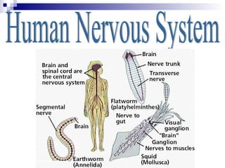
5. Central Nervous system (CNS)
Cerebrum part of the forebrain, associated with higher functions,
including language and abstract thought
Cerebellum concerned with fine motor coordination and body
movement, posture, and balance;
Medulla The region of the brain that, controls heart rate, constriction
and dilation of blood vessels, respiration, and digestion.
Spinal Cord receives sensory information and sends output motor signals;
with the brain, forms the central nervous system.
Peripheral nervous system
Somatic Nervous system -neurons that connect the CNS to skeletal
muscles (voluntary), the skin & sense organs
Autonomic nervous system Serves the internal organs of the body,
Usually no voluntary control. Ex. breathing
Malfunctions: Alzheimer’s, Parkinson’s, Multiple Sclerosis, stroke…
9. Nucleus
More information
Dendrites
axon
Synapse
Myelin Sheath
Terminal Branch
10.
11.
12.
13. How many neurons (nerve cells) are in the brain? How big are they?
It is estimated that there are 100 billion (100,000,000,000) neurons
in the human brain. To get an idea of how many 100 billion is, think
of this:
Assume that you were going to count all 100 billion cells at a rate of
1 cell per second. How long would it take you to count all 100 billion
cells? Calculations say it would take about 3,171 years!!!.
14.
15.
16. Central Nervous System
brain spinal cord
Peripheral Nervous System
cranial nerves spinal nerves
sensory fibers motor fibers
somatic nervous autonomic nervous
system (to skeletal system (to smooth
muscles) muscle, cardiac
muscle, and glands)
sympathetic parasympathetic
division division
17. Peripheral Nervous System
Any neurons outside of the CNS.
Sensory Division –
bringsin stimuli from external environment
monitors status of internal environment
Motor Division –
Somatic – carries signals to skeletal muscles
Autonomic – coordinates functions of organs and
helps maintain homeostasis.
Sympathetic and Parasympathetic
Oftenhave opposite effects on the body.
One system stimulates, the other inhibits
23. Main Parts of the Eye
Cornea – light enters, transparent layer of cells
Aqueous Humor – in anterior chamber, light passes
through
Iris – colored part; regulates size of pupil
Pupil – black dot; opening; light enters inner eye
Lens – behind pupil; focuses light onto retina
Vitreous Humor – fluid behind lens
Retina – has photoreceptors
Rods – black and white; extremely sensitive to light
Cones – color, less sensitive to light. Concentrated in the
FOVEA.
Optic Nerve – Carries impulses to the brain.
Sclera and Choroid – outer layers of the eye
25. Outer Ear
Auricle (Pinna) – visible part of the ear.
Collects sound
Auditory Canal – sound enters,
ear canal
Tympanum(eardrum) -
membrane. Vibrates
26. Middle Ear
Hammer(malleus) – bone…attached to
eardrum…accepts vibrations and
passes them to
Anvil(incus) – bone – accepts
vibrations and passes them to
Stirrup(stapes) = bone – accepts
vibrations and passes them to the Oval
Window
27. Inner Ear
Oval Window – attached to stirrup…
creates pressure waves in the cochlea
Cochlea - snail like. Fluid filled. Lined
with tiny sensory cilia that produce
nerve impulse…….taken to the…
Auditory Nerve(cochlear nerve) – takes
impulse to the brain
29. BALANCE
Semicircular Canals – helps CNS maintain
balance and your sense of equilibrium.
Filled with fluid…lined with ciliary sensory
nerves.
When your head changes position, the
hairs sense and send impulses to the
brain.
30. TASTE AND SMELL
Smell – chemoreceptor in the lining of
the nasal passages.
Taste – taste buds on the tongue.
Sensitivity to taste varies on the area of
the tongue.
Sweet, sour, bitter, salty.
31.
32.
33. Touch
Not found in one particular place.
All skin regions are sensitive to touch.
Temperature (Hot and Cold), Pain,
Pressure, light touch.
Greatest concentration of touch
receptors: face, hand, toes