Polio Final Presentation

Dear Doctor, Its humbling that you liked the presentation and would like to use it for your purpose. Kindly find your requested presentation attached with this email. The shortlink for your future reference is http://go.drankush.com/PolioFinal We would always appreciate if you would place this reference as a due credit in your work and while sharing for others use. Ankush, Amroskar S, Bhamaikar V, Barreto J. "Polio Final Presentation" Accessed from http://go.drankush.com/PolioFinal ----------------------------------------------------- As we near eradication of this dreaded disease - "POLIO", we would like to share the following presentation we made for our Pediatrics seminar in 2012. Best attempts have been made to cover most of the topic, keeping the size under 100 slides. Hope you like it. Ankush Shahin Amroskar Varsha Bhamaikar Joyce Barreto

Recommended
More Related Content
What's hot
What's hot (20)
Viewers also liked
Similar to Polio Final Presentation
Similar to Polio Final Presentation (20)
Recently uploaded
Recently uploaded (20)
Polio Final Presentation
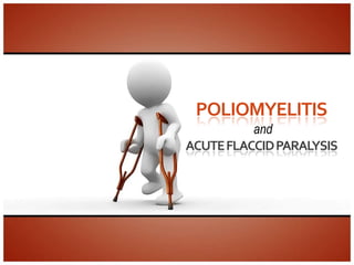
- 1. POLIOMYELITIS and ACUTE FLACCID PARALYSIS
- 3. Introduction Synonyms Infantile paralysis Definition Acute viral infectious disease Caused by enterovirus Route – feco – oral Greek poliós - "grey" myelós - "spinal cord"
- 4. History Timeline Events ANCIENT EGYPT An egyptian mummy 3.,700 B.C with probable polio found. 1,209 B.C Mummy Giptah with an equinus foot Eighteenth century First known 1789 description by underwood Ninteenth century First epidemic of 1834 polio in island of St. Helena
- 5. History Timeline Events 1855 First description by Duchenne of the pathalogical process Twentieth century Transmission of polio to 1908 a monkey by Landsteiner 1949 Growth of virus on tissue culture 1951 Three types of polio virus isolated and identified 1954 First large scale trial of Salk 1958 First general use of Sabin
- 6. Epidemiology Incidence (2011) India: 5 cases- 1 Wild polio, 4 VAPP (2010) Global: 332 confirmed cases India: 42 confirmed cases Last case detected in Goa in 1997
- 9. Agent Poliovirus Structure: Group- group IV ((+)ssRNA) Genus- Enterovirus Family- Picornaviridae 3 serotypes- type 1, type2, type 3 Composed of an RNA genome and a protein capsid. Resistance: In feces – for months at 40 C & years at -200 C Inactivated by heat and chlorination
- 10. Agent Host range Natural infection occurs only in humans Mode of transmission Feco-oral route In early stage of disease- through inhalation or entry through conjunctiva of droplets of respiratory secretion of patient. Period of communicability 7 to 10 days before and after the onset of symptoms
- 11. Host Age Most vulnerable- 6 months to three years Sex M: F ratio 3:1 Immunity First 6 months maternal antibody Acquired through infection with the wild virus Immunization
- 12. Environment Seasonal More during rainy season Environmental sources of infection Contaminated water and food Flies Overcrowding and poor sanitation
- 13. Pathogenesis Incubation period: 7-10 days (4- 35 days) Feco-oral Inhalational Portal of entry- Local Minor/Primary mouth multiplication Viremia infects the pharynx and intestinal mucosa. Gains entry by Multiplies in To spinal cord Major/Secondary neurons and brain Viremia binding to an immunoglobulin-like receptor, known as the poliovirus receptor or Lesions are mostly in anterior horns of DESTROYS CD155, on the cell spinal cord causing THEM! membrane flaccid paralysis
- 14. Pathogenesis Portal of entry- Local Minor/Primary mouth multiplication Viremia Epithelial cells of GIT Multiplies in To spinal cord Major/Secondary neurons and brain Viremia Lymphatic tissue- from tonsils to Lesions are mostly DESTROYS in anterior horns of Peyer’s spinal cord causing flaccid paralysis THEM! patches
- 15. Pathogenesis Spreads to Portal of entry- Local Minor/Primary mouth multiplication Viremia regional lymph nodes Multiplies in To spinal cord Major/Secondary Enters blood neurons and brain Viremia stream - primary viremia Lesions are mostly in anterior horns of DESTROYS spinal cord causing flaccid paralysis THEM!
- 16. Pathogenesis Multiplies in Portal of entry- Local Minor/Primary reticulo- mouth multiplication Viremia endothelial systems Multiplies in To spinal cord Major/Secondary Enters blood neurons and brain Viremia stream again - secondary Lesions are mostly viremia in anterior horns of DESTROYS spinal cord causing flaccid paralysis THEM!
- 17. Pathogenesis Carried to spinal cord & brain Portal of entry- Local Minor/Primary mouth multiplication Viremia Multiplies in To spinal cord Major/Secondary Bloodstream Direct neurons and brain Viremia (Tonsillectomy trauma IM injections fatigue ) Lesions are mostly in anterior horns of DESTROYS spinal cord causing flaccid paralysis THEM!
- 18. Pathogenesis Multiplies in neurons Degeneration of Portal of entry- Local Minor/Primary mouth multiplication Viremia Nissl’s body (chromatolysis) Nuclear changes Multiplies in To spinal cord Major/Secondary follows neurons and brain Viremia When degeneration irreversible Lesions are mostly in anterior horns of DESTROYS phagocytosed by spinal cord causing flaccid paralysis THEM! leucocytes or macrophages
- 19. Pathogenesis Lesions are in anterior horn of spinal cord Flaccid paralysis Portal of entry- Local Minor/Primary mouth multiplication Viremia Can cause encephalitis involving • Brainstem Multiplies in To spinal cord Major/Secondary • Motor & neurons and brain Viremia Premotor areas of cerebral cortex Lesions are mostly in anterior horns of DESTROYS spinal cord causing flaccid paralysis THEM!
- 20. Pathogenesis Portal of entry- Local Minor/Primary mouth multiplication Viremia Multiplies in To spinal cord Major/Secondary neurons and brain Viremia Lesions are mostly in anterior horns of DESTROYS spinal cord causing flaccid paralysis THEM!
- 21. Clinical Features Abortive polio Inapparent (90- 95%) Non- paralytic Infection aseptic Apparent (5- meningitis 10%) Paralytic poliomyelitis Polio encephalitis
- 22. Abortive polio 4 – 8% of infections Minor illness Symptoms low grade fever sore throat vomiting abdominal pain Loss of appetite malaise Recovery – complete, no paralysis
- 23. Non paralytic aseptic meningitis 1- 2 % of infections Symptoms headache nausea vomiting pain and stiffness of back and legs
- 24. Non paralytic aseptic meningitis Signs Tripod sign Kiss the knee test Head drop sign Neck rigidity Recovery within 2 – 10 days
- 25. Tripod sign
- 26. Head drop sign Method Hand placed under patient’s shoulder and trunk is raised Observation Head lags behind limply
- 27. Kiss the knee test Method knees kept down child asked to kiss his knees Observation Cannot do the maneuver due to stiffness spine May draw up the knees sharply
- 28. Neck rigidity Method In uncooperative child-place childs head beyond the edge of table Observation True involuntary neck rigidity persists Voluntary stiffening of muscles disappears
- 29. Paralytic poliomyelitis 0.5 – 1% of infections 2 PHASES - Minor Major Minor- same as abortive polio Major- muscle pain ,spasm and return of fever Followed by rapid onset flaccid paralysis complete within 72hrs
- 31. Paralytic poliomyelitis Spinal paralytic poliomyelitis Paralytic Bulbar polio Poliomyelitis Bulbo-spinal polio
- 32. Spinal paralytic poliomyelitis Most common 80% of cases Results from lower motor neuron lesion of anterior horn cells of spinal cord Affects muscles of legs, arms and/or trunk Severe cases – quadriplegia , paralysis of trunk abdominal and thoracic muscles
- 33. Spinal paralytic poliomyelitis Paralysis – asymmetrical ( legs > arms), descending paralysis Muscles – floppy Reflexes diminished Sensation normal Residual paralysis after 60 days
- 35. Bulbar polio 2% of cases Life threatening Cranial nerve lesion - vagus Symptoms o Nasal twang and hoarseness of voice o Nasal regurgitation o Dyspnea o Dysphagia o Child refuses to feed o Secretions accumulate in pharynx - aspiration
- 36. Bulbar polio • Involvement of respiratory centre - Shallow , irregular respiration Vasomotor centre - BP rises then falls • pulse – rapid weak thready • Skin – dusky red mottled • Restless , confused and comatose
- 37. Bulbo-spinal poliomyelitis 20% cases Combination of spinal paralytic and bulbar polio
- 38. Polio Encephalitis Occurs in rare cases Symptoms Irritability Delirium Disorientation Tremors Convulsions Paralysis is of upper motor neuron type
- 39. Residual paralysis Acute phase of illness lasts for 0-4weeks Recovery –variable At 60 days mild to severe residual paresis Maximum recovery – first 6 months Slow recovery upto 2 yrs After 2 yrs post polio residual paralysis persists throughout life
- 40. Diagnosis History Clinical examination Stool examination CSF examination Serological tests
- 41. Stool examination Collection of sample Two samples 24 hr apart Within 14 days of onset of paralysis 8-10 grams or thumb size Collected in a clean wide mouth bottle – plastic or glass with screw cap Sample stored below 8°C No dessication or leakage till received at WHO Accredited Lab If paralysis detected after 2 wks sample taken upto 60 days from onset
- 42. Stool examination Contact sampling Done when child has died without adequate stool sampling 5 children in close contact with the child are taken Single stool sample collected
- 43. CSF examination Characteristics Observations Appearance Clear / slightly turbid Cells Leucocytosis (mainly lymphocytes) Proteins Normal / slightly raised glucose Normal
- 44. Serological tests 3 types of antibodies Neutralizing antibodies (IgG) Antibodies to C antigen (IgM) Anti-D antibodies Complement fixation test – detects IgM and Anti-D antibodies Identifies exposure to poliovirus not for type- specific diagnosis Less often employed
- 45. Differential diagnosis • Most common GB syndrome Transverse myelitis • Others Traumatic neuritis Meningitis Encephalitis Toxin – diphtheria and botulism
- 46. TREATMENT
- 47. Treatment Symptomatic and supportive Rest in bed Relief of pain and spasm of muscles Neutral positioning of the limbs Physiotherapy Good nursing
- 48. Bed Rest Essential during acute phase Physical activity & trauma increases risk of paralytic polio Posture to be changed every 2-3 hrs. Child to be placed on stomach for short periods each day, to prevent pneumonia
- 49. Optimum position for limbs Hip – slight flexion Knee – 5 degree flexion Foot – 90 degree support against the sole Pain Relief Sister Kenny’s treatment Hot moist packs applied to the muscles to relieve pain and spasm analgesics
- 50. Physiotherapy Method • Joints & paralysed muscles – moved passively through full range • For 10 min , 2-3 times/day Benefits • Prevents deformities and contracture • Promote development of muscle power in non-paralysed muscles
- 51. Physiotherapy
- 52. Good nursing Team approach is essential Nursing staff is an imp part Diet Nutritious , balanced & wholesome In non paralytic polio- normal diet In paralytic Fed by Ryles tube Calories/kg body wt.
- 53. Good Nursing In dysphagia pt. nursed in prone position with foot end raised – gravity drainage of pooled secretions in pharynx Or intermittent suction Tracheostomy Respiratory failure – assisted respiration with mechanical ventilator
- 54. Treatment Indications for hospitalization Paralysis of upper limbs <3 days duration Progression of paralysis Bulbar involvement Respiratory distress Marked drowsiness Complications
- 55. Rehabilitation
- 56. Rehabilitation Physical Emotional and Psychological Social
- 57. Rehabilitation Emotional support to the child helps prepare himself for better adjustment in life despite the handicap
- 59. Complications Myocarditis Hypertension Pulmonary edema Pneumonia Urinary tract infections Skeletal deformities - equinus foot scoliosis osteoporosis bone fractures Compression neuropathy
- 60. Prognosis Non paralytic cases – complete recovery Paralytic polio – permanent weakness in 2/3rd cases Worse – older children sudden onset of illness with high fever
- 61. Post – polio syndrome Observed in people who had polio during their childhood. Affects about 25-50 % of the polio survivors. More common in females General fatigue muscular weakness joint pains & breathing problems are seen in affected
- 62. PREVENTION
- 63. Immunisation • History • Sabin’s Live Polio Vaccine I. Preparation II. Storage and transport III. Administration IV. Dosage V. Development of Immunity VI. Advantages and Disadvantages VII. Complications and Contraindications • Salk’s Killed Polio Vaccine I. Preparation II. Dosage • Sabin Vs Salk • Pulse Polio Immunization
- 64. History • Earliest vaccines- a. Crude suspensions of spinal cord from infected monkeys -Ineffective b. Inactivated with Formalin (Brodie and Park) -Often dangerous Ricinoleate (Kolmer) causing vaccination poliomyelitis • By 1953 a. Salk had developed a killed vaccine b. Almost simultaneously, Koprowsky, Cox and Sabin independently developed live attenuated vaccines
- 65. Sabin’s Live Polio Vaccine • Sabin’s attenuated strains are employed • Developed by plaque selection in MKTC • Preparation a. Attenuated strains grown in MKTC b. Stringent precautions to ensure freedom from SV40 and B virus. c. Use of molar MgCl2 or sucrose stabilises the vaccine against heat inactivation
- 66. Sabin’s Live Polio Vaccine • Criteria for selection a. Should not be neurovirulent as tested by intraspinal inoculation in monkeys b. Should be able to set up intestinal infection following feeding & induce immune response c. Should be stable & not acquire neurovirulence after serial enteric passage d. Should posses stable genetic markers enabling differentiation from wild virulent strians.
- 67. Sabin’s Live Polio Vaccine Genetic Markers: • D Marker • Rct 40 • MS • Mcbride’s intratypic antigenic marker Molecular Epidemiological Methods: • Monoclonal antibodies specific to vaccine strains • Oligonucleotide finger printing • Nucleic acid sequencing
- 68. Sabin’s Live Polio Vaccine • Storage i. Stabilised vaccine: 1 year at 4 °C 1 month at room temperature ii. Non-stabilised vaccine: -20 °C in deep freeze (In the freezer compartment of refrigerator) • During transport, keep the vaccine under i. Dry ice (solid carbon dioxide) ii. Freezing mixture (equal quantities of wet ice and ammonium chloride) • At vaccination clinic i. Shouldn’t be frozen and thawed repeatedly deleterious effect on potency ii. Keep vaccine in ice during administration
- 69. Sabin’s Live Polio Vaccine • OPV in India, trivalent, contains a. Type 1- 1 lakh TC ID 50 b. Type 2- 2 lakh TC ID 50 per 0.5 ml c. Type 3- 3 lakh TC ID 50 (2 drops in India) • Administration- 2 drops • Use the dropper supplied a. Tilt the child’s back b. Gently squeeze the cheeks/ pinch the nose make the mouth open c. Let the drops fall from the dropper onto the tongue.
- 70. Sabin’s Live Polio Vaccine • National Immunization Schedule Age Dose At birth OPV-0 At 6 weeks OPV-1 At 10 weeks OPV-2 At 14 weeks OPV-3 16-24 months OPV
- 71. Sabin’s Live Polio Vaccine • Indian Academy of Paediatrics recommendation Age Dose At birth OPV-0 At 6 weeks OPV-1+IPV At 10 weeks OPV-2+IPV At 14 weeks OPV-3+IPV 16-24 MONTHS OPV+IPV 5 years OPV
- 72. Development of Immunity Infects intestinal epithelial cells Replicates transported to Peyer’s patches Stimulates production of IgA antibodies Secondary multiplication d subsequent viremia (LOCAL IMMUNITY) Spreads to other parts of body Prevents infection of GIT with wild strains Production of circulating antibodies Vaccine progeny excreted in feces Prevents dissemination of virus to nervous system Non-immunized persons immunized Prevents paralytic polio (SYSTEMIC IMMUNITY) HERD IMMUNITY
- 73. Sabin’s Live Polio Vaccine • Advantages i. Oral easily admin no need of highly trained personnel ii. Induces both humoral and systemic immunity iii. Antibodies quickly produced* iv. Vaccinees excrete virus herd immunity v. Useful in epidemics vi. Relatively inexpensive • Disadvantages i. Instability at high temperatures ii. Frequent vaccine failures even with fully potent vaccines iii. Very small residual neurovirulence in OPV
- 74. Sabin’s Live Polio Vaccine • Complications a. Mutation (esp. type 3[1] /2[2]) b. WHO estimated the risk of i. vaccine-associated paralysis : 1 case/million vaccinees ii. Risk of close contact of vaccinee : 1 case/5 million doses of vaccine developing paralytic polio Contraindications a. Immunocompromised individuals leukemics, malignacy, those receiving corticosteroids. b. Pregnant mothers OPV should be delayed until after pregnancy unless immediate protection is required, when IPV is indicated. c. Premature Babies
- 75. ALERT! DIARRHOEA NOT A CONTRAINDICATION But a dose of OPV given at that time shouldn't be considered as part of the series and should receive another at earliest opportunity.
- 76. Salk’s Killed Polio Vaccine Formalin inactivated preparation Three types of polio virus grown in monkey kidney tissue culture(MKTC) •Procedure for Preparation 3 types of PVs Adequate titre Inactivated with Standard virulent grown separately filtered to remove formalin at 37°C strains used in MKTC debris and clumps FOR 12-15 DAYS Stringent tests to Further tests for Three types are ensure complete safety and Issued for use further pooled inactivation potency
- 77. Salk’s Killed Polio Vaccine • 1954 nationwide field trial (USA)- 80 -90% protection • 1955 – ‘Cutter incident’; over 100 cases of paralytic poliomyelitis occurred in vaccines and their contacts following insufficiently inactivated vaccine.
- 78. Salk’s Killed Polio Vaccine • Injectable Polio Vaccine (IPV) a. 1st dose given at 6 weeks. b. Immunity sustained by booster doses every 3-5 years thereafter c. Vaccination of choice among HIV, other immunocompromised states, pregnant mothers. • Enhanced potency IPV a. Produced in human diploid cells b. Two s.c. Does, 4-8 weeks apart, third may be 6-12 months later. c. Better seroconversion
- 79. Sabin Vs Salk Feature Sabin’s Vaccine (live) Salk’s Vaccine (killed) Strain Live attenuated virus Killed formalised vaccine Administration Oral (preferred in mass Injectable (adv. can be campaigns) given with DPT) Factors affecting Diarrhoeal disease preventing Not affected by these efficacy colonisation by vaccine virus factors Safety Safe Safe But, cases of vaccine induced paralysis reported
- 80. Sabin Vs Salk Feature Sabin’s Vaccine (live) Salk’s Vaccine (killed) Economical More Less (because, virus content is 10,000 times more, hence costlier) Nature of Local and systemic Only systemic, no immunity intestinal immunity No herd immunity Duration Life long Periodic booster doses required Use in epidemic Ideal Not very ideal- May promote multiplication on 7 dissemination of wild virus.
- 81. Pulse Polio Immunization Largest public health campaign ever conducted in a single country • Occurs as two rounds 4-6 wks apart during low transmission season of polio- Nov to Feb • First round- 9th Dec ‘95 and 20th Jan ‘96 • Sudden, simultaneous, mass administration of OPV on a single day • To all children 0-5 years • Regardless of previous immunization • Extra doses which supplement • Do not replace the doses during routine immunization • Children/infants should receive all their schedules OPV doses.
- 82. WE NEED JUST ONE MORE THING TO END POLIO FOREVER “YOU”
- 83. Acute Flaccid Paralysis
- 84. Introduction Case definition • Child less than 15 yrs with acute onset flaccid paralysis for which no obvious cause is found • Acute - onset paralysis < 4 wks • Flaccid - floppy or limp paralysis • Background rate of AFP • One case of AFP per year for every one lakh population of children less than 15 years
- 85. Conditions causing AFP Site Conditions Muscle Myoglobinuric myopathy Hypokalemic paralysis Toxic paralysis Myopathy of intensive care Neuromuscular junction Myasthenia gravis Botulism Hypermagnesemia Peripheral nerve Guillian barre syndrome Diphtheric neuropathy Porphyria Lead neuropathy Hypophosphatemia Cobalamin deficiency Anterior horn cells Poliomyelitis Other enteroviruses
- 86. Differential Diagnosis of AFP Feature Poliomyelitis G.B. Syndrome Transverse Traumatic myelitis neuritis History Progression to 24-48 hrs Hours to days Hours to 4 days Hours to 4 days full paralysis Fever onset High always No Present before No present at onset paralysis of paralysis Bladder Absent Transient Present Never dysfunction
- 87. Feature Poliomyelitis G.B. Syndrome Transverse Traumatic myelitis neuritis Differential Diagnosis of AFP Examination Flaccidity Acute, Acute, Acute, Acute, assymetric assymetrical, symmetrical, symmetrical, proximal distal, ascending lower limb Muscle tone Diminished Diminished Diminished in Diminished lower limbs Deep tendon Decrease or Absent Absent early, Decreased or reflexes absent hyperreflexia late absent Sensation Severe myalgia Cramps, tingling, Anesthesia of Pain in gluteal and backache, no hypo anesthesia lower limb with region sensory changes of palms and sensory level soles Cranial nerves Only when Often present Absent Absent bulbar and affecting nerves bulbo-spinal VII,IX,X,XI,XII Respiratory Only when bulbo In severe cases Sometimes Absent insufficiency and bulbo-spinal
- 88. Feature Poliomyelitis G.B. Syndrome Transverse Traumatic myelitis neuritis Differential Diagnosis of AFP Investigations CSF examination High Less than 10 Cellular or Normal leucocytosis; leucocytes: high acellular; normal normal or slightly protein or slighly increased protein increased protein EMG at three Abnormal Normal Normal May show weeks abnormality Nerve Normal Abnormal, Normal Abnormal conduction demylination or velocity at 3 axonal damage weeks Sequlae at 3 months Severe, Symmetrical Diplegia, atrophy Moderate assymetrical atrophy of distal after years, atrophy in the atrophy; skeletal muscles recovery recovery in affected limb deformity appear in milder cases milder cases late
- 89. AFP Surveillance • All cases to be reported • All reported cases classified as polio non polio • High sensitivity of reporting will ensure detection of all cases resulting in control measures to interrupt transmission • Two critical indicators of quality of surveillance are - a. rate of non polio AFP b. proportion of AFP cases with two adequate stools collected within 14 days of onset of paralysis
- 90. Case investigation • Stool sample collection • Stool sample result • 60 days follow up • Special investigations if indicated • Final classification
- 91. Stool examination Collection of sample Two samples 24 hr apart Within 14 days of onset of paralysis 8-10 grams or thumb size Collected in a clean wide mouth bottle – plastic or glass with screw cap Sample stored below 8 C No dessication or leakage till received at WHO Accredited Lab If paralysis detected after 2 wks sample taken upto 60 days from onset
- 92. Stool examination Contact sampling Done when child has died without adequate stool sampling 5 children in close contact with the child are taken Single stool sample collected
- 93. Virological AFP Compatible Wild polio Confirm virus expert Residual review weakness, died lost to follow up discard AFP Inadequate or No no specimen residual discard No wild virus weakness 2 adequate stool specimen discard
- 94. Clinical AFP Lost follow confirm up Residual paralysis confirm AFP Follow-up at 60 days Positive for confirm wild polio virus No residual paralysis discard
- 95. Sequence of action to be taken after detecting a case of AFP Acute onset of paralysis Within 48 hrs Investigate the suspected case Discard the case of traumatic / electrolyte imbalance Within 3 days 2 adequate stool specimen National lab to collected report within 28 days Outbreak response immunization and active surveillance Intratyping 60 days follow up (before 70 Final classification of days) case within 90 days
- 96. Bibliography Textbooks 1. M.D RMK, Stanton BF, Geme JS, Schor N, Behrman RE. Nelson Textbook of Pediatrics: Expert Consult Premium Edition - Enhanced Online Features and Print. Elsevier - Health Sciences Division; 2011. 2. Parthasarathy et al. Textbook of Pediatrics IAP. Jaypee Brothers Publishers; 2005. 3. O.P. Ghai et al. Essential Pediatrics. CBS Publishers & Distributors; 2009. 4. Park JE. Textbook of preventive and social medicine: a treatise on community health. Banarsidas Bhanot; 1972. 5. Fauci AS, Eugene B, M.D SLH, M.D DLL, J J, Joseph L. Harrison’s principles of internal medicine. McGraw-Hill; 2008.
- 97. Bibliography • Online Resources • AFP Statistics: i. National Polio Surveillance Project http://www.npspindia.org/ • Image Courtesy: i. Rotary International Foundation http://www.rotary.org ii. Bill and Melinda Gates foundation http://www.gatesfoundation.org/ iii. Global Polio Eradication Initiative http://www.polioeradication.org/
- 98. Contact • For distribution and discussion: Dr. Ankush Goa Medical College, Goa drankush1989@gmail.com
Editor's Notes
- JI C, Cohen JI. Harrison’s Principles of Internal Medicine Chapter 175: Enteroviruses and Reoviruses. McGraw-Hill Professional; 2004. p. pp.&nbsp;1144.Chamberlin SL NB (eds ), Chamberlin SL, Narins B (eds.). The Gale Encyclopedia of Neurological Disorders. Detroit: Thomson Gale; 2005.
- Rationale Risk of VAPP is extremely low as child receives OPV at a time when he is protected by VAPP by maternal antibodies.Subsequently he is protected from VAPP by IPV
- Ghai’s Essential Paediatrics, Pradeep Seth, Surjit Singh, AditiSinhaImmunity and ImmunizationPg 167Park’s Textbook of Preventive and Social Medicine, K. Park, Epidemiology of Communicable diseases-Poliomyelitis Pg 181
- Salk D. Eradication of Poliomyelitis in the United States. II. Experience with Killed Poliovirus Vaccine. Review of Infectious Diseases. 1980 Mar 1;2(2):243 -257. Lapinleimu K, Stenvik M. Experiences with polio vaccination and herd immunity in Finland. Dev. Biol. Stand. 1981;47:241-246.