SlideShare a Scribd company logo
1 of 45
Radiological anatomy of the spine: 
Dr/ ABD ALLAH NAZEER. MD.
C-spine normal anatomy - Open mouth view.
Normal AP and Lateral Views.
Thoracic Spine - AP & Lateral View.
L.S. Spine.
Oblique Views.
Extension and flexion x-rays.
CT of the Cervical Spine: Anatomy. 
CT of the Cervical Spine, 
axial reconstruction. Level 1. 
Image 1. 1,Intervertebral 
disc C3-C4. 2, Vertebral 
body (C3). 3, Uncinate 
process C4. 4, Transverse 
process of C4. 5, Lamina. 
6, Superior articular 
process of C4.
CT of the Cervical 
Spine, coronal 
reconstruction. Level 1. 
Image 2.Arrow, 
Atlantooccipital 
joint. 1, Transverse 
process of C4 (right 
side). 2, Uncinate 
process C4.
CT of the Cervical 
Spine, sagittal 
reconstruction. Level 1. 
Image 3 1, Anterior 
arch of l‘ atlas (C1). 2, 
Intervertebral space 
(disc) C3-C4. 3, 
Vertebral foramen. 4, 
Spinous process of C2. 
5, Posterior arch of C1. 
6, Vertebral body C5.
CT of the Thoracic Spine: Anatomy.
1-vertebral body, 2-neural foramen, 3-articular 
pillar 4-lamina, 5-spinous process.
CT of the Lumbo-sacral Spine: Anatomy. 
CT of lumbar Spine, axial 
reconstruction. Level 1. 
Image 1. 1, Vertebral 
body (L4). 2, Transverse 
process. 3, Facet joint. 4, 
Spinous process. 5, 
Inferior articular process 
L3. 6, Superior articular 
process of L4. 7, 
Intervertebral disc L3-L4.
CT of lumbar Spine, 
coronal reconstruction. 
Level 1. 
Image 2. 1, 
Transverse process 
L1. 2, Lamina. 3, 
Inferior articular 
process of L3. 4, 
Superior articular 
process of L2.
CT of lumbar Spine, 
sagittal 
reconstruction. 
Level 1. 
Image 3. 1, Spinous 
process de L2. S, 
Sacrum. L1, Vertebral 
body, L1. L2, 
Vertebral body, L2.
SPINAL MR IMAGING TECHNIQUES: 
Sagittal and axial magnetic resonance images should be 
acquired through the cervical, thoracic, and lumbar segments 
of the spine, as they are generally considered complementary, 
and imaging the spine in only one plane may result in 
misinterpretation. The addition of coronal images may also be 
useful, especially in patients with scoliosis. Stacked axial 
images and/or angled images through the discs can be 
obtained, often useful when the indication for imaging is pain, 
degenerative change, and/or radiculopathy.1 Although 
imaging in the axial plane is a matter of personal preference, 
using only angled axial images through the discs may be 
inadequate, as portions of the spinal canal will not be imaged 
axially. Slice thickness from 3 to 4 mm is generally optimal for 
imaging of the spine. Axial gradient-echo images through the 
cervical spine are typically 2 mm Thick.
Sagittal T2-weighted image, 
cervical spine. 1,Clivus; 2, Atlanto-occipital 
ligament; 3, Anterior 
longitudinal ligament; 4, Anterior 
arch C1; 5, Superior fascicle of 
cruciform ligament/tectorial 
membrane; 6, Apical ligament; 7, 
Transverse ligament (of cruciform 
ligament); 8, Posterior arch C1; 9, 
Posterior occipital atlantal 
membrane; 10, Nuchal ligament; 
11, Semispinalis capitis muscle; 
12, Cervical spinal cord; 13, 
Posterior longitudinal 
ligament/anterior thecal sac dura; 
14, Posterior dural sac; 15, 
Interspinous ligament; 16, Gray 
matter along central canal; 17, 
Supraspinous ligament; 18, 
Ligamentum flavum; 19, Dental 
synchondrosis (disc anlage).
Coronal T1-weighted 
image, craniocervical 
junction. 1, Alar 
ligament; 2, 
Transverse ligament 
(of cruciform 
ligament); 3, Apical 
ligament; 4, Dens; 5, 
Lateral mass C1; 6, 
Occipital condyle; 7, 
Jugular tubercle of 
occipital bone; 8, 
Hypoglossal canal; 9, 
Uncinate process C3; 
10, Vertebral artery.
Coronal T1- 
weighted image, 
cervical spine. 1, 
Uncinate 
processes; 2, 
Segmental spinal 
veins and 
nerve roots.
Parasagittal T1-weighted 
image, cervical spine. 1, 
Hypoglossal canal; 2, Occipital 
condyle; 3, Lateral mass C1; 4, 
Vertebral artery; 5, Rectus 
capitis posterior major muscle; 
6, Obliquus capitis inferior 
muscle; 7, Articular pillar C2; 8, 
C3 dorsal root ganglion; 9, 
Longuscoli (cervicis) muscle; 
10, Vertebral artery; 11, 
Apophyseal (facet) joint C3-4; 
12, Multifidis muscle; 13, 
Semispinalis cervicis muscle; 
14, Splenius capitis muscle; 15, 
Trapezius muscle; 16, Superior 
articular process; 17, Inferior 
articular process.
Coronal oblique T1- 
weighted image, 
cervical spine. 1, 
Cervical spinal cord; 
2, Subarachnoid 
space; 3, Dorsal root 
ganglion; 4, Superior 
foraminal vein; 5, 
Vertebral artery.
A) Axial T2-weighted image, lower cervical spine. 1, Uncinate process C7; 2, Superior 
articular process C7; 3, Apophyseal (facet) joint; 4, Inferior articular process C6; 5, 
Foraminal vein; 6, Ligamentum flavum/cortex of lamina; 7, Dorsal rootlet C7; 8, 
Uncovertebral joint; 9, Ventral rootlets C7. (B) Axial T2-weighted image, mid cervical 
spine. 1, Dorsal root ganglion; 2, Vertebral artery; 3, Posterior longitudinal 
ligament/anterior thecal sac dura; 4, Longus colli muscle; 5, Internal jugular vein; 6, 
Dorsal rootlets; 7, Lamina; 8, Sternocleidomastoid muscle; 9, Longissimus capitis muscle; 
10, Levator scapulae muscle; 11, Semispinalis colli muscle; 12, Semispinalis capitis 
muscle; 13, Splenius capitis muscle; 14, Trapezius muscle; 15, Nuchal ligament.
Sagittal T1-weighted 
image, thoracic spine. 
1, Thoracic spinal 
cord; 2, Subarachnoid 
space; 3, Posterior 
epidural fat; 4, 
Ligamentum flavum; 
5, Transversospinalis 
(multifidus) muscle; 
6, Spinous process; 7, 
Epidural vein; 8, 
Supraspinous 
ligament..
Parasagittal T2-weighted image, 
thoracic spine. 1, Posterior thecal 
sac dura; 2, Posterior epidural 
fat; 3, Ligamentum flavum. 
Sagittal T2-weighted image, thoracic spine. 1, 
Thoracic spinal cord; 2, Subarachnoid space; 3, 
Ligamentum flavum; 4, Transversospinalis 
(multifidus) muscle; 5, Spinous process; 6, 
Supraspinous ligament; 7, Basivertebral vein; 
8, Conus medullaris; 9, Cauda equina.
Sagittal T2-weighted image demonstrating 
CSF pulsation artifact within the posterior 
aspect of the thecal sac (asterisk). 
Artifact degraded sagittal T2-weighted 
image, thoracic spine. Truncation artifacts 
(asterisk). Truncation artifact simulating 
spinal cord syrinx (solid white arrow).
Parasagittal T2: thoracic 
neural foramen. 1, 
Foraminal vein; 2, 
Thoracic paravertebral 
intercostal vein and 
artery; 3, Foraminal 
nerve root; 4, Superior 
articular process; 5, 
Inferior articular process; 
6, Facet joint; 7, Pars 
interarticularis; 8, 
Pedicle; 9, Ligamentum 
flavum; 10, Erector 
spinae muscle group; 11, 
Trapezius muscle.
Axial T2-weighted image, thoracic spine. 1, Costovertebral joint; 2, 
Head of rib; 3, Ligamentum flavum; 4, Pedicle; 5, Lamina; 6, Transverse 
process; 7, Spinous process; 8, Costotransverse joint; 9, Tubercle of rib; 
10, Hemiazygous vein; 11, Posterior longitudinal ligament.
Axial T2-weighted image, thoracic spine. 1, Aorta; 2, Hemiazygous vein; 3, Azygous vein; 
4, Foraminal veins; 5, Thoracic intercostal vein; 6, Dorsal root ganglion; 7, Basivertebral 
veins, slow flow; 8, Posterior longitudinal ligament; 9, CSF flow artifacts; 10, 
Transversospinalis (multifidis) muscle; 11, Longissimus dorsi muscle; 12, Trapezius muscle.
Sagittal T1-weighted 
image, lumbar spine. 1, 
Spinal cord; 2, Conus 
medullaris; 3, Cauda 
equina; 4,Subarachnoid 
space; 5, Posterior 
epidural fat; 6, 
Ligamentum flavum; 7, 
Interspinous ligament; 8, 
Supraspinous ligament; 
9, Basivertebral venous 
plexus; 10, Epidural 
venous plexus; 11, 
Anterior epidural fat; 
12,Aorta.
Parasagittal T1-weighted 
image, lumbar spine. 1, 
Lumbar vein; 2, Lumbar 
artery; 3, Inferior 
foraminal veins; 4, 
Dorsal root ganglia; 5, 
Superior foraminal veins; 
6, Facet joints; 7, 
Transversospinalis 
(multifidis) muscle; 8, 
Erector spinae muscle 
group; 9, Thoracolumbar 
fascia, posterior layer.
Axial T1-weighted image, lumbar spine at L5-S1. 1, Psoas muscle; 2, L5 nerve 
root, ventral ramus; 3, L5 nerve root, dorsal ramus; 4, Ligamentum flavum; 5, 
Subarachnoid space; 6, Nerve roots of cauda equina; 7, Facet joint; 8, Iliolumbar 
ligament (signifies L5 vertebral level); 9, Left external iliac vein; 10, Left external 
iliac artery; 11, Right external iliac artery; 12, Right external iliac vein; 13, 
Transversospinalis (multifidis) muscle; 14, Erector spinae muscle group.
Thank You.

More Related Content

What's hot

Presentation1.pptx, radiological anatomy of the thigh and leg.
Presentation1.pptx, radiological anatomy of the thigh and leg.Presentation1.pptx, radiological anatomy of the thigh and leg.
Presentation1.pptx, radiological anatomy of the thigh and leg.
Abdellah Nazeer
 
Presentation1.pptx, radiological anatomy of the neck.
Presentation1.pptx, radiological anatomy of the neck.Presentation1.pptx, radiological anatomy of the neck.
Presentation1.pptx, radiological anatomy of the neck.
Abdellah Nazeer
 
Presentation2.pptx wrist joint.
Presentation2.pptx wrist joint.Presentation2.pptx wrist joint.
Presentation2.pptx wrist joint.
Abdellah Nazeer
 
Presentation1.pptx, ultrasound examination of the hip joint
Presentation1.pptx, ultrasound examination of the hip jointPresentation1.pptx, ultrasound examination of the hip joint
Presentation1.pptx, ultrasound examination of the hip joint
Abdellah Nazeer
 

What's hot (20)

Presentation1.pptx, radiological anatomy of the thigh and leg.
Presentation1.pptx, radiological anatomy of the thigh and leg.Presentation1.pptx, radiological anatomy of the thigh and leg.
Presentation1.pptx, radiological anatomy of the thigh and leg.
 
Dr Harshil radio spine
Dr Harshil radio spineDr Harshil radio spine
Dr Harshil radio spine
 
Presentation1.pptx, radiological anatomy of the neck.
Presentation1.pptx, radiological anatomy of the neck.Presentation1.pptx, radiological anatomy of the neck.
Presentation1.pptx, radiological anatomy of the neck.
 
Imaging of shoulder - Dr. Vishal Sankpal
Imaging of shoulder - Dr. Vishal SankpalImaging of shoulder - Dr. Vishal Sankpal
Imaging of shoulder - Dr. Vishal Sankpal
 
Mri – spine
Mri – spineMri – spine
Mri – spine
 
Larynx anatomy CT and MRI
Larynx anatomy CT and MRILarynx anatomy CT and MRI
Larynx anatomy CT and MRI
 
Orbit imaging anatomy
Orbit imaging anatomyOrbit imaging anatomy
Orbit imaging anatomy
 
CT Angiography Lower Limb
CT Angiography Lower LimbCT Angiography Lower Limb
CT Angiography Lower Limb
 
Radiology of the Elbow Joint. Dr. Sumit Sharma
Radiology of the Elbow Joint. Dr. Sumit SharmaRadiology of the Elbow Joint. Dr. Sumit Sharma
Radiology of the Elbow Joint. Dr. Sumit Sharma
 
Retroperitoneal masses radiology
Retroperitoneal masses radiologyRetroperitoneal masses radiology
Retroperitoneal masses radiology
 
radiology Spotters mixed bag
radiology Spotters mixed bagradiology Spotters mixed bag
radiology Spotters mixed bag
 
CSF cisterns
CSF cisternsCSF cisterns
CSF cisterns
 
Hip joint
Hip jointHip joint
Hip joint
 
Presentation2.pptx wrist joint.
Presentation2.pptx wrist joint.Presentation2.pptx wrist joint.
Presentation2.pptx wrist joint.
 
CT Cervical Spine
CT Cervical SpineCT Cervical Spine
CT Cervical Spine
 
Plain X-ray SKULL
Plain X-ray SKULLPlain X-ray SKULL
Plain X-ray SKULL
 
Larynx anatomy ct and mri
Larynx anatomy ct and mriLarynx anatomy ct and mri
Larynx anatomy ct and mri
 
CT anatomy of Neck Spaces RV
CT anatomy of Neck Spaces RVCT anatomy of Neck Spaces RV
CT anatomy of Neck Spaces RV
 
Radiographic evaluation of shoulder
Radiographic evaluation of shoulderRadiographic evaluation of shoulder
Radiographic evaluation of shoulder
 
Presentation1.pptx, ultrasound examination of the hip joint
Presentation1.pptx, ultrasound examination of the hip jointPresentation1.pptx, ultrasound examination of the hip joint
Presentation1.pptx, ultrasound examination of the hip joint
 

Viewers also liked

Clinical spinal anatomy for students v2
Clinical spinal anatomy for students v2Clinical spinal anatomy for students v2
Clinical spinal anatomy for students v2
ess_online
 
radiology.Spine 1.(dr.ameer)
radiology.Spine 1.(dr.ameer)radiology.Spine 1.(dr.ameer)
radiology.Spine 1.(dr.ameer)
student
 
Biomechanics of lumbar spine
Biomechanics of lumbar spineBiomechanics of lumbar spine
Biomechanics of lumbar spine
Venus Pagare
 
Head neck spine face.r f12
Head neck spine face.r f12Head neck spine face.r f12
Head neck spine face.r f12
hschuyler
 
LBP - Diagnostic Radiology UPR
LBP - Diagnostic Radiology UPRLBP - Diagnostic Radiology UPR
LBP - Diagnostic Radiology UPR
E ML
 
Spinal and-spinal-cord284
Spinal and-spinal-cord284Spinal and-spinal-cord284
Spinal and-spinal-cord284
cute_girl89
 
Examination of spine
Examination of spineExamination of spine
Examination of spine
group7usmkk
 
Understanding disorders of the spine| anatomy of the spine | chronic back pain
Understanding disorders of the spine| anatomy of the spine | chronic back painUnderstanding disorders of the spine| anatomy of the spine | chronic back pain
Understanding disorders of the spine| anatomy of the spine | chronic back pain
Dr. Donald Corenman, M.D., D.C.
 
Lower Spine MRI (1) - עותק
Lower Spine MRI (1) - עותקLower Spine MRI (1) - עותק
Lower Spine MRI (1) - עותק
Gal Shai
 
Nomenclature in spine mri
Nomenclature in spine mriNomenclature in spine mri
Nomenclature in spine mri
Hieder Al-Shami
 

Viewers also liked (20)

Anatomy of spine
Anatomy of spineAnatomy of spine
Anatomy of spine
 
Clinical spinal anatomy for students v2
Clinical spinal anatomy for students v2Clinical spinal anatomy for students v2
Clinical spinal anatomy for students v2
 
radiology.Spine 1.(dr.ameer)
radiology.Spine 1.(dr.ameer)radiology.Spine 1.(dr.ameer)
radiology.Spine 1.(dr.ameer)
 
Anatomy of Vertebral Column
Anatomy of Vertebral ColumnAnatomy of Vertebral Column
Anatomy of Vertebral Column
 
The spine
The spineThe spine
The spine
 
Biomechanics of lumbar spine
Biomechanics of lumbar spineBiomechanics of lumbar spine
Biomechanics of lumbar spine
 
Head neck spine face.r f12
Head neck spine face.r f12Head neck spine face.r f12
Head neck spine face.r f12
 
LBP - Diagnostic Radiology UPR
LBP - Diagnostic Radiology UPRLBP - Diagnostic Radiology UPR
LBP - Diagnostic Radiology UPR
 
Spinal and-spinal-cord284
Spinal and-spinal-cord284Spinal and-spinal-cord284
Spinal and-spinal-cord284
 
54 DAVID SUTTON PICTURES NEURORADIOLOGY OF SPINE
54 DAVID SUTTON PICTURES NEURORADIOLOGY OF SPINE54 DAVID SUTTON PICTURES NEURORADIOLOGY OF SPINE
54 DAVID SUTTON PICTURES NEURORADIOLOGY OF SPINE
 
Anatomy of spine and autonomic ns
Anatomy of spine and autonomic nsAnatomy of spine and autonomic ns
Anatomy of spine and autonomic ns
 
Examination of spine
Examination of spineExamination of spine
Examination of spine
 
Understanding disorders of the spine| anatomy of the spine | chronic back pain
Understanding disorders of the spine| anatomy of the spine | chronic back painUnderstanding disorders of the spine| anatomy of the spine | chronic back pain
Understanding disorders of the spine| anatomy of the spine | chronic back pain
 
Lower Spine MRI (1) - עותק
Lower Spine MRI (1) - עותקLower Spine MRI (1) - עותק
Lower Spine MRI (1) - עותק
 
Spine
SpineSpine
Spine
 
Vertebral Column And Contents Of The Vertebral Canal
Vertebral Column And Contents Of The Vertebral CanalVertebral Column And Contents Of The Vertebral Canal
Vertebral Column And Contents Of The Vertebral Canal
 
Radiology of the spine and musc final 2012
Radiology of the spine and musc final 2012Radiology of the spine and musc final 2012
Radiology of the spine and musc final 2012
 
Nomenclature in spine mri
Nomenclature in spine mriNomenclature in spine mri
Nomenclature in spine mri
 
X-ray spine
X-ray spineX-ray spine
X-ray spine
 
Lumbar and sacral Biomechanics
Lumbar and sacral BiomechanicsLumbar and sacral Biomechanics
Lumbar and sacral Biomechanics
 

Similar to Presentation1.pptx, normal spinal anatomy.

Spine And Thorax 1
Spine And Thorax 1Spine And Thorax 1
Spine And Thorax 1
jo Han
 
Diaphragm and chest wall anatomy with some clinical correlates
 Diaphragm and chest wall anatomy with some clinical correlates Diaphragm and chest wall anatomy with some clinical correlates
Diaphragm and chest wall anatomy with some clinical correlates
Adugna Dagne
 
cervicalvertebra17-4-19-190528145645 (1).pdf
cervicalvertebra17-4-19-190528145645 (1).pdfcervicalvertebra17-4-19-190528145645 (1).pdf
cervicalvertebra17-4-19-190528145645 (1).pdf
ShiriShir
 

Similar to Presentation1.pptx, normal spinal anatomy. (20)

Muscle Attachment Table (Compartment Wise)
Muscle Attachment Table (Compartment Wise)Muscle Attachment Table (Compartment Wise)
Muscle Attachment Table (Compartment Wise)
 
human-anatomy-synopsis-axilla-upper-limb.pdf
human-anatomy-synopsis-axilla-upper-limb.pdfhuman-anatomy-synopsis-axilla-upper-limb.pdf
human-anatomy-synopsis-axilla-upper-limb.pdf
 
Diagnostic Imaging of Temporal bone
Diagnostic Imaging of Temporal boneDiagnostic Imaging of Temporal bone
Diagnostic Imaging of Temporal bone
 
Anatomy of upper limb
Anatomy of upper limbAnatomy of upper limb
Anatomy of upper limb
 
Mri anatomy of knee Dr. Muhammad Bin Zulfiqar
Mri anatomy of knee Dr. Muhammad Bin ZulfiqarMri anatomy of knee Dr. Muhammad Bin Zulfiqar
Mri anatomy of knee Dr. Muhammad Bin Zulfiqar
 
Brachial plexus of nerves.pptx
Brachial plexus of nerves.pptxBrachial plexus of nerves.pptx
Brachial plexus of nerves.pptx
 
anatomyofupperlimb-180420055830.pdf
anatomyofupperlimb-180420055830.pdfanatomyofupperlimb-180420055830.pdf
anatomyofupperlimb-180420055830.pdf
 
Complete Summary Of the Head and Neck Anatomy
Complete Summary Of the Head and Neck AnatomyComplete Summary Of the Head and Neck Anatomy
Complete Summary Of the Head and Neck Anatomy
 
Human Physiology & Pathophysiology UNIT 3.pdf
Human Physiology & Pathophysiology  UNIT 3.pdfHuman Physiology & Pathophysiology  UNIT 3.pdf
Human Physiology & Pathophysiology UNIT 3.pdf
 
Human Osteology
Human OsteologyHuman Osteology
Human Osteology
 
Spine And Thorax 1
Spine And Thorax 1Spine And Thorax 1
Spine And Thorax 1
 
Craniovertebral junction
Craniovertebral junction Craniovertebral junction
Craniovertebral junction
 
Carotid 360°
Carotid 360°Carotid 360°
Carotid 360°
 
head-and-neck-radiographs.ppt for Ent residents
head-and-neck-radiographs.ppt for Ent residentshead-and-neck-radiographs.ppt for Ent residents
head-and-neck-radiographs.ppt for Ent residents
 
head-and-neck-radiographs.ppt for Ent residents
head-and-neck-radiographs.ppt for Ent residentshead-and-neck-radiographs.ppt for Ent residents
head-and-neck-radiographs.ppt for Ent residents
 
Diaphragm and chest wall anatomy with some clinical correlates
 Diaphragm and chest wall anatomy with some clinical correlates Diaphragm and chest wall anatomy with some clinical correlates
Diaphragm and chest wall anatomy with some clinical correlates
 
motaz seminar 3.pptx
motaz seminar   3.pptxmotaz seminar   3.pptx
motaz seminar 3.pptx
 
Muskuloskltone
MuskuloskltoneMuskuloskltone
Muskuloskltone
 
cervicalvertebra17-4-19-190528145645 (1).pdf
cervicalvertebra17-4-19-190528145645 (1).pdfcervicalvertebra17-4-19-190528145645 (1).pdf
cervicalvertebra17-4-19-190528145645 (1).pdf
 
Shoulder Region
Shoulder RegionShoulder Region
Shoulder Region
 

More from Abdellah Nazeer

Presentation1, radiological imaging of lateral hindfoot impingement.
Presentation1, radiological imaging of lateral hindfoot impingement.Presentation1, radiological imaging of lateral hindfoot impingement.
Presentation1, radiological imaging of lateral hindfoot impingement.
Abdellah Nazeer
 
Presentation2, radiological anatomy of the liver and spleen.
Presentation2, radiological anatomy of the liver and spleen.Presentation2, radiological anatomy of the liver and spleen.
Presentation2, radiological anatomy of the liver and spleen.
Abdellah Nazeer
 
Presentation1, artifacts and pitfalls of the wrist and elbow joints.
Presentation1, artifacts and pitfalls of the wrist and elbow joints.Presentation1, artifacts and pitfalls of the wrist and elbow joints.
Presentation1, artifacts and pitfalls of the wrist and elbow joints.
Abdellah Nazeer
 
Presentation1, artifact and pitfalls of the knee, hip and ankle joints.
Presentation1, artifact and pitfalls of the knee, hip and ankle joints.Presentation1, artifact and pitfalls of the knee, hip and ankle joints.
Presentation1, artifact and pitfalls of the knee, hip and ankle joints.
Abdellah Nazeer
 
Presentation1, radiological imaging of artifact and pitfalls in shoulder join...
Presentation1, radiological imaging of artifact and pitfalls in shoulder join...Presentation1, radiological imaging of artifact and pitfalls in shoulder join...
Presentation1, radiological imaging of artifact and pitfalls in shoulder join...
Abdellah Nazeer
 
Presentation1, radiological imaging of internal abdominal hernia.
Presentation1, radiological imaging of internal abdominal hernia.Presentation1, radiological imaging of internal abdominal hernia.
Presentation1, radiological imaging of internal abdominal hernia.
Abdellah Nazeer
 
Presentation11, radiological imaging of ovarian torsion.
Presentation11, radiological imaging of ovarian torsion.Presentation11, radiological imaging of ovarian torsion.
Presentation11, radiological imaging of ovarian torsion.
Abdellah Nazeer
 
Presentation1, new mri techniques in the diagnosis and monitoring of multiple...
Presentation1, new mri techniques in the diagnosis and monitoring of multiple...Presentation1, new mri techniques in the diagnosis and monitoring of multiple...
Presentation1, new mri techniques in the diagnosis and monitoring of multiple...
Abdellah Nazeer
 
Presentation1, radiological application of diffusion weighted mri in neck mas...
Presentation1, radiological application of diffusion weighted mri in neck mas...Presentation1, radiological application of diffusion weighted mri in neck mas...
Presentation1, radiological application of diffusion weighted mri in neck mas...
Abdellah Nazeer
 
Presentation1, radiological application of diffusion weighted images in breas...
Presentation1, radiological application of diffusion weighted images in breas...Presentation1, radiological application of diffusion weighted images in breas...
Presentation1, radiological application of diffusion weighted images in breas...
Abdellah Nazeer
 
Presentation1, radiological application of diffusion weighted images in abdom...
Presentation1, radiological application of diffusion weighted images in abdom...Presentation1, radiological application of diffusion weighted images in abdom...
Presentation1, radiological application of diffusion weighted images in abdom...
Abdellah Nazeer
 
Presentation1, radiological application of diffusion weighted imges in neuror...
Presentation1, radiological application of diffusion weighted imges in neuror...Presentation1, radiological application of diffusion weighted imges in neuror...
Presentation1, radiological application of diffusion weighted imges in neuror...
Abdellah Nazeer
 

More from Abdellah Nazeer (20)

Muculoskeletal Pediatic Imaging..pptx
Muculoskeletal Pediatic Imaging..pptxMuculoskeletal Pediatic Imaging..pptx
Muculoskeletal Pediatic Imaging..pptx
 
Presentation1, Ultrasound of the bowel loops and the lymph nodes..pptx
Presentation1, Ultrasound of the bowel loops and the lymph nodes..pptxPresentation1, Ultrasound of the bowel loops and the lymph nodes..pptx
Presentation1, Ultrasound of the bowel loops and the lymph nodes..pptx
 
Presentation1 Short cases MD..pptx
Presentation1 Short cases MD..pptxPresentation1 Short cases MD..pptx
Presentation1 Short cases MD..pptx
 
Presentation1, MD MCQ Cases..pptx
Presentation1, MD MCQ Cases..pptxPresentation1, MD MCQ Cases..pptx
Presentation1, MD MCQ Cases..pptx
 
Presentation1, Short Cases Quiz..pptx
Presentation1, Short Cases Quiz..pptxPresentation1, Short Cases Quiz..pptx
Presentation1, Short Cases Quiz..pptx
 
Presentation1, radiological imaging of lateral hindfoot impingement.
Presentation1, radiological imaging of lateral hindfoot impingement.Presentation1, radiological imaging of lateral hindfoot impingement.
Presentation1, radiological imaging of lateral hindfoot impingement.
 
Presentation2, radiological anatomy of the liver and spleen.
Presentation2, radiological anatomy of the liver and spleen.Presentation2, radiological anatomy of the liver and spleen.
Presentation2, radiological anatomy of the liver and spleen.
 
Presentation1, artifacts and pitfalls of the wrist and elbow joints.
Presentation1, artifacts and pitfalls of the wrist and elbow joints.Presentation1, artifacts and pitfalls of the wrist and elbow joints.
Presentation1, artifacts and pitfalls of the wrist and elbow joints.
 
Presentation1, artifact and pitfalls of the knee, hip and ankle joints.
Presentation1, artifact and pitfalls of the knee, hip and ankle joints.Presentation1, artifact and pitfalls of the knee, hip and ankle joints.
Presentation1, artifact and pitfalls of the knee, hip and ankle joints.
 
Presentation1, radiological imaging of artifact and pitfalls in shoulder join...
Presentation1, radiological imaging of artifact and pitfalls in shoulder join...Presentation1, radiological imaging of artifact and pitfalls in shoulder join...
Presentation1, radiological imaging of artifact and pitfalls in shoulder join...
 
Presentation1, radiological imaging of internal abdominal hernia.
Presentation1, radiological imaging of internal abdominal hernia.Presentation1, radiological imaging of internal abdominal hernia.
Presentation1, radiological imaging of internal abdominal hernia.
 
Presentation11, radiological imaging of ovarian torsion.
Presentation11, radiological imaging of ovarian torsion.Presentation11, radiological imaging of ovarian torsion.
Presentation11, radiological imaging of ovarian torsion.
 
Presentation1, musculoskeletal anatomy.
Presentation1, musculoskeletal anatomy.Presentation1, musculoskeletal anatomy.
Presentation1, musculoskeletal anatomy.
 
Presentation1, new mri techniques in the diagnosis and monitoring of multiple...
Presentation1, new mri techniques in the diagnosis and monitoring of multiple...Presentation1, new mri techniques in the diagnosis and monitoring of multiple...
Presentation1, new mri techniques in the diagnosis and monitoring of multiple...
 
Presentation1, radiological application of diffusion weighted mri in neck mas...
Presentation1, radiological application of diffusion weighted mri in neck mas...Presentation1, radiological application of diffusion weighted mri in neck mas...
Presentation1, radiological application of diffusion weighted mri in neck mas...
 
Presentation1, radiological application of diffusion weighted images in breas...
Presentation1, radiological application of diffusion weighted images in breas...Presentation1, radiological application of diffusion weighted images in breas...
Presentation1, radiological application of diffusion weighted images in breas...
 
Presentation1, radiological application of diffusion weighted images in abdom...
Presentation1, radiological application of diffusion weighted images in abdom...Presentation1, radiological application of diffusion weighted images in abdom...
Presentation1, radiological application of diffusion weighted images in abdom...
 
Presentation1, radiological application of diffusion weighted imges in neuror...
Presentation1, radiological application of diffusion weighted imges in neuror...Presentation1, radiological application of diffusion weighted imges in neuror...
Presentation1, radiological application of diffusion weighted imges in neuror...
 
Presentation1, mr physics.
Presentation1, mr physics.Presentation1, mr physics.
Presentation1, mr physics.
 
Presentation1. ct physics.
Presentation1. ct physics.Presentation1. ct physics.
Presentation1. ct physics.
 

Presentation1.pptx, normal spinal anatomy.

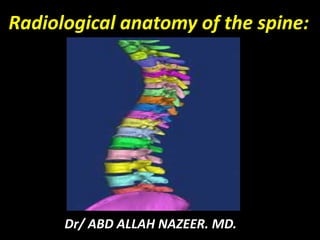
  • 1. Radiological anatomy of the spine: Dr/ ABD ALLAH NAZEER. MD.
  • 2.
  • 3.
  • 4.
  • 5.
  • 6. C-spine normal anatomy - Open mouth view.
  • 7.
  • 8. Normal AP and Lateral Views.
  • 9.
  • 10.
  • 11. Thoracic Spine - AP & Lateral View.
  • 12.
  • 14.
  • 17. CT of the Cervical Spine: Anatomy. CT of the Cervical Spine, axial reconstruction. Level 1. Image 1. 1,Intervertebral disc C3-C4. 2, Vertebral body (C3). 3, Uncinate process C4. 4, Transverse process of C4. 5, Lamina. 6, Superior articular process of C4.
  • 18. CT of the Cervical Spine, coronal reconstruction. Level 1. Image 2.Arrow, Atlantooccipital joint. 1, Transverse process of C4 (right side). 2, Uncinate process C4.
  • 19. CT of the Cervical Spine, sagittal reconstruction. Level 1. Image 3 1, Anterior arch of l‘ atlas (C1). 2, Intervertebral space (disc) C3-C4. 3, Vertebral foramen. 4, Spinous process of C2. 5, Posterior arch of C1. 6, Vertebral body C5.
  • 20. CT of the Thoracic Spine: Anatomy.
  • 21.
  • 22. 1-vertebral body, 2-neural foramen, 3-articular pillar 4-lamina, 5-spinous process.
  • 23. CT of the Lumbo-sacral Spine: Anatomy. CT of lumbar Spine, axial reconstruction. Level 1. Image 1. 1, Vertebral body (L4). 2, Transverse process. 3, Facet joint. 4, Spinous process. 5, Inferior articular process L3. 6, Superior articular process of L4. 7, Intervertebral disc L3-L4.
  • 24. CT of lumbar Spine, coronal reconstruction. Level 1. Image 2. 1, Transverse process L1. 2, Lamina. 3, Inferior articular process of L3. 4, Superior articular process of L2.
  • 25. CT of lumbar Spine, sagittal reconstruction. Level 1. Image 3. 1, Spinous process de L2. S, Sacrum. L1, Vertebral body, L1. L2, Vertebral body, L2.
  • 26. SPINAL MR IMAGING TECHNIQUES: Sagittal and axial magnetic resonance images should be acquired through the cervical, thoracic, and lumbar segments of the spine, as they are generally considered complementary, and imaging the spine in only one plane may result in misinterpretation. The addition of coronal images may also be useful, especially in patients with scoliosis. Stacked axial images and/or angled images through the discs can be obtained, often useful when the indication for imaging is pain, degenerative change, and/or radiculopathy.1 Although imaging in the axial plane is a matter of personal preference, using only angled axial images through the discs may be inadequate, as portions of the spinal canal will not be imaged axially. Slice thickness from 3 to 4 mm is generally optimal for imaging of the spine. Axial gradient-echo images through the cervical spine are typically 2 mm Thick.
  • 27.
  • 28.
  • 29.
  • 30. Sagittal T2-weighted image, cervical spine. 1,Clivus; 2, Atlanto-occipital ligament; 3, Anterior longitudinal ligament; 4, Anterior arch C1; 5, Superior fascicle of cruciform ligament/tectorial membrane; 6, Apical ligament; 7, Transverse ligament (of cruciform ligament); 8, Posterior arch C1; 9, Posterior occipital atlantal membrane; 10, Nuchal ligament; 11, Semispinalis capitis muscle; 12, Cervical spinal cord; 13, Posterior longitudinal ligament/anterior thecal sac dura; 14, Posterior dural sac; 15, Interspinous ligament; 16, Gray matter along central canal; 17, Supraspinous ligament; 18, Ligamentum flavum; 19, Dental synchondrosis (disc anlage).
  • 31. Coronal T1-weighted image, craniocervical junction. 1, Alar ligament; 2, Transverse ligament (of cruciform ligament); 3, Apical ligament; 4, Dens; 5, Lateral mass C1; 6, Occipital condyle; 7, Jugular tubercle of occipital bone; 8, Hypoglossal canal; 9, Uncinate process C3; 10, Vertebral artery.
  • 32. Coronal T1- weighted image, cervical spine. 1, Uncinate processes; 2, Segmental spinal veins and nerve roots.
  • 33. Parasagittal T1-weighted image, cervical spine. 1, Hypoglossal canal; 2, Occipital condyle; 3, Lateral mass C1; 4, Vertebral artery; 5, Rectus capitis posterior major muscle; 6, Obliquus capitis inferior muscle; 7, Articular pillar C2; 8, C3 dorsal root ganglion; 9, Longuscoli (cervicis) muscle; 10, Vertebral artery; 11, Apophyseal (facet) joint C3-4; 12, Multifidis muscle; 13, Semispinalis cervicis muscle; 14, Splenius capitis muscle; 15, Trapezius muscle; 16, Superior articular process; 17, Inferior articular process.
  • 34. Coronal oblique T1- weighted image, cervical spine. 1, Cervical spinal cord; 2, Subarachnoid space; 3, Dorsal root ganglion; 4, Superior foraminal vein; 5, Vertebral artery.
  • 35. A) Axial T2-weighted image, lower cervical spine. 1, Uncinate process C7; 2, Superior articular process C7; 3, Apophyseal (facet) joint; 4, Inferior articular process C6; 5, Foraminal vein; 6, Ligamentum flavum/cortex of lamina; 7, Dorsal rootlet C7; 8, Uncovertebral joint; 9, Ventral rootlets C7. (B) Axial T2-weighted image, mid cervical spine. 1, Dorsal root ganglion; 2, Vertebral artery; 3, Posterior longitudinal ligament/anterior thecal sac dura; 4, Longus colli muscle; 5, Internal jugular vein; 6, Dorsal rootlets; 7, Lamina; 8, Sternocleidomastoid muscle; 9, Longissimus capitis muscle; 10, Levator scapulae muscle; 11, Semispinalis colli muscle; 12, Semispinalis capitis muscle; 13, Splenius capitis muscle; 14, Trapezius muscle; 15, Nuchal ligament.
  • 36. Sagittal T1-weighted image, thoracic spine. 1, Thoracic spinal cord; 2, Subarachnoid space; 3, Posterior epidural fat; 4, Ligamentum flavum; 5, Transversospinalis (multifidus) muscle; 6, Spinous process; 7, Epidural vein; 8, Supraspinous ligament..
  • 37. Parasagittal T2-weighted image, thoracic spine. 1, Posterior thecal sac dura; 2, Posterior epidural fat; 3, Ligamentum flavum. Sagittal T2-weighted image, thoracic spine. 1, Thoracic spinal cord; 2, Subarachnoid space; 3, Ligamentum flavum; 4, Transversospinalis (multifidus) muscle; 5, Spinous process; 6, Supraspinous ligament; 7, Basivertebral vein; 8, Conus medullaris; 9, Cauda equina.
  • 38. Sagittal T2-weighted image demonstrating CSF pulsation artifact within the posterior aspect of the thecal sac (asterisk). Artifact degraded sagittal T2-weighted image, thoracic spine. Truncation artifacts (asterisk). Truncation artifact simulating spinal cord syrinx (solid white arrow).
  • 39. Parasagittal T2: thoracic neural foramen. 1, Foraminal vein; 2, Thoracic paravertebral intercostal vein and artery; 3, Foraminal nerve root; 4, Superior articular process; 5, Inferior articular process; 6, Facet joint; 7, Pars interarticularis; 8, Pedicle; 9, Ligamentum flavum; 10, Erector spinae muscle group; 11, Trapezius muscle.
  • 40. Axial T2-weighted image, thoracic spine. 1, Costovertebral joint; 2, Head of rib; 3, Ligamentum flavum; 4, Pedicle; 5, Lamina; 6, Transverse process; 7, Spinous process; 8, Costotransverse joint; 9, Tubercle of rib; 10, Hemiazygous vein; 11, Posterior longitudinal ligament.
  • 41. Axial T2-weighted image, thoracic spine. 1, Aorta; 2, Hemiazygous vein; 3, Azygous vein; 4, Foraminal veins; 5, Thoracic intercostal vein; 6, Dorsal root ganglion; 7, Basivertebral veins, slow flow; 8, Posterior longitudinal ligament; 9, CSF flow artifacts; 10, Transversospinalis (multifidis) muscle; 11, Longissimus dorsi muscle; 12, Trapezius muscle.
  • 42. Sagittal T1-weighted image, lumbar spine. 1, Spinal cord; 2, Conus medullaris; 3, Cauda equina; 4,Subarachnoid space; 5, Posterior epidural fat; 6, Ligamentum flavum; 7, Interspinous ligament; 8, Supraspinous ligament; 9, Basivertebral venous plexus; 10, Epidural venous plexus; 11, Anterior epidural fat; 12,Aorta.
  • 43. Parasagittal T1-weighted image, lumbar spine. 1, Lumbar vein; 2, Lumbar artery; 3, Inferior foraminal veins; 4, Dorsal root ganglia; 5, Superior foraminal veins; 6, Facet joints; 7, Transversospinalis (multifidis) muscle; 8, Erector spinae muscle group; 9, Thoracolumbar fascia, posterior layer.
  • 44. Axial T1-weighted image, lumbar spine at L5-S1. 1, Psoas muscle; 2, L5 nerve root, ventral ramus; 3, L5 nerve root, dorsal ramus; 4, Ligamentum flavum; 5, Subarachnoid space; 6, Nerve roots of cauda equina; 7, Facet joint; 8, Iliolumbar ligament (signifies L5 vertebral level); 9, Left external iliac vein; 10, Left external iliac artery; 11, Right external iliac artery; 12, Right external iliac vein; 13, Transversospinalis (multifidis) muscle; 14, Erector spinae muscle group.