
Recommended
More Related Content
What's hot
What's hot (20)
Similar to Radioiodine ablation of normal remnants after less-than-total thyroidectomy for cancer.
Similar to Radioiodine ablation of normal remnants after less-than-total thyroidectomy for cancer. (20)
More from Herbert Klein
More from Herbert Klein (11)
Recently uploaded
Recently uploaded (20)
Radioiodine ablation of normal remnants after less-than-total thyroidectomy for cancer.
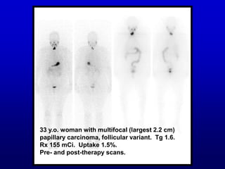
- 1. 33 y.o. woman with multifocal (largest 2.2 cm) papillary carcinoma, follicular variant. Tg 1.6. Rx 155 mCi. Uptake 1.5%. Pre- and post-therapy scans.
- 2. 16 months later, Thyrogen-stimulated scan was negative with Tg <0.5.
- 5. When is ablate not ablate? “Thyroid ablation refers to the use of radioiodine to destroy the remaining normal thyroid tissue after less-than-total thyroidectomy (intended or otherwise) [as distinct from therapy].” Sweeney DC et al cited in ref. 3, Woodrum et al (2005)
- 6. 43 y.o. man with papillary carcinoma, multifocal, extension to skeletal muscle and metastasis to multiple lymph nodes. TSH 58. Tg 22.4. Uptake 15%. 24- and 48-hour I-123 scans.
- 7. One year later, no detectable lesions, Tg <0.5.
- 8. Q. When is ablate not ablate? A. When the initial treatment attacks cancer. ♦ Assertion: The initial I-131 dose in thyroid cancer, often called an ablative dose, may in fact be given in the presence of cancer and likely treats the cancer. After all, if we are treating because the cancer might recur, there is surely a finite chance of cancer cells being present at the time of the first treatment, even when not visible on the scan.
- 9. CPT code book, 2003: 79030 Radiopharmaceutical ablation of gland for thyroid carcinoma. 79035 Radiopharmaceutical therapy for metastases of thyroid carcinoma. 2006: Only 79005: Radiopharmaceutical therapy, by oral administration. Others were deleted. Some authors use “ablate” in quotes.
- 10. Objectives: 1. Review the rationale for initial post-surgical I-131 treatment for differentiated thyroid cancer (DTC) 2. Review the various initial presentations on radioiodine imaging after surgery for DTC 3. Review considerations in choosing the dose for initial post-surgical I-131 treatment in DTC
- 11. Bibliography: 1. Sisson JC. Applying the radioactive eraser: I-131 to ablate normal thyroid tissue in patients from whom thyroid cancer has been resected (editorial). J Nucl Med. 1983;24:743-745. 2. Mazzaferri EL. Thyroid remnant 131I ablation for papillary and follicular thyroid carcinoma. Thyroid. 1997;7:265-271. 3, Woodrum DT, Gauger PG. Role of 131I in the treatment of well differentiated thyroid cancer. J Surg Oncol. 2005:89:114-121. 4. Doi SAR, Woodhouse NJY. Ablation of the thyroid remnant and 131I dose in differentiated thyroid cancer. Clin Endocrinol. 2000;52:764-773. 5. National Comprehensive Cancer Network Thyroid Carcinoma Panel Members. NCCN Clinical Practice Guidelines in Oncology. Thyroid Carcinoma. 6. The American Thyroid Association Guidelines Taskforce. Management guidelines for patients with thyroid nodules and differentiated thyroid cancer. Thyroid. 2006;16:109-142. http://www.ncc.org/professionals/physician gls/PDF/thyroid.pdf 2006
- 12. Rationale for initial therapy, adapted from Ref. 5, NCCN Guidelines (2006) 1. There is evidence that outcomes are better. 2. Attack metastases that are visible on the initial scan. 3. Ablate normal tissue destined to become malignant, 4. Attack residual malignancy a. microscopic in remnant b. remote from remnant, e.g. tiny foci c. outside remnant and obscured by uptake in remnant
- 13. Rationale for initial therapy, cont. 5. Demonstration of unsuspected malignancy on post-therapy scan (10-26%), which alters disease stage and affects patient management. 6. Simplified patient follow-up, because elimination of “thyroid bed” uptake eliminates mis- interpretation of it as disease. 7. Remnant ablation eliminates normal tissue as a source of Tg production, which facilitates identification of patients who are free of disease and promotes early identification of those with residual cancer. 8. Elimination of normal tissue may eliminate the nidus for continued confounding anti-Tg antibody production.
- 14. 44 y.o. woman, previous history of irradiation, with “multicentric papillary carcinoma exhibiting follicular differentiation”, multiple positive regional lymph nodes and infiltration of perinodal tissue. Rx 194 mCi. Most lesions disappeared, but she required subsequent treatment.
- 15. E.H. 44 y.o. M s/p thyrx, multifocal diffuse sclerosing variant of pap. adenoca with active vascular and lymphatic permeation, intraglandular metastasis, and +LN’s. Tg 160. Rx 202 mCi. Post –Rx 6 d.
- 16. 53 y.o. man, well differentiated papillary ca, encapsulated follicular variant, ~5 cm. Relatively low thyroid bed activity; abnormal focus upper midline ant. thorax. Tg = 0.8. Rx 202 mCi. Pre- and post-Rx scans.
- 17. This was 10.3 weeks after CT with contrast. Urinary iodine was 346µg/24 h (<400 considered important, under 50 desirable). 8 months later, scan negative, Tg <0.5, urinary iodine 86 µg/24 h.
- 18. 54 y.o. man, S/P total laryngectomy (squamous cell ca), total thyroidectomy and lymph node dissection. Papillary ca multifocal (largest 2.1 cm), focal angiolymphatic invasion, positive LN’s. Tg <0.5. No therapy given. 24-hour urinary iodine: specimen lost.
- 19. 45 y.o. man with single 2.1 x 2.0 cm papillary ca. Scanned and treated (100 mCi) with Thyrogen stimulation. Tg not known before treatment.
- 20. 40 y.o. woman with papillary carcinoma with follicular elements and positive LN’s, whose TSH didn’t rise. Reported to have had thyroidectomy elsewhere. Treated with I-131, 78 mCi, to ablate normal tissue.
- 21. 35 y.o. woman with papillary ca with follicular elements, with direct extension into adjacent soft tissue and not amenable to total removal, and 1 pos. LN, prior pos. lung biopsy. Scan (I-131, 3.9 mCi): activity in neck and lungs. Neck uptake 26%. Borderline hyperthyroid: TSH low! Repeated for dosimetry 2 months later; TSH elevated (thought 2˚ to 4 mCi I-131 dose!). Rx 250 mCi. Needed subsequent treatments.
- 22. A young woman scanned 5 years after initial treatment.
- 23. Same patient scanned after therapeutic dose.
- 24. When TSH doesn’t go up (after withdrawal) ♦ Too much normal tissue ♦ Functioning tumor (euthyroid or hyperthyroid) ♦ Patient kept taking hormone ♦ Sluggish pituitary When there is no tissue seen on scan ♦ Really total thyroidectomy ♦ Iodine load
- 25. Ref. 1. Sisson (1983). Applying the Radioactive Eraser: I-131 to Ablate…: “Although continued depiction of thyroid cancer is ominous, it does not necessarily follow that a blank scintigram is ideal. The menace of residual portions of normal thyroid glands has not been established, and their elimination must be controversial…30 mCi… succeeded in 81% of the patients…But complete… ablation of all normal tissue is not required to achieve …high [TSH]…If the remnant…is already small,… benefits from its removal are not readily perceived… To ablate or not to ablate is a question that will haut us for some time to come.”
- 26. Inference: Some may not need ablation, and incomplete ablation (e.g. with a low dose) may be tolerable in others. Tg not discussed in this editorial.
- 27. Ref. 2. Mazzaferri (1997). 1004 patients with DTC followed for median 14.7 years or more. Tumor recurrence was about 3-fold lower (p<0.001) and fewer patients developed distant metastases (p<0.002) after ablation than thyroid hormone alone or no therapy. There were fewer cancer deaths after ablation ((p<0.001); this difference occurred only in patients ≥ 40 y.o. These effects are not apparent in patients with isolated tumors < 1.5 cm that are not metastatic to regional lymph nodes or invading the thyroid capsule.
- 28. Years After Initial Therapy Cumulative Deaths (%) 0 5 10 15 20 25 30 35 16 14 12 10 8 6 4 2 0 No medical therapy Thyroid hormone only P<0.001 P<0.05 131 l Remnant ablation Cancer death rate after thyroid remnant ablation, thyroid hormone therapy alone, or no postoperative medical therapy.
- 29. Comments on “ablation” benefit ♦ No mention of patients whose initial scans showed metastases. Where do they fit into the picture? ♦ Ref. 5, NCCN Guidelines: “…Long-term evaluation of recurrence risk after adjuvant radioiodine may be confounded by the… possibility that patients who receive adjuvant therapy may be more likely to undergo more intensive follow-up testing”
- 30. Comments on “ablation” benefit (cont.) ♦ Ref. 6, ATA Guidelines (2006): “[Some] studies show no…benefit of [ablation], at least among the majority of patients with papillary…carcinoma…Lower risk patients do not show evidence for benefit…No prospective studies have been performed…”
- 31. Ref. 3, Woodrum et al (2005): ♦ “The future of [DTC] treatment will like involve an approach where patients are selected for postoperative radioiodine therapy based on individual risk assessment…” ♦ This is a good recent review in which Sisson is thanked for his critical review of the text, 22 years after ref. 1.
- 32. Ref. 4, Doi et al (2000): Meta-analysis. High dose was more efficient than low dose for remnant ablation, and a higher proportion of those with a near-total resection than with a less complete resection achieved ablation.
- 33. Ref. 5, NCCN Guidelines (2006): “[Some] experts advocate that the whole-body [radioiodine] diagnostic scan may alter therapy, for example: (1) when unsuspected metastases are identified, or (2) when an unexpectedly large remnant is identified that requires additional surgery or a reduction in radioiodine dosage…123I does not carry a risk of stunning…Empiric administration of radioiodine without a diagnostic scan is not routinely recommended by the panel.” Comment: Lack of uptake may trigger suspicion of an iodine load.
- 34. Considerations in I-131 dose choice: Risk factors include gender, age, size and multifocality of tumor, aggressive histology, molecular markers, elevated thyroglobulin, nodal metastases, extrathyroidal or vascular invasion Examples: ♦ Low risk patient, e.g. 35-y.o. woman with single 1.5 cm lesion of papillary cancer, no evidence of spread, no radiation history, Tg <0.5 Low dose or no Rx ♦ High risk patient: e.g. 60-y.o. man with multifocal papillary carcinoma, largest 2.0 cm, one positive LN, Tg = 20 (even though only remnants on scan) High dose
- 35. Considerations in I-131 dose choice (cont.): • I-131 has risks • Tg may be falsely elevated for several weeks by injury from surgery.
- 36. Suggested guidelines for dose decisions: 1. Metastases evident on diagnostic scan High dose, 150-200 mCi, consider dosimetry 2. Other high risk High dose, 150-200 mCi 3. Low risk, definite low-grade thyroid remnant uptake, low Tg Low dose, 30-100 mCi—or no Rx 4. Low risk, low risk, remnant or Tg may interfere with detection Low dose, 30-100 mCi
- 37. Suggested guidelines for dose decisions (cont.): 5. Scan negative, Tg low No Rx. Consider urine iodide 6. Scan negative, Tg elevated Consider Rx. Consider urine iodide 7. Only remnants visible, very high Tg Rx (conventional wisdom), but this could already be a herald of non- iodine-avid lesions
- 38. Pet peeves ♦ “Ablation” is misleading. ♦ “Ablation” to the point of seeing zero activity is probably a pointless fetish. ♦ Ref. 4, Doi et al (200): “Residual thyroid may compete with recurrent or metastatic thyroid cancer for radioiodine uptake.” Tissue in the neck will not inhibit detection of a metastasis in the femur, so long as the TSH is elevated.
- 39. "Why did the ibis cross the road?"