AACR 2014 Abstract# 3730: A quick and cost effective 12-cell line panel assay to predict drug activity in human tumor xenograft models
1. A quick and cost effective 12-cell line panel assay to predict drug activity in human tumor
xenograft models
Abstract #3730
Michael J. Roberts1, Tommie A. Gamble1, Richard D. May1 Murray Stackhouse1 Kristy L. Berry1, Andrew D. Penman1, Robert J. Rooney2, Yulia Maxuitenko1 Michael S. Koratich1
1Southern Research Institute, Birmingham, AL; 2Genome Explorations Inc., Memphis, TN
2000 Ninth Avenue South ● Birmingham, AL 35205 ● www.SouthernResearch.org ● 1 (800) 967-6774 (USA) ● 1 (205) 581-2000
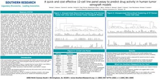
Figure 1: Unsupervised Hierarchical Clustering of 57 Human
Tumor Cell Lines and 43 Human Tumor Xenograft ModelsThe procedure to identify and develop an anti-cancer drug first involves testing drug
candidates in cell lines followed by human tumor xenograft models, usually selected
based upon the histotype of the cell lines in which the drug showed optimal activity.
Many drugs fail at this stage, as activity in cell lines does not often correlate with activity
in xenograft models. This is not surprising, as we have previously shown that gene
expression in xenograft models does not necessarily correlate with the cell line from
which it was derived. In an attempt to improve the success rate of drugs tested in
xenograft models, we have developed a fast and cost effective 12-panel human tumor
cell line assay that represents the genetic diversity of all our xenograft models and
several different cancer histotypes. Affymetrix genomic analysis was performed on 100
human tumor xenograft and cell line models. The genomic profiles obtained underwent
Unsupervised Hierarchical Cluster Analysis to group models with similar genetic profiles.
This analysis resulted in 12 distinct clusters; a representative cell line was chosen from
each cluster. Stocks of each representative cell line were frozen and tested to ensure
exponential growth immediately upon thawing, resulting in no waiting time for drug
testing. It follows that if a candidate drug shows activity in one or more of these
representative cell lines, other cell lines and/or xenograft models in the same cluster can
also be tested. As the cell lines and xenograft models within the same cluster will have a
similar genetic profile, the chances of success should thus be increased. To test the
effectiveness of this approach, we used our database to further develop an internal
compound. SRI-20900 had been tested previously in the CCRF-CEM and CAKI-1
xenograft models. The compound showed no activity in CCRF-CEM cells, but excellent
activity in CAKI-1 cells. These models were in completely different clusters. So, based on
these data, we tested the compound in the SKOV-3 and IGROV-1 xenograft models, as
these clustered closely to the CAKI-1 model. The compound showed excellent activity in
both SKOV-3 and IGROV-1 models. Although these data provide proof of principle,
further work needs to be done by testing targeted compounds in the 12-cell line panel,
followed by testing in xenograft models within the same cluster as the cell lines that
show optimal activity. In addition, it would follow that a xenograft model within the same
cluster as an inactive cell line should also be tested. We hope to start these studies early
in 2014.
The novel Southern Research Institute Nucleoside SRI-20900 had previously been shown
to be active in the CAKI-1 human renal Xenograft model but inactive in the CCRF-CEM
human leukemia xenograft model.
The cluster analysis illustrated in Figure 1 showed that the human SKOV-3 and IGROV-1
ovarian xenograft models clustered closely with the CAKI-1 renal model.
The nucleoside SRI-20900 was active in both the SKOV-3 and IGROV-1 ovarian
xenograft models.
A cluster analysis was therefore performed using only the human tumor cell line models,
and this analysis is illustrated in Figure 2.
A cell line was chosen from each of the 12 clusters to develop a simple in vitro assay
encompassing the genetic diversity across all our models and representing several
different phenotypes.
Figure 2: Unsupervised Hierarchical Clustering of 57 Human
Tumor Cell Lines
Figure 3: 12-Cell Line Panel
Introduction
Results
Southern Research has developed a cost effective in vitro model enabling more
compounds to be tested earlier in the drug development process and hence increasing
the success rate.
If wide ranging activity across all models is observed this would be similar to seeing
wide ranging activity in the NCI-60 panel.
The in vitro model covers the entire genetic variability of our models and several
different phenotypes thus enabling selective activity to be more easily identified.
Southern will search our genetic database for models that express your target of interest
and conduct testing using those models
Following in vitro screening of your compound, Southern will suggest other in vitro
and/or in vivo models with a similar genetic profile
Following in vivo screening, Southern will suggest other in vivo models with a similar
genetic profile, often expanding the potential of your drug to be utilized in other
histotypes
What Can Southern Research Do For You?
CFPAC-1
MCF-7
MX-1
OVCAR-3
SKOV-3
IGROV-1
ZR-75-1
H322M
U251
A431
BxPC-3
Colo-205
OVCAR-5
HT29
HEPG2
SW620
HCT-116
DLD-1
HCT-15
UISO-BCA-1
MALME-3M
SK-MEL-28
UACC62
MDA-MB-435
SK-MEL-2
MiaPaca-2
MES-SA
H522
A2780/DDPt
A2780/S
K-562
H82
A549/pac
Caki-1
A498
RXF-393
LOX-IMV1
PC-3
NCI/ADR-RES
MDA-MB-231
PANC-1
DU145
SF-295
UACC257
H460
A549
A549/cis
H69
NCI-H69/cis
NCI-H69/pac
CCRF-CEM
MOLT-4
RPMI-8226
HL-60
AS283
RL
cellline
cellline
cellline
cellline
cellline
cellline
cellline
cellline
cellline
cellline
cellline
cellline
cellline
cellline
cellline
cellline
cellline
cellline
cellline
cellline
cellline
cellline
cellline
cellline
cellline
cellline
cellline
cellline
cellline
cellline
cellline
cellline
cellline
cellline
cellline
cellline
cellline
cellline
cellline
cellline
cellline
cellline
cellline
cellline
cellline
cellline
cellline
cellline
cellline
cellline
cellline
cellline
cellline
cellline
cellline
cellline
Pancreatic
BreastEstrogenDependent
Breast
Ovarian
Ovarian
Ovarian
Breast
Lungnon-SmallCell
Glioblastoma
SkinEpidermoid
Pancreatic
Colon
Ovarian
Colon
Liver
Colon
Colon
Colon
Colon
Breast
SkinMelanoma
SkinMelanoma
SkinMelanoma
SkinMelanoma
SkinMelanoma
Pancreatic
Uterine
Lungnon-SmallCell
Ovarian
Ovarian
Leukemia
LungSmallCell
Lung
Kidney
Kidney
Kidney
SkinMelanoma
Prostate
Ovarian
Breast
Pancreatic
Prostate
Glioblastoma
SkinMelanoma
LungLargeCell
Lung
Lung
LungSmallCell
LungSmallCell
LungSmallCell
Leukemia
Leukemia
Leukemia
Leukemia
Lymphoma
Lymphoma
MiaPaca-2
CFPAc-1
CFPAc-1
LOX-IMV1
SW620
U251
RPMI-8226
RPMI-8226
AS283
CCRF-CEM
CCRF-CEM
MOLt-4
MOLt-4
HL-60
HL-60
AS283
RL
RL
H69
H69
NCI-H69/cis
NCI-H69/pac
UACC62
UACC62
SK-MEL-2
SK-MEL-2
MDA-MB-435
MALME-3M
SK-MEL-28
UACC257
A549/pac
Caki-1
A498
RXF-393
SF-295
U251
H460
DU145
A549
A549/cis
Pc-3
Pc-3
PANc-1
PANc-1
MDA-MB-231
MDA-MB-231
LOX-IMV1
NCI/ADR-RES
SKOV-3
SKOV-3
IGROV-1
IGROV-1
RXF-393
H82
Caki-1
H460
A498
Colo-205
Colo-205
OVCAR-5
HCt-15
HT29
HCt-15
DLD-1
DLD-1
HT29
OVCAR-5
SW620
HCt-116
HCt-116
MX-1
NCI/ADR-RES
BxPc-3
BxPc-3
SR475
A431
A431
H322M
H322M
OVCAR-3
UISO-BCA-1
ZR-75-1
UISO-BCA-1
MES-SA
LnCaP
LnCaP
MCF-7
MCF-7
MX-1
A549
A2780/S
A2780/S
A2780/DDPt
A2780/DDPt
HEPG2
K-562
H82
H522
MES-SA
MiaPaca-2
xenografttumor
cellline
xenografttumor
xenografttumor
xenografttumor
xenografttumor
cellline
xenografttumor
xenografttumor
xenografttumor
cellline
cellline
xenografttumor
cellline
xenografttumor
cellline
cellline
xenografttumor
xenografttumor
cellline
cellline
cellline
cellline
xenografttumor
cellline
xenografttumor
cellline
cellline
cellline
cellline
cellline
cellline
cellline
cellline
cellline
cellline
cellline
cellline
cellline
cellline
cellline
xenografttumor
cellline
xenografttumor
cellline
xenografttumor
cellline
cellline
cellline
xenografttumor
cellline
xenografttumor
xenografttumor
xenografttumor
xenografttumor
xenografttumor
xenografttumor
xenografttumor
cellline
xenografttumor
xenografttumor
xenografttumor
cellline
xenografttumor
cellline
cellline
cellline
cellline
cellline
xenografttumor
xenografttumor
xenografttumor
cellline
xenografttumor
xenografttumor
xenografttumor
cellline
cellline
xenografttumor
cellline
cellline
cellline
xenografttumor
xenografttumor
cellline
xenografttumor
xenografttumor
cellline
cellline
xenografttumor
cellline
xenografttumor
cellline
xenografttumor
cellline
cellline
cellline
cellline
cellline
cellline
Pancreatic
Pancreatic
Pancreatic
SkinMelanoma
Colon
Glioblastoma
Leukemia
Leukemia
Lymphoma
Leukemia
Leukemia
Leukemia
Leukemia
Leukemia
Leukemia
Lymphoma
Lymphoma
Lymphoma
LungSmallCell
LungSmallCell
LungSmallCell
LungSmallCell
SkinMelanoma
SkinMelanoma
SkinMelanoma
SkinMelanoma
SkinMelanoma
SkinMelanoma
SkinMelanoma
SkinMelanoma
Lung
Kidney
Kidney
Kidney
Glioblastoma
Glioblastoma
LungLargeCell
Prostate
Lung
Lung
Prostate
Prostate
Pancreatic
Pancreatic
Breast
Breast
SkinMelanoma
Ovarian
Ovarian
Ovarian
Ovarian
Ovarian
Kidney
LungSmallCell
Kidney
LungLargeCell
Kidney
Colon
Colon
Ovarian
Colon
Colon
Colon
Colon
Colon
Colon
Ovarian
Colon
Colon
Colon
Breast
Ovarian
Pancreatic
Pancreatic
NeckSquamousCell
SkinEpidermoid
SkinEpidermoid
Lungnon-SmallCell
Lungnon-SmallCell
Ovarian
Breast
Breast
Breast
Uterine
Prostate
Prostate
BreastEstrogenDependent
BreastEstrogenDependent
Breast
Lung
Ovarian
Ovarian
Ovarian
Ovarian
Liver
Leukemia
LungSmallCell
Lungnon-SmallCell
Uterine
Pancreatic
Cell Line Phenotype
CFPAC-1 Pancreatic
MCF-7 Breast
IGROV-1 Ovarian
Colo205 Colon
DLD-1 Colon
UACC-62 Melanoma
MiaPaca-2 Pancreatic
A498 Kidney
PC-3 Prostate
A549 Lung
NCI-H69 Small Cell Lung
RL Lymphoma
A compound showing anti-tumor activity in
one or more of the 12-panel cell lines should
be tested in other human tumor cell lines or
human tumor xenograft models with a similar
genetic background identified from Figure 2.
Ubitquitous activity across all cell lines would
suggest a ubitquitous target.
Observed activity in a particular cell line
doesn’t necessarily mean the corresponding
xenograft model should be tested.
The CFPAC-1 cell line model is genetically
similar to the CFPAC-1 xenograft model.
The MiaPaca-2 cell line model is not
genetically similar to the MiaPaca-2
xenograft model.