SlideShare a Scribd company logo
1 of 72
MR DIFFUSION ,
PERFUSION and
ARTIFACTS
DR. YASNA KIBRIA
MD RESIDENT ,PHASE-A
Department of RADIOLOGY and IMAGING
BSMMU
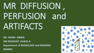
DIFFUSION
• Diffusion means random movement
of the water protons.
• The process by which water protons
diffuse randomly in the space is
called BROWNIAN MOTION.
• The difference in the mobility of
water molecules between tissues
gives the contrast in DWI and helps
to characterize tissues and
pathology.
ISOTROPIC DIFFUSION
• Possibility of water protons moving in
any one particular direction is equal to
the probability that it will move in any
other direction.
• Isotropy= uniformity in all direction .
• Isotropic diffusion forms the basis for
routine DWI.
ANISOTROPIC DIFFUSION
• In anisotropic diffusion , water
molecules have preferred
direction of movement.
• Water protons move more easily
in some direction than other.
• Anisotropic diffusion forms the
basis for DTI or Tactography.
HOW do we acquire DWIs?
• The “stejskal-Tanner pulsed gradient
spin echo sequence “ –was the first
experimental sequence described for
acquisition of DWI.
• It is a T2-w spin echo sequence with
diffusion gradients applied before
and after the 180 degree pulse.
THE b VALUE
• The b value indicates the magnitude of diffusion weighting provided by the
diffusion gradient. It also indicates sensitivity of the sequence to the diffusion.
• Expressed in second/mm2.
• The b value increases with diffusion gradient strength, duration and time
between application of the two gradients.
• As the b value increases the signal from water molecule reduces.
• At high b value(b=1000) only tissues with very high T2 relaxation time or those
with restricted diffusion will have high signal.
DIFFUSION TRACE
• Isotropic diffusion is the basis of DWI.
• However, there are some anisotropy
of water molecules in the tissues .
• To reduce this anisotropy ,the image
with higher b value like b=1000 is
acquired in three directions along X,Y,Z
axes.
• Diffusion changes along all 3 axes are
then averaged out to get a “TRACE “
diffusion image.
CLINICAL APPLICATIONS OF DWI
NEUROIMAGING applications :
1. Stroke
2. Epidermoid vs arachnoid cyst
3. Abscess vs simple cystic lesion
4. DWI in brain tumors
DWI in BODY IMAGING:
• Relatively new
• Lower b value is used
• Mainly focused on tumor imaging & assessing treatment response
• Staging of the tumors & lymphomas
DIFFUSION TENSOR IMAGING
• DTI is based on the
anisotropic diffusion of water
molecules.
• Tensor Is the mathematical
formalism used to model
anisotropic diffusion.
TECHNIQUE OF DTI :
• MR scanner axes X ,Y, Z are never perfectly parallel to white matter tracts at
every point in the image.
• In DTI, images are acquired in at least 6 , usually 12-24 directions instead of 3
in the usual.
• Various maps used to
indicate orientation of
fibres :
FA ( Fractional Anisotropy)
RA (Regional Anisotropy )
VA (Volume ratio )
USES
• Diffusion Tensor measures
the magnitude of the ADC in
preferred direction of water
and also perpendicular to
the direction.
• The resultant image show
white matter tracts very well
• Hence, this technique is also
called as TRACTOGRAPHY.
CLINICAL applications
• assess the deformation of white matter by tumors - deviation,
infiltration, destruction of white matter
• delineate the anatomy of immature brains
• pre-surgical planning
• Alzheimer disease - detection of early disease
• schizophrenia
• Basic colors can tell the observer how the
fibers are oriented in a 3D coordinate
system, this is termed an "anisotropic
map". The software could encode the
colors in this way:
• Red indicates directions in the X axis: right
to left or left to right.
• Green indicates directions in
the Y axis: posterior to anterior or
from anterior to posterior.
• Blue indicates directions in the Z axis: foot-
to-head direction or vice versa.
MR PERFUSION
• PERFUSION :
Refers to the passage of blood from an
arterial supply to venous drainage
through the microcirculation.
Perfusion is necessary for the nutritive
supply to the tissues & for clearance of
products of metabolism.
It can be affected by various diseases.so
measuring changes in perfusion can help
in diagnosis of certain diseases.
PRINCIPLES
• Paramagnetic substance like Gadolinium causes
shortening of both T1 & T2.
• T1 shortening results into increased signal
intensity.
• T2 shortening results into signal drop or
blackening.
• Gd based contrast agent passes through
microvasculature in high concentration
decrease in signal in surrounding tissues from
magnetic susceptibility induced shortening of
T2* relaxation time.
• This signal drop is proportional to the
perfusion.
• More the number of microvasculature/small
vessels per voxel-more will be the signal drop.
Technique of MR PERFUSION with exogenous
contrast agent (DSC ):
• A dose of 0.1 mmol/kg of Gd based
CONTRAST AGENT is injected
intravenously using power injector at
the rate of 5 ml/sec .
• Fast T2* weighted EPI sequence is run
to catch first pass of the contrast
through microcirculation.
• This sequence typically acquire 15-20
slices covering entire brain in 1-2
seconds.
• From raw data images various color
maps are constructed using
software.
• These maps include :
1. rCBV: relative Cerebral Blood
Volume
2. CBF:Cerebral Blood Flow
3. TTP: Time to Peak
4. MTT: Mean Transit Time
Routine contrast
enhancement
Perfusion imaging
1. Sequence T1 weighted imaging T2* weighted EPI sequence
2. Signal change Increase in signal intensity Drop in signal intensity
3. Mechanism Gd causes reduction in T1
relaxation time
Gd causes reduction in T2 or
T2* relaxation time &
magnetic susceptibility
4. Detects Break in the BBB leading to
leakage of Gd
Gd in the microvasculature
(capillaries).
Thus gives information about
number of small vessels
(vascularity ) & perfusion of
the tissue.
PERMEABILITY OR LEAKINESS
• Increased permeability or leakiness
because of break in BBB results in
accumulation of Gd based contrast in
extravascular space.
• T1 enhancing effects of this extravascular
Gd may predominate to counteract the T2
signal lowering effect of intravascular Gd,
resulting in falsely low rCBV values.
• To reduce permeability induced effects on
rCBV include mathematical calculation of
PERMEABILITY or K2 maps.
MR PERFUSION in STROKE
• DWI & PWI together are very
effective in detection of early
ischemia.
• The mismatch between PW &
DW represents potentially
salvageable tissue(PENUMBRA).
• Small mismatch has a good
outcome.
MR PERFUSION in brain TUMORs
• MR perfusion can be useful in-
1. Grading tumors like gliomas
2. In guiding biopsies
3. Differentiating between therapy induced necrosis & recurrent /residual
tumors.
OTHER clinical uses: CNS vasculitis
GHOSTS /MOTION ARTIFACTS
• Ghosts are replica of something in the image.
• Ghosts are produced by body part moving
along a gradient during pulse sequence
resulting into phase mismapping.
AXIS :almost always along PHASE encoding
gradient.
CORRECTION :
1. Phase encoding axis swap
2. Saturation band
3. Respiratory compensation
4. ECG gating for cardiac motion
ALIASING /WRAPAROUND
• In aliasing, anatomy that exists outside FOV
appears within the image & on the opposite side.
• When FOV is smaller than the anatomy being
imaged , aliasing occurs.
AXIS :can occur along any axis-frequency ,phase,
slice selection gradient.
CORRECTION:
1. Frequency wrap :low pass filters.
2. Phase wrap : increasing FOV along phase
encoding gradient.
CHEMICAL SHIFT related Artifacts :
Because of different chemical environment protons in water & fat
precess at different frequencies.
TRUNCATION ARTIFACTS (edge ,
Gibbs’ & ringing artifacts )
• Truncation artifacts produce low intensity
band running through high intensity area.
• The artifacts caused by under sampling of
data so that interfaces of high & low signal
are incorrectly presented on the image.
AXIS : Phase encoding
CORRECTION :
Increase the number of phase encoding steps.
Ex- 256x256 matrix instead of 256 x 128.
MAGNETIC SUSCEPTIBILITY ARTIFACTS
• Some tissues magnetize to different
degree than other , resulting into
differences in precessional frequency
& phase.
• This causes dephasing at the interface
of these tissues & signal loss.
AXIS : frequency & phase encoding.
CORRECTION :
1. Use of SE sequence
2. Remove all metals
GOOD effects of Magnetic
Susceptibility Artifacts:
1. Used to diagnose haemorrhage ,
hemosiderin deposition &
calcification.
2. Forms the basis of post-contrast
T2* weighted MR PERFUSION studies.
3. Used to quantify myocardial &
liver iron overload.
ZIPPER ARTIFACTS
• Is a line with alternating bright &
dark pixels propagating along
frequency encoding gradient.
• Caused by stimulated echo that
have missed phase encoding.
CORRECTION :
Site of the leak should be located
& corrected.
Straight LINES
• Regularly spaced straight lines
through MR image is caused by
spike in K-space (bad data point
in K-space)
• Spike can be result from loose
electrical connectivity &
breakdown of interconnections
in RF coils.
SHADING ARTIFACTS
• In shading artifacts , image has
uneven contrast with loss of signal
intensity in one part of image.
• Uneven excitation of the nuclei due
to RF pulses applied at flip angle
other than 90 & 180 degree ,
abnormal loading of the
coil,inhomogeneity of the magnetic
field.
CORRECTION :
1. Load the coil correctly
2. Shimming to reduce magnetic field
inhomogeneity.
CLINICAL importance of OTHER MR
SEQUENCES:
 T1 WEIGHTED IMAGES :
Subacute haemorrhage
Fat containing structures
Anatomical details
 T2 WEIGHTED IMAGES:
Edema
Demyelination
Infarction
Chronic haemorrhage
 STIR :
Bone marrow imaging
Orbital imaging
SI joint imaging
 FLAIR :
Peri-lesional edema
Brain infarction
Sub-arachnoid haemorrhage
Syrinx /cysts in spinal cord
PD weighted images:
Detection of joint & muscle diseases &
injuries.
Well differentiation of GM & WM in brain.
MR Diffusion, Perfusion and Artifacts Guide

More Related Content

What's hot

Perfusion imaging: CT, MRI, Nuclear Medicine- Avinesh Shrestha
Perfusion imaging: CT, MRI, Nuclear Medicine- Avinesh ShresthaPerfusion imaging: CT, MRI, Nuclear Medicine- Avinesh Shrestha
Perfusion imaging: CT, MRI, Nuclear Medicine- Avinesh ShresthaAvinesh Shrestha
 
Perfusion and dynamic contrast enhanced mri
Perfusion and dynamic contrast enhanced mriPerfusion and dynamic contrast enhanced mri
Perfusion and dynamic contrast enhanced mrifahad shafi
 
Gradient echo pulse sequence swastik
Gradient echo pulse sequence  swastikGradient echo pulse sequence  swastik
Gradient echo pulse sequence swastikBipul Poudel
 
magnetic resonance angiography
magnetic resonance angiographymagnetic resonance angiography
magnetic resonance angiographyqavi786
 
CT ITS BASIC PHYSICS
CT ITS BASIC PHYSICSCT ITS BASIC PHYSICS
CT ITS BASIC PHYSICSDEEPAK
 
K space and parallel imaging
K space and parallel imagingK space and parallel imaging
K space and parallel imagingASHIK E H
 
MRI brain; Basics and Radiological Anatomy
MRI brain; Basics and Radiological AnatomyMRI brain; Basics and Radiological Anatomy
MRI brain; Basics and Radiological AnatomyImran Rizvi
 
MR ANGIOGRAPHY PHYSICS
MR ANGIOGRAPHY PHYSICSMR ANGIOGRAPHY PHYSICS
MR ANGIOGRAPHY PHYSICSAvinashDahatre
 
Image reconstruction in computed tomography
Image reconstruction in computed tomographyImage reconstruction in computed tomography
Image reconstruction in computed tomographyManojzz Bhatta
 
Contrast enhanced ultrasound
Contrast enhanced ultrasoundContrast enhanced ultrasound
Contrast enhanced ultrasoundNeeraj Patange
 
MRI spin echo pulse sequences
MRI spin echo pulse sequencesMRI spin echo pulse sequences
MRI spin echo pulse sequencesShiva Prakash
 
Diffusion weighted imaging
Diffusion  weighted imagingDiffusion  weighted imaging
Diffusion weighted imagingSharath Raj
 
Fat suppression imaging
Fat suppression imagingFat suppression imaging
Fat suppression imagingRoshan Shah
 
Basic and advanced mri imaging sequences in brain
Basic and advanced mri imaging sequences in brainBasic and advanced mri imaging sequences in brain
Basic and advanced mri imaging sequences in brainDev Lakhera
 
Magnetic Resonance Diffusion
Magnetic Resonance DiffusionMagnetic Resonance Diffusion
Magnetic Resonance DiffusionRahman Ud Din
 

What's hot (20)

Perfusion imaging: CT, MRI, Nuclear Medicine- Avinesh Shrestha
Perfusion imaging: CT, MRI, Nuclear Medicine- Avinesh ShresthaPerfusion imaging: CT, MRI, Nuclear Medicine- Avinesh Shrestha
Perfusion imaging: CT, MRI, Nuclear Medicine- Avinesh Shrestha
 
Mri inversion
Mri inversionMri inversion
Mri inversion
 
Perfusion and dynamic contrast enhanced mri
Perfusion and dynamic contrast enhanced mriPerfusion and dynamic contrast enhanced mri
Perfusion and dynamic contrast enhanced mri
 
CTV and MRV
CTV and MRVCTV and MRV
CTV and MRV
 
Gradient echo pulse sequence swastik
Gradient echo pulse sequence  swastikGradient echo pulse sequence  swastik
Gradient echo pulse sequence swastik
 
magnetic resonance angiography
magnetic resonance angiographymagnetic resonance angiography
magnetic resonance angiography
 
CT ITS BASIC PHYSICS
CT ITS BASIC PHYSICSCT ITS BASIC PHYSICS
CT ITS BASIC PHYSICS
 
K space and parallel imaging
K space and parallel imagingK space and parallel imaging
K space and parallel imaging
 
MRI brain; Basics and Radiological Anatomy
MRI brain; Basics and Radiological AnatomyMRI brain; Basics and Radiological Anatomy
MRI brain; Basics and Radiological Anatomy
 
MR ANGIOGRAPHY PHYSICS
MR ANGIOGRAPHY PHYSICSMR ANGIOGRAPHY PHYSICS
MR ANGIOGRAPHY PHYSICS
 
Image reconstruction in computed tomography
Image reconstruction in computed tomographyImage reconstruction in computed tomography
Image reconstruction in computed tomography
 
Imaging in acute stroke
Imaging in acute strokeImaging in acute stroke
Imaging in acute stroke
 
BASIC MRI SEQUENCES
BASIC MRI SEQUENCESBASIC MRI SEQUENCES
BASIC MRI SEQUENCES
 
Contrast enhanced ultrasound
Contrast enhanced ultrasoundContrast enhanced ultrasound
Contrast enhanced ultrasound
 
MRI spin echo pulse sequences
MRI spin echo pulse sequencesMRI spin echo pulse sequences
MRI spin echo pulse sequences
 
Ct physics – II
Ct physics – IICt physics – II
Ct physics – II
 
Diffusion weighted imaging
Diffusion  weighted imagingDiffusion  weighted imaging
Diffusion weighted imaging
 
Fat suppression imaging
Fat suppression imagingFat suppression imaging
Fat suppression imaging
 
Basic and advanced mri imaging sequences in brain
Basic and advanced mri imaging sequences in brainBasic and advanced mri imaging sequences in brain
Basic and advanced mri imaging sequences in brain
 
Magnetic Resonance Diffusion
Magnetic Resonance DiffusionMagnetic Resonance Diffusion
Magnetic Resonance Diffusion
 

Similar to MR Diffusion, Perfusion and Artifacts Guide

Diffusion tensor imaging in Neurology
Diffusion tensor imaging in NeurologyDiffusion tensor imaging in Neurology
Diffusion tensor imaging in NeurologyOsama Ragab
 
DWI/ ADC MRI principles/ applications in veterinary medicine
DWI/ ADC MRI principles/ applications  in veterinary medicineDWI/ ADC MRI principles/ applications  in veterinary medicine
DWI/ ADC MRI principles/ applications in veterinary medicineRobert Cruz
 
Diffusion Weighted Imaging- Avinesh Shrestha
Diffusion Weighted Imaging- Avinesh ShresthaDiffusion Weighted Imaging- Avinesh Shrestha
Diffusion Weighted Imaging- Avinesh ShresthaAvinesh Shrestha
 
MRI basics - How to read and understand MRI sequences
MRI basics - How to read and understand MRI sequencesMRI basics - How to read and understand MRI sequences
MRI basics - How to read and understand MRI sequencesRamesh Babu
 
MRI (Magnetic resonance imaging)
MRI (Magnetic resonance imaging)MRI (Magnetic resonance imaging)
MRI (Magnetic resonance imaging)Summu Thakur
 
Thorax cardio nsclc yw hang
Thorax cardio nsclc yw hangThorax cardio nsclc yw hang
Thorax cardio nsclc yw hangJFIM
 
Spinal cord tractography
Spinal cord tractographySpinal cord tractography
Spinal cord tractographyPratikDhabalia
 
Power Doppler,MRS,SWI.pptx
Power Doppler,MRS,SWI.pptxPower Doppler,MRS,SWI.pptx
Power Doppler,MRS,SWI.pptxChaudharyAnjali3
 
DIFFUSION TENSOR IMAGING_MFD .pptx
DIFFUSION TENSOR IMAGING_MFD       .pptxDIFFUSION TENSOR IMAGING_MFD       .pptx
DIFFUSION TENSOR IMAGING_MFD .pptxkeerthikrishna41
 
Imaging of acute stroke , Interventions
Imaging of acute stroke  , InterventionsImaging of acute stroke  , Interventions
Imaging of acute stroke , InterventionsArif S
 
Contrast sensitivity
Contrast sensitivityContrast sensitivity
Contrast sensitivitySuman Sen
 
Advances in neuro-imaging
Advances in neuro-imaging Advances in neuro-imaging
Advances in neuro-imaging Osama Ragab
 
Diffusion tensor imaging in evaluation of epilepsy ppt.pptx
Diffusion tensor imaging in evaluation of epilepsy ppt.pptxDiffusion tensor imaging in evaluation of epilepsy ppt.pptx
Diffusion tensor imaging in evaluation of epilepsy ppt.pptxaasrithakotha2
 
OPTICAL COHERENCE TOMOGRAPHY ANGIOGRAPHY
OPTICAL COHERENCE TOMOGRAPHY  ANGIOGRAPHY  OPTICAL COHERENCE TOMOGRAPHY  ANGIOGRAPHY
OPTICAL COHERENCE TOMOGRAPHY ANGIOGRAPHY Shruti Laddha
 

Similar to MR Diffusion, Perfusion and Artifacts Guide (20)

Diffusion tensor imaging in Neurology
Diffusion tensor imaging in NeurologyDiffusion tensor imaging in Neurology
Diffusion tensor imaging in Neurology
 
DWI/ ADC MRI principles/ applications in veterinary medicine
DWI/ ADC MRI principles/ applications  in veterinary medicineDWI/ ADC MRI principles/ applications  in veterinary medicine
DWI/ ADC MRI principles/ applications in veterinary medicine
 
Diffusion Weighted Imaging- Avinesh Shrestha
Diffusion Weighted Imaging- Avinesh ShresthaDiffusion Weighted Imaging- Avinesh Shrestha
Diffusion Weighted Imaging- Avinesh Shrestha
 
MRI basics - How to read and understand MRI sequences
MRI basics - How to read and understand MRI sequencesMRI basics - How to read and understand MRI sequences
MRI basics - How to read and understand MRI sequences
 
DIFFUSION & PERFUSION r MRI.ppt
DIFFUSION  &   PERFUSION  r MRI.pptDIFFUSION  &   PERFUSION  r MRI.ppt
DIFFUSION & PERFUSION r MRI.ppt
 
dwi dti.pptx
dwi dti.pptxdwi dti.pptx
dwi dti.pptx
 
MRI (Magnetic resonance imaging)
MRI (Magnetic resonance imaging)MRI (Magnetic resonance imaging)
MRI (Magnetic resonance imaging)
 
Thorax cardio nsclc yw hang
Thorax cardio nsclc yw hangThorax cardio nsclc yw hang
Thorax cardio nsclc yw hang
 
Spinal cord tractography
Spinal cord tractographySpinal cord tractography
Spinal cord tractography
 
Power Doppler,MRS,SWI.pptx
Power Doppler,MRS,SWI.pptxPower Doppler,MRS,SWI.pptx
Power Doppler,MRS,SWI.pptx
 
DIFFUSION TENSOR IMAGING_MFD .pptx
DIFFUSION TENSOR IMAGING_MFD       .pptxDIFFUSION TENSOR IMAGING_MFD       .pptx
DIFFUSION TENSOR IMAGING_MFD .pptx
 
MRI Brain
MRI BrainMRI Brain
MRI Brain
 
MRI PERFUSION.pdf
MRI PERFUSION.pdfMRI PERFUSION.pdf
MRI PERFUSION.pdf
 
Imaging of acute stroke , Interventions
Imaging of acute stroke  , InterventionsImaging of acute stroke  , Interventions
Imaging of acute stroke , Interventions
 
Contrast sensitivity
Contrast sensitivityContrast sensitivity
Contrast sensitivity
 
Diffusion Final
Diffusion FinalDiffusion Final
Diffusion Final
 
Advances in neuro-imaging
Advances in neuro-imaging Advances in neuro-imaging
Advances in neuro-imaging
 
Neuroimaging
NeuroimagingNeuroimaging
Neuroimaging
 
Diffusion tensor imaging in evaluation of epilepsy ppt.pptx
Diffusion tensor imaging in evaluation of epilepsy ppt.pptxDiffusion tensor imaging in evaluation of epilepsy ppt.pptx
Diffusion tensor imaging in evaluation of epilepsy ppt.pptx
 
OPTICAL COHERENCE TOMOGRAPHY ANGIOGRAPHY
OPTICAL COHERENCE TOMOGRAPHY  ANGIOGRAPHY  OPTICAL COHERENCE TOMOGRAPHY  ANGIOGRAPHY
OPTICAL COHERENCE TOMOGRAPHY ANGIOGRAPHY
 

More from yasna kibria

Diffusion tensor imaging .pptx
Diffusion tensor imaging .pptxDiffusion tensor imaging .pptx
Diffusion tensor imaging .pptxyasna kibria
 
Parkes Weber syndrome.pptx
Parkes Weber syndrome.pptxParkes Weber syndrome.pptx
Parkes Weber syndrome.pptxyasna kibria
 
Basic MRI in hepatobiliary surgery.pptx
Basic MRI in hepatobiliary surgery.pptxBasic MRI in hepatobiliary surgery.pptx
Basic MRI in hepatobiliary surgery.pptxyasna kibria
 
Ventricular Septal Defects
Ventricular Septal DefectsVentricular Septal Defects
Ventricular Septal Defectsyasna kibria
 
Basics of MRI Physics
Basics of MRI PhysicsBasics of MRI Physics
Basics of MRI Physicsyasna kibria
 

More from yasna kibria (8)

Vasculitis.pptx
Vasculitis.pptxVasculitis.pptx
Vasculitis.pptx
 
MRS.pptx
MRS.pptxMRS.pptx
MRS.pptx
 
Diffusion tensor imaging .pptx
Diffusion tensor imaging .pptxDiffusion tensor imaging .pptx
Diffusion tensor imaging .pptx
 
Parkes Weber syndrome.pptx
Parkes Weber syndrome.pptxParkes Weber syndrome.pptx
Parkes Weber syndrome.pptx
 
Basic MRI in hepatobiliary surgery.pptx
Basic MRI in hepatobiliary surgery.pptxBasic MRI in hepatobiliary surgery.pptx
Basic MRI in hepatobiliary surgery.pptx
 
Ventricular Septal Defects
Ventricular Septal DefectsVentricular Septal Defects
Ventricular Septal Defects
 
Basics of MRI Physics
Basics of MRI PhysicsBasics of MRI Physics
Basics of MRI Physics
 
Rickets
RicketsRickets
Rickets
 

Recently uploaded

Hierarchy of management that covers different levels of management
Hierarchy of management that covers different levels of managementHierarchy of management that covers different levels of management
Hierarchy of management that covers different levels of managementmkooblal
 
Types of Journalistic Writing Grade 8.pptx
Types of Journalistic Writing Grade 8.pptxTypes of Journalistic Writing Grade 8.pptx
Types of Journalistic Writing Grade 8.pptxEyham Joco
 
DATA STRUCTURE AND ALGORITHM for beginners
DATA STRUCTURE AND ALGORITHM for beginnersDATA STRUCTURE AND ALGORITHM for beginners
DATA STRUCTURE AND ALGORITHM for beginnersSabitha Banu
 
ENGLISH 7_Q4_LESSON 2_ Employing a Variety of Strategies for Effective Interp...
ENGLISH 7_Q4_LESSON 2_ Employing a Variety of Strategies for Effective Interp...ENGLISH 7_Q4_LESSON 2_ Employing a Variety of Strategies for Effective Interp...
ENGLISH 7_Q4_LESSON 2_ Employing a Variety of Strategies for Effective Interp...JhezDiaz1
 
Blooming Together_ Growing a Community Garden Worksheet.docx
Blooming Together_ Growing a Community Garden Worksheet.docxBlooming Together_ Growing a Community Garden Worksheet.docx
Blooming Together_ Growing a Community Garden Worksheet.docxUnboundStockton
 
Introduction to AI in Higher Education_draft.pptx
Introduction to AI in Higher Education_draft.pptxIntroduction to AI in Higher Education_draft.pptx
Introduction to AI in Higher Education_draft.pptxpboyjonauth
 
Enzyme, Pharmaceutical Aids, Miscellaneous Last Part of Chapter no 5th.pdf
Enzyme, Pharmaceutical Aids, Miscellaneous Last Part of Chapter no 5th.pdfEnzyme, Pharmaceutical Aids, Miscellaneous Last Part of Chapter no 5th.pdf
Enzyme, Pharmaceutical Aids, Miscellaneous Last Part of Chapter no 5th.pdfSumit Tiwari
 
Painted Grey Ware.pptx, PGW Culture of India
Painted Grey Ware.pptx, PGW Culture of IndiaPainted Grey Ware.pptx, PGW Culture of India
Painted Grey Ware.pptx, PGW Culture of IndiaVirag Sontakke
 
Alper Gobel In Media Res Media Component
Alper Gobel In Media Res Media ComponentAlper Gobel In Media Res Media Component
Alper Gobel In Media Res Media ComponentInMediaRes1
 
EPANDING THE CONTENT OF AN OUTLINE using notes.pptx
EPANDING THE CONTENT OF AN OUTLINE using notes.pptxEPANDING THE CONTENT OF AN OUTLINE using notes.pptx
EPANDING THE CONTENT OF AN OUTLINE using notes.pptxRaymartEstabillo3
 
Employee wellbeing at the workplace.pptx
Employee wellbeing at the workplace.pptxEmployee wellbeing at the workplace.pptx
Employee wellbeing at the workplace.pptxNirmalaLoungPoorunde1
 
AmericanHighSchoolsprezentacijaoskolama.
AmericanHighSchoolsprezentacijaoskolama.AmericanHighSchoolsprezentacijaoskolama.
AmericanHighSchoolsprezentacijaoskolama.arsicmarija21
 
How to Make a Pirate ship Primary Education.pptx
How to Make a Pirate ship Primary Education.pptxHow to Make a Pirate ship Primary Education.pptx
How to Make a Pirate ship Primary Education.pptxmanuelaromero2013
 
18-04-UA_REPORT_MEDIALITERAСY_INDEX-DM_23-1-final-eng.pdf
18-04-UA_REPORT_MEDIALITERAСY_INDEX-DM_23-1-final-eng.pdf18-04-UA_REPORT_MEDIALITERAСY_INDEX-DM_23-1-final-eng.pdf
18-04-UA_REPORT_MEDIALITERAСY_INDEX-DM_23-1-final-eng.pdfssuser54595a
 
Proudly South Africa powerpoint Thorisha.pptx
Proudly South Africa powerpoint Thorisha.pptxProudly South Africa powerpoint Thorisha.pptx
Proudly South Africa powerpoint Thorisha.pptxthorishapillay1
 
Difference Between Search & Browse Methods in Odoo 17
Difference Between Search & Browse Methods in Odoo 17Difference Between Search & Browse Methods in Odoo 17
Difference Between Search & Browse Methods in Odoo 17Celine George
 
Pharmacognosy Flower 3. Compositae 2023.pdf
Pharmacognosy Flower 3. Compositae 2023.pdfPharmacognosy Flower 3. Compositae 2023.pdf
Pharmacognosy Flower 3. Compositae 2023.pdfMahmoud M. Sallam
 

Recently uploaded (20)

Hierarchy of management that covers different levels of management
Hierarchy of management that covers different levels of managementHierarchy of management that covers different levels of management
Hierarchy of management that covers different levels of management
 
Types of Journalistic Writing Grade 8.pptx
Types of Journalistic Writing Grade 8.pptxTypes of Journalistic Writing Grade 8.pptx
Types of Journalistic Writing Grade 8.pptx
 
DATA STRUCTURE AND ALGORITHM for beginners
DATA STRUCTURE AND ALGORITHM for beginnersDATA STRUCTURE AND ALGORITHM for beginners
DATA STRUCTURE AND ALGORITHM for beginners
 
ENGLISH 7_Q4_LESSON 2_ Employing a Variety of Strategies for Effective Interp...
ENGLISH 7_Q4_LESSON 2_ Employing a Variety of Strategies for Effective Interp...ENGLISH 7_Q4_LESSON 2_ Employing a Variety of Strategies for Effective Interp...
ENGLISH 7_Q4_LESSON 2_ Employing a Variety of Strategies for Effective Interp...
 
Blooming Together_ Growing a Community Garden Worksheet.docx
Blooming Together_ Growing a Community Garden Worksheet.docxBlooming Together_ Growing a Community Garden Worksheet.docx
Blooming Together_ Growing a Community Garden Worksheet.docx
 
Introduction to AI in Higher Education_draft.pptx
Introduction to AI in Higher Education_draft.pptxIntroduction to AI in Higher Education_draft.pptx
Introduction to AI in Higher Education_draft.pptx
 
Enzyme, Pharmaceutical Aids, Miscellaneous Last Part of Chapter no 5th.pdf
Enzyme, Pharmaceutical Aids, Miscellaneous Last Part of Chapter no 5th.pdfEnzyme, Pharmaceutical Aids, Miscellaneous Last Part of Chapter no 5th.pdf
Enzyme, Pharmaceutical Aids, Miscellaneous Last Part of Chapter no 5th.pdf
 
Painted Grey Ware.pptx, PGW Culture of India
Painted Grey Ware.pptx, PGW Culture of IndiaPainted Grey Ware.pptx, PGW Culture of India
Painted Grey Ware.pptx, PGW Culture of India
 
Alper Gobel In Media Res Media Component
Alper Gobel In Media Res Media ComponentAlper Gobel In Media Res Media Component
Alper Gobel In Media Res Media Component
 
Model Call Girl in Tilak Nagar Delhi reach out to us at 🔝9953056974🔝
Model Call Girl in Tilak Nagar Delhi reach out to us at 🔝9953056974🔝Model Call Girl in Tilak Nagar Delhi reach out to us at 🔝9953056974🔝
Model Call Girl in Tilak Nagar Delhi reach out to us at 🔝9953056974🔝
 
EPANDING THE CONTENT OF AN OUTLINE using notes.pptx
EPANDING THE CONTENT OF AN OUTLINE using notes.pptxEPANDING THE CONTENT OF AN OUTLINE using notes.pptx
EPANDING THE CONTENT OF AN OUTLINE using notes.pptx
 
Employee wellbeing at the workplace.pptx
Employee wellbeing at the workplace.pptxEmployee wellbeing at the workplace.pptx
Employee wellbeing at the workplace.pptx
 
AmericanHighSchoolsprezentacijaoskolama.
AmericanHighSchoolsprezentacijaoskolama.AmericanHighSchoolsprezentacijaoskolama.
AmericanHighSchoolsprezentacijaoskolama.
 
How to Make a Pirate ship Primary Education.pptx
How to Make a Pirate ship Primary Education.pptxHow to Make a Pirate ship Primary Education.pptx
How to Make a Pirate ship Primary Education.pptx
 
Model Call Girl in Bikash Puri Delhi reach out to us at 🔝9953056974🔝
Model Call Girl in Bikash Puri  Delhi reach out to us at 🔝9953056974🔝Model Call Girl in Bikash Puri  Delhi reach out to us at 🔝9953056974🔝
Model Call Girl in Bikash Puri Delhi reach out to us at 🔝9953056974🔝
 
18-04-UA_REPORT_MEDIALITERAСY_INDEX-DM_23-1-final-eng.pdf
18-04-UA_REPORT_MEDIALITERAСY_INDEX-DM_23-1-final-eng.pdf18-04-UA_REPORT_MEDIALITERAСY_INDEX-DM_23-1-final-eng.pdf
18-04-UA_REPORT_MEDIALITERAСY_INDEX-DM_23-1-final-eng.pdf
 
Proudly South Africa powerpoint Thorisha.pptx
Proudly South Africa powerpoint Thorisha.pptxProudly South Africa powerpoint Thorisha.pptx
Proudly South Africa powerpoint Thorisha.pptx
 
TataKelola dan KamSiber Kecerdasan Buatan v022.pdf
TataKelola dan KamSiber Kecerdasan Buatan v022.pdfTataKelola dan KamSiber Kecerdasan Buatan v022.pdf
TataKelola dan KamSiber Kecerdasan Buatan v022.pdf
 
Difference Between Search & Browse Methods in Odoo 17
Difference Between Search & Browse Methods in Odoo 17Difference Between Search & Browse Methods in Odoo 17
Difference Between Search & Browse Methods in Odoo 17
 
Pharmacognosy Flower 3. Compositae 2023.pdf
Pharmacognosy Flower 3. Compositae 2023.pdfPharmacognosy Flower 3. Compositae 2023.pdf
Pharmacognosy Flower 3. Compositae 2023.pdf
 

MR Diffusion, Perfusion and Artifacts Guide

  • 1. MR DIFFUSION , PERFUSION and ARTIFACTS DR. YASNA KIBRIA MD RESIDENT ,PHASE-A Department of RADIOLOGY and IMAGING BSMMU
  • 2. DIFFUSION • Diffusion means random movement of the water protons. • The process by which water protons diffuse randomly in the space is called BROWNIAN MOTION. • The difference in the mobility of water molecules between tissues gives the contrast in DWI and helps to characterize tissues and pathology.
  • 3.
  • 4.
  • 5. ISOTROPIC DIFFUSION • Possibility of water protons moving in any one particular direction is equal to the probability that it will move in any other direction. • Isotropy= uniformity in all direction . • Isotropic diffusion forms the basis for routine DWI.
  • 6. ANISOTROPIC DIFFUSION • In anisotropic diffusion , water molecules have preferred direction of movement. • Water protons move more easily in some direction than other. • Anisotropic diffusion forms the basis for DTI or Tactography.
  • 7.
  • 8. HOW do we acquire DWIs? • The “stejskal-Tanner pulsed gradient spin echo sequence “ –was the first experimental sequence described for acquisition of DWI. • It is a T2-w spin echo sequence with diffusion gradients applied before and after the 180 degree pulse.
  • 9.
  • 10.
  • 11.
  • 12.
  • 13.
  • 14.
  • 15.
  • 16. THE b VALUE • The b value indicates the magnitude of diffusion weighting provided by the diffusion gradient. It also indicates sensitivity of the sequence to the diffusion. • Expressed in second/mm2. • The b value increases with diffusion gradient strength, duration and time between application of the two gradients. • As the b value increases the signal from water molecule reduces. • At high b value(b=1000) only tissues with very high T2 relaxation time or those with restricted diffusion will have high signal.
  • 17.
  • 18.
  • 19.
  • 20.
  • 21.
  • 22.
  • 23.
  • 24.
  • 25.
  • 26.
  • 27.
  • 28. DIFFUSION TRACE • Isotropic diffusion is the basis of DWI. • However, there are some anisotropy of water molecules in the tissues . • To reduce this anisotropy ,the image with higher b value like b=1000 is acquired in three directions along X,Y,Z axes. • Diffusion changes along all 3 axes are then averaged out to get a “TRACE “ diffusion image.
  • 29.
  • 30.
  • 31. CLINICAL APPLICATIONS OF DWI NEUROIMAGING applications : 1. Stroke 2. Epidermoid vs arachnoid cyst 3. Abscess vs simple cystic lesion 4. DWI in brain tumors DWI in BODY IMAGING: • Relatively new • Lower b value is used • Mainly focused on tumor imaging & assessing treatment response • Staging of the tumors & lymphomas
  • 32.
  • 33.
  • 34.
  • 35.
  • 36.
  • 37.
  • 38. DIFFUSION TENSOR IMAGING • DTI is based on the anisotropic diffusion of water molecules. • Tensor Is the mathematical formalism used to model anisotropic diffusion.
  • 39. TECHNIQUE OF DTI : • MR scanner axes X ,Y, Z are never perfectly parallel to white matter tracts at every point in the image. • In DTI, images are acquired in at least 6 , usually 12-24 directions instead of 3 in the usual.
  • 40.
  • 41. • Various maps used to indicate orientation of fibres : FA ( Fractional Anisotropy) RA (Regional Anisotropy ) VA (Volume ratio )
  • 42. USES • Diffusion Tensor measures the magnitude of the ADC in preferred direction of water and also perpendicular to the direction. • The resultant image show white matter tracts very well • Hence, this technique is also called as TRACTOGRAPHY.
  • 43. CLINICAL applications • assess the deformation of white matter by tumors - deviation, infiltration, destruction of white matter • delineate the anatomy of immature brains • pre-surgical planning • Alzheimer disease - detection of early disease • schizophrenia
  • 44. • Basic colors can tell the observer how the fibers are oriented in a 3D coordinate system, this is termed an "anisotropic map". The software could encode the colors in this way: • Red indicates directions in the X axis: right to left or left to right. • Green indicates directions in the Y axis: posterior to anterior or from anterior to posterior. • Blue indicates directions in the Z axis: foot- to-head direction or vice versa.
  • 45.
  • 46.
  • 47. MR PERFUSION • PERFUSION : Refers to the passage of blood from an arterial supply to venous drainage through the microcirculation. Perfusion is necessary for the nutritive supply to the tissues & for clearance of products of metabolism. It can be affected by various diseases.so measuring changes in perfusion can help in diagnosis of certain diseases.
  • 48.
  • 49. PRINCIPLES • Paramagnetic substance like Gadolinium causes shortening of both T1 & T2. • T1 shortening results into increased signal intensity. • T2 shortening results into signal drop or blackening. • Gd based contrast agent passes through microvasculature in high concentration decrease in signal in surrounding tissues from magnetic susceptibility induced shortening of T2* relaxation time. • This signal drop is proportional to the perfusion. • More the number of microvasculature/small vessels per voxel-more will be the signal drop.
  • 50. Technique of MR PERFUSION with exogenous contrast agent (DSC ): • A dose of 0.1 mmol/kg of Gd based CONTRAST AGENT is injected intravenously using power injector at the rate of 5 ml/sec . • Fast T2* weighted EPI sequence is run to catch first pass of the contrast through microcirculation. • This sequence typically acquire 15-20 slices covering entire brain in 1-2 seconds. • From raw data images various color maps are constructed using software. • These maps include : 1. rCBV: relative Cerebral Blood Volume 2. CBF:Cerebral Blood Flow 3. TTP: Time to Peak 4. MTT: Mean Transit Time
  • 51.
  • 52. Routine contrast enhancement Perfusion imaging 1. Sequence T1 weighted imaging T2* weighted EPI sequence 2. Signal change Increase in signal intensity Drop in signal intensity 3. Mechanism Gd causes reduction in T1 relaxation time Gd causes reduction in T2 or T2* relaxation time & magnetic susceptibility 4. Detects Break in the BBB leading to leakage of Gd Gd in the microvasculature (capillaries). Thus gives information about number of small vessels (vascularity ) & perfusion of the tissue.
  • 53. PERMEABILITY OR LEAKINESS • Increased permeability or leakiness because of break in BBB results in accumulation of Gd based contrast in extravascular space. • T1 enhancing effects of this extravascular Gd may predominate to counteract the T2 signal lowering effect of intravascular Gd, resulting in falsely low rCBV values. • To reduce permeability induced effects on rCBV include mathematical calculation of PERMEABILITY or K2 maps.
  • 54.
  • 55.
  • 56. MR PERFUSION in STROKE • DWI & PWI together are very effective in detection of early ischemia. • The mismatch between PW & DW represents potentially salvageable tissue(PENUMBRA). • Small mismatch has a good outcome.
  • 57.
  • 58. MR PERFUSION in brain TUMORs • MR perfusion can be useful in- 1. Grading tumors like gliomas 2. In guiding biopsies 3. Differentiating between therapy induced necrosis & recurrent /residual tumors. OTHER clinical uses: CNS vasculitis
  • 59.
  • 60.
  • 61.
  • 62. GHOSTS /MOTION ARTIFACTS • Ghosts are replica of something in the image. • Ghosts are produced by body part moving along a gradient during pulse sequence resulting into phase mismapping. AXIS :almost always along PHASE encoding gradient. CORRECTION : 1. Phase encoding axis swap 2. Saturation band 3. Respiratory compensation 4. ECG gating for cardiac motion
  • 63. ALIASING /WRAPAROUND • In aliasing, anatomy that exists outside FOV appears within the image & on the opposite side. • When FOV is smaller than the anatomy being imaged , aliasing occurs. AXIS :can occur along any axis-frequency ,phase, slice selection gradient. CORRECTION: 1. Frequency wrap :low pass filters. 2. Phase wrap : increasing FOV along phase encoding gradient.
  • 64. CHEMICAL SHIFT related Artifacts : Because of different chemical environment protons in water & fat precess at different frequencies.
  • 65. TRUNCATION ARTIFACTS (edge , Gibbs’ & ringing artifacts ) • Truncation artifacts produce low intensity band running through high intensity area. • The artifacts caused by under sampling of data so that interfaces of high & low signal are incorrectly presented on the image. AXIS : Phase encoding CORRECTION : Increase the number of phase encoding steps. Ex- 256x256 matrix instead of 256 x 128.
  • 66. MAGNETIC SUSCEPTIBILITY ARTIFACTS • Some tissues magnetize to different degree than other , resulting into differences in precessional frequency & phase. • This causes dephasing at the interface of these tissues & signal loss. AXIS : frequency & phase encoding. CORRECTION : 1. Use of SE sequence 2. Remove all metals
  • 67. GOOD effects of Magnetic Susceptibility Artifacts: 1. Used to diagnose haemorrhage , hemosiderin deposition & calcification. 2. Forms the basis of post-contrast T2* weighted MR PERFUSION studies. 3. Used to quantify myocardial & liver iron overload.
  • 68. ZIPPER ARTIFACTS • Is a line with alternating bright & dark pixels propagating along frequency encoding gradient. • Caused by stimulated echo that have missed phase encoding. CORRECTION : Site of the leak should be located & corrected.
  • 69. Straight LINES • Regularly spaced straight lines through MR image is caused by spike in K-space (bad data point in K-space) • Spike can be result from loose electrical connectivity & breakdown of interconnections in RF coils.
  • 70. SHADING ARTIFACTS • In shading artifacts , image has uneven contrast with loss of signal intensity in one part of image. • Uneven excitation of the nuclei due to RF pulses applied at flip angle other than 90 & 180 degree , abnormal loading of the coil,inhomogeneity of the magnetic field. CORRECTION : 1. Load the coil correctly 2. Shimming to reduce magnetic field inhomogeneity.
  • 71. CLINICAL importance of OTHER MR SEQUENCES:  T1 WEIGHTED IMAGES : Subacute haemorrhage Fat containing structures Anatomical details  T2 WEIGHTED IMAGES: Edema Demyelination Infarction Chronic haemorrhage  STIR : Bone marrow imaging Orbital imaging SI joint imaging  FLAIR : Peri-lesional edema Brain infarction Sub-arachnoid haemorrhage Syrinx /cysts in spinal cord PD weighted images: Detection of joint & muscle diseases & injuries. Well differentiation of GM & WM in brain.