2. band of fibrous tissue within the nucleus. “Normal”
does not give regard to the clinical context and does
not include aging, adaptive, developmental, or degen-
erative changes, which may be clinically “normal”
25%
90°
Normal disk
FIG 2. Schematic of normal disk considered as a 360-degree arc.
FIG 3. Congenital developmental variant of disk which has undergone
morphologic change secondary to scoliosis in this case.
Generalized
disk bulge
25%
90°
FIG 4. Generalized disk bulge. Disk displacement involves Ͼ50%
(180°) of ring apophyses.
central canal zone
central zone
(right or left)
or
subarticular zone
nucleus pulposusannulus fibrosus
foraminal zone
(pedicle zone)
extra-foraminal zone
(far lateral zone)
FIG 5. Normal disk consists of the nucleus pulposus and surrounding
annulus fibrosus. Disk hernias can be further described in its zonal
location.
FIG 6. Annular tears are seen as high intensity nucleus pulposus
existing through areas of separation between the dark annular fibers.
Intravertebral hernias are also seen.
TABLE 2. Subcategories of degenerative/traumatic lesions
Annular tear/fissure
Herniation (extrusion/protrusion)
Degeneration (desiccation, spondylosis deformans,
intervertebral osteochondrosis)
Curr Probl Diagn Radiol, May/June 2010 119
3. 90°
Focal Hernia
25%
90°
Broad-based hernia
25%
FIG 7. Localized displacement of disk can be a focal (Ͻ25%) or broad-based (25-50%) hernia.
90°
Protrusion
25%
or
FIG 8. Protrusion type hernia.
120 Curr Probl Diagn Radiol, May/June 2010
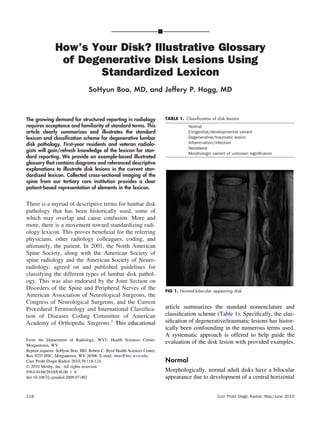
4. (Fig 1).1
The normal disk can be considered as a
360-degree arc divided into 4 quadrants (Fig 2).1
Congenital Developmental Variant
These abnormal disks have undergone morphologic
changes to adapt to abnormal growth of the spine such
as in scoliosis or spondylolisthesis (Fig 3).
There is generalized displacement of disk beyond
the endplates that constitutes a disk bulge. By conven-
tion, bulging disks involve Ͼ50% (180°) of the ring
apophyses (Fig 4).1
By this standard lexicon, a bulge
is not a hernia.
Degenerative/Traumatic
Degenerative/traumatic change in the disk is a broad
category that again is purely descriptive and identifies
and does not imply any relationship to the clinical
context, pathology, or need for treatment. Several
subcategories of degenerative/traumatic lesions are
identified in Table 2.
90°
Extrusion
25%
or
FIG 9. Extrusion type hernias.
Curr Probl Diagn Radiol, May/June 2010 121
5. Generalized
disk bulge
25%
Identify displacement
of disk.
(generalized vs.. localized)
Is
displacement > 50%
(180 degrees)
of the edge of the ring
apophyses?
YES
NO
Is displacement
< 25%?
Are the widest
edges of displaced disk,
in any plane, less than
the distance
between the edges of
the hernia base?
Localized
Hernia
Focal Broad-based
Focal
Extrusion
Focal Protrusion
Are the widest
edges of displaced
disk,
in any plane, greater
than the distance
between the edges of
the hernia base?
Broad-based
Extrusion
Broad-based
Protrusion
ONSEY
YES YESNO NO
90°
Broad-based
hernia 25%
90°
Focal
hernia
25%
Generalized
bulge
90°
FIG 10. Algorithm for analysis of disk displacement.
122 Curr Probl Diagn Radiol, May/June 2010
6. Annular Tears
Annular tears or fissures describe breaks or separation
between the dark annular fibers that extend concentri-
cally, transversely, or radially, allowing the more high
intensity nucleus to exit through (Figs 5 and 6).2,3
Intravertebral hernia is also seen in Fig 6.
Disk Hernias
By convention, a hernia is defined as localized dis-
placement of disk material, whether it be nucleus,
cartilage, annular fibers, etc, beyond the intervertebral
disk space. This interspace is defined by the vertebral
body endplates and ring apophyses, exclusive of os-
teophytes.
Localized displacement of disk can be categorized
as focal or broad-based. It is usually best appreciated
on axial imaging (Fig 7). However, the displacement
of disk may occur in any plane. Visualization in all
planes must occur to properly categorize herniations.
Furthermore, disk herniations can be defined as
protrusions vs extrusions. These are typically easily
defined on sagittal images, but again, the definition
must be applied in any plane. The base of the hernia is
the widest part in a protrusion (Fig 8), whereas the
herniated disk is wider than the base in an extrusion
(Fig 9A-C). We provide an algorithm for analysis of
disk displacement (Fig 10).
Degeneration
Degeneration is a broad description that can include
changes seen with apparent desiccation (Fig 11) or
fibrosis of disk, disk space narrowing, vacuum gas
phenomena, diffuse generalized bulging disk, endplate
sclerosis, and osteophytosis.1,2
When these changes
occur, they can be subcategorized as spondylosis
FIG 11. Degenerative change from disk desiccation.
FIG 12. Spondylosis deformans.
FIG 13. Intervertebral osteochondrosis.
Curr Probl Diagn Radiol, May/June 2010 123
7. deformans2
(Fig 12), typically seen with aging, and/or
intervertebral osteochondrosis2
(Fig 13), which is a
more pronounced pathologic change within the disk.
REFERENCES
1. Fardon DF, Milette PC. Nomenclature and classification of
lumbar disc pathology: Recommendations of the Combined
Task Forces of the North American Spine Society, American
Society of Spine Radiology, and American Society of Neu-
roradiology. Spine 2001;26:E93-E113.
2. Grossman RI, Yousem DM. Neuroradiology: The Requisites,
2nd edition. St Louis, MO: Mosby, 2003.
3. Harnsberger HR, et al. Diagnostic and Surgical Imaging
Anatomy: Brain, Head and Neck, Spine. Salt Lake City, UT:
Amirsys, 2006.
124 Curr Probl Diagn Radiol, May/June 2010