SlideShare a Scribd company logo
1 of 13
Download to read offline
Plating of the Distal Radius
Arvind D. Nana, MD, Atul Joshi, MD, and David M. Lichtman, MD
Abstract
Distal radius fractures constitute up
to 15% of all extremity fractures.1
Patients include both active elderly
individuals and younger persons in-
volved in high-energy trauma. Con-
sequently, restoration of wrist func-
tion to close to preinjury levels is of
concern to patients in both groups.
Anatomic restoration of the distal
radius and ulna is of major impor-
tance in achieving normal wrist func-
tion; open reduction has become in-
creasingly useful in reaching this goal.
The advantages of plating include ac-
curate restoration of bony anatomy,
stable internal fixation, a decreased
period of immobilization, and early
return of wrist function. Recent ad-
vances in plate design can help re-
duce or eliminate additional steps to
augment stability, such as bone void
filler, supplemental pins, and exter-
nal fixation. Athorough understand-
ing of plating alternatives is necessary
to tailor treatment methods appropri-
ately.
Anatomy
Distal Radius
The distal radius consists of three
independent articular surfaces—the
scaphoid facet, lunate facet, and sig-
moid notch (Fig. 1). The scaphoid fac-
et is part of the lateral distal radius,
which includes the radial styloid. The
medial aspect of the distal radius con-
sists of the lunate facet and sigmoid
notch.Thesigmoidnotchisnearlyper-
pendicular to the lunate facet and ar-
ticulates with the distal ulna to form
the distal radioulnar joint (DRUJ). The
close proximity of the sigmoid notch
to the lunate facet implies that any in-
jury to the medial aspect of the distal
radius,whetherintra-articularorextra-
articular, involves the DRUJ.
The lunate facet and its strong lig-
amentous attachments to the proxi-
mal carpal row and the ulnar styloid
form the medial complex of the dis-
tal radius. The carpus is nearly always
displaced with the volar and/or dor-
sal medial fragment of the distal ra-
diusbecauseoftheexceptionallystrong
ligaments of the medial complex.2
Distal Ulna and Distal
Radioulnar Joint
The head of the ulna articulates
with the sigmoid notch of the distal
radius and abuts the triangular fibro-
cartilage complex (TFCC) and the ul-
nar carpus. Because the base of the
ulna styloid (fovea) is the insertion
point for much of the TFCC and im-
portant ulnocarpal ligaments, its in-
tegrity is critical to the stability of the
DRUJ.3
Dr. Nana is Staff Physician, Department of Or-
thopaedic Surgery, JPS Health Network, Fort
Worth, TX. Dr. Joshi is Resident, Department of
Orthopaedic Surgery, JPS Health Network. Dr.
Lichtman is Chairman/Director, Department of
Orthopaedic Surgery, JPS Health Network.
None of the following authors or the departments
with which they are affiliated has received anything
of value from or owns stock in a commercial com-
pany or institution related directly or indirectly
to the subject of this article: Dr. Nana, Dr. Joshi,
and Dr. Lichtman.
Reprint requests: Dr. Nana, JPS Health Network,
1500 South Main Street, Fort Worth, TX 76104.
Copyright 2005 by the American Academy of
Orthopaedic Surgeons.
Distal radius fractures are common injuries that can be treated by a variety of meth-
ods. Restoration of the distal radius anatomy within established guidelines yields
the best short- and long-term results. Guidelines for acceptable reduction are (1)
radial shortening <5 mm, (2) radial inclination >15°, (3) sagittal tilt on lateral pro-
jection between 15° dorsal tilt and 20° volar tilt, (4) intra-articular step-off <2 mm
of the radiocarpal joint, and (5) articular incongruity <2 mm of the sigmoid notch
of the distal radius. Treatment options range from closed reduction and immobili-
zation to open reduction with plates and screws; options are differentiated based on
their ability to reinforce and stabilize the three columns of the distal radius and ulna.
Plating allows direct restoration of the anatomy, stable internal fixation, a decreased
period of immobilization, and early return of wrist function. Buttress plates reduce
and stabilize vertical shear intra-articular fractures through an antiglide effect, where-
as conventional and locking plates address metaphyseal comminution and/or pre-
serve articular congruity/reduction. With conventional and locking plates, intra-
articular fractures are directly reduced; with buttress plates, the plate itself helps
reduce the intra-articular fracture. Complications associated with plating include
tendon irritation or rupture and the need for plate removal.
J Am Acad Orthop Surg 2005;13:159-171
Vol 13, No 3, May/June 2005 159
Columnar Classification of the
Distal Radius and Ulna
Conceptually, the distal radius and
ulna may be divided into three col-
umns based on the anatomy (Fig. 1,
inset);thiscolumnarclassificationthen
can be used to guide treatment plans.
The distal radius is divided into the
lateral and medial columns, which an-
atomically correlate with the scaphoid
facetandlunatefacet,respectively.The
medial column of the distal radius is
further subdivided into dorsal medi-
al and volar medial columns. The lat-
eral, dorsal medial, and volar medi-
al columns correspond with Melone’s2
system for classifying intra-articular
distal radius fractures. The ulnar col-
umn represents the ulnar styloid and
the TFCC.
Pathophysiology
In most activities of daily living, the
dorsum of the distal radius is subject
to tensile forces, whereas the volar
surface is subject to compression. This
is reflected in the bony architecture
of the distal radius, with its strong
volar buttressing cortex and thinner
cancellous dorsal surface. When the
wrist is subjected to a nonphysiolog-
ic load, as in a dorsally directed com-
pression force (eg, a fall on the out-
stretched hand), predictable fracture
patterns result.
Melone2 aptly described the four
major components of intra-articular
distal radius fractures as radial sty-
loid, dorsal medial, volar medial, and
shaft fragments (Fig. 1). The transverse
coronal split between the dorsal and
volar medial distal radius fragments
can be difficult to assess on plain ra-
diographs.4 In many ways, the com-
binations of displaced Melone com-
ponentsreflectvariantsoffracturetypes
andclassificationdescribedinthepast.
For example, isolated radial styloid
displacement represents a chauffeur’s
fracture. Volar medial displacement
describes Barton’s fracture; displace-
ment of the dorsal medial fragment
represents the common reversed Bar-
ton’s or dorsal compression fracture.
Displacementofallarticularfragments
as a unit is comparable to the classic
extra-articular Colles fracture (dorsal)
or to Smith’s fracture (volar).
Associated Injuries
Associated injuries must be consid-
ered in any comprehensive treatment
plan for patients with distal radius and
ulna fractures. Up to 68% of distal ra-
dius fractures are associated with soft-
tissue injuries, such as partial or com-
plete tears of the TFCC, scapholunate
ligament, and/or lunotriquetral lig-
ament.5,6 Fractures involving the lu-
nate facet (the medial aspect of the dis-
tal radius) and fractures of the radial
styloid cause the most intracarpal in-
juries because of the strong ligamen-
tous interconnections between the dis-
tal radius and carpus.5 Intracarpal
ligament disruption does not have to
be repaired unless gross instability is
noted on postreduction radiographs
(eg, scapholunate diastasis). The 4- to
6-week immobilization necessary for
distal radius healing is usually suf-
ficient for ligament healing in nondis-
placed injuries.
Radiographic and clinical exami-
nations of the injured wrist after sur-
gical reduction are helpful in evalu-
ating the integrity of the DRUJ and
TFCC, both components of the ulnar
column. TFCC tears are also common,
particularly when the medial column
of the distal radius and/or the ulnar
styloid is intact. A fracture of the ul-
nar styloid base and significant dis-
placement (>2 mm) of an ulnar sty-
loid fracture increase the risk of DRUJ
instability.7 Treatment of the ulnar col-
umn is focused on restoring DRUJ
stability by closed, percutaneous, or
open treatment of the ulnar styloid
and the TFCC.
Imaging Studies
Posteroanterior (PA) and lateral ra-
diographs are necessary for every pa-
tient with a distal radius injury. The
lateral view of the wrist places the an-
terior surface of pisiform between the
volar surface of the scaphoid (the
scaphoid tuberosity) and the anteri-
or surface of the capitate.8
Treatment options for distal radi-
us fractures are based on the initial
injury as well as on radiographs made
after closed reduction. If the initial
injury position is within acceptable
guidelines9 (Table 1) for the patient’s
functional requirements, any loss of
reduction is usually insignificant as
Figure 1 Distal radius anatomy. The fracture line between the volar medial and dorsal me-
dial columns extends into the sigmoid notch and thus must also be evaluated on postreduc-
tion radiographs. (Reproduced with permission from Trumble TE, Culp RW, Hanel DP, Geissler
WB, Berger RA: Intra-articular fractures of the distal aspect of the radius. Instr Course Lect
1999;48:465-480.) Inset, Columnar classification of distal radius and ulna.
Plating of the Distal Radius
160 Journal of the American Academy of Orthopaedic Surgeons
long as the fracture is adequately im-
mobilized and protected. Converse-
ly, unacceptable initial displacement
often means that loss of reduction af-
ter nonsurgical treatment may require
surgical intervention.
The initial postinjury lateral view
of the distal radius will demonstrate
either the more common dorsal dis-
placement or the less frequent volar
displacement of the distal radius col-
umns. The direction of initial displace-
ment on the lateral radiograph gen-
erally correlates with the side of both
greatest cortical comminution and re-
quired initial treatment. For example,
if the lateral view shows dorsal dis-
placement of the lunate facet, then the
dorsal surface will have the greatest
cortical fragmentation, and reduction
will initially be focused dorsally.
Postreduction or traction radio-
graphs also are useful in assessing
whether the distal radius fracture is
intra-articular or extra-articular. The
extent of cortical comminution is
more easily visualized on postre-
duction radiographs than on initial
postinjury radiographs.11 All plain ra-
diographs of distal radius fractures
should include evaluation of radial
inclination, radial length, ulnar vari-
ance, radial tilt,9 articular step-off or
gap, sigmoid notch step-off or gap,
DRUJ subluxation/dislocation, and
the presence and extent of displace-
ment of ulnar styloid fractures (Fig.
2). Because of individual variability,
appropriate radiographs of the unin-
jured contralateral wrist are recom-
mended to evaluate the patient’s nor-
mal anatomy and corresponding
radiologic measurements.
Plain radiographs may be insuffi-
cient to assess the extent of intra-
articular and extra-articular fragmen-
tation and injury to the DRUJ. Axial
computed tomography (CT) scans
with sagittal and coronal plane recon-
structions can aid in visualizing die-
punch fractures, volar rim fractures,
and scaphoid facet fractures.
Treatment of Distal Radius
Fractures
All patients with distal radius frac-
tures warrant a trial of closed reduc-
tion and plaster immobilization; ex-
Table 1
Guidelines for Acceptable Reduction of Distal Radius Fractures*
1. Radial shortening <5 mm at the DRUJ compared with the contralateral
side
2. Radial inclination on posteroanterior radiographs >15°
3. Sagittal tilt on the lateral projection between 15° dorsal tilt and 20° volar
tilt
4. Intra-articular step-off or gap <2 mm of the radiocarpal joint10
5. Articular incongruity <2 mm of the sigmoid notch of the distal radius
*These guidelines must be individualized to accommodate each patient’s functional
activity levels and general medical status.
Adapted with permission from Graham TJ: Surgical correction of malunited fractures
of the distal radius. J Am Acad Orthop Surg 1997;5:270-281.
Figure 2 The various angles to assess in distal radius fractures. A, Radial inclination (RI;
normal, 22°). B, Radial length (RL; normal, 12 mm). C, Ulnar variance (UV; normal, 0 to −2
mm). D, Radial tilt (RT; normal, 11° volar). (Reproduced with permission from Graham TJ:
Surgical correction of malunited fractures of the distal radius. J Am Acad Orthop Surg 1997;
5:270-281.)
Arvind D. Nana, MD, et al
Vol 13, No 3, May/June 2005 161
ceptions include those with open
injuries, those who cannot tolerate im-
mobilization, and those with volar or
dorsalverticalshearfractures(Barton’s
fracture). Multiple trauma is also an
indication for early surgical interven-
tion. If the fracture can be reduced to
within acceptable guidelines (Table 1)
and maintained in satisfactory align-
ment by plaster sugar-tong splint im-
mobilization, it is considered to be sta-
ble.Anunstablefractureischaracterized
by inadequate reduction of a displaced
fracture fragment or by the inability
to maintain reduction after closed frac-
turemanipulation.Dependingonfunc-
tional expectations, surgical treatment
may be considered for unstable frac-
tures involving the medial column of
the distal radius (Fig. 3).
Determining short-term stability
after closed reduction is difficult and
is based largely on personal experi-
ence in the treatment of distal radius
fractures. If the surgeon thinks that
a fracture is unstable and may present
an unacceptable risk to the patient,
then early open treatment with fix-
ation should be considered. Success-
ful closed treatment requires attention
to detail by the physician as well as
complete patient compliance with
postreduction protocols.
Normal wrist and forearm motion
subjects the dorsal metaphysis of the
distal radius to both tensile and com-
pressive forces, but the volar surface
transmits greater compressive forc-
es.8 Restoration of these biomechan-
ical relationships is necessary to es-
tablish a stable reduction of the distal
radius fracture. The first step toward
a stable reduction is to create a sta-
ble volar buttress. In distal radius
fractures with dorsal comminution,
volar integrity is reestablished by ac-
curate cortical apposition of the large
volar metaphyseal fragments. With
comminuted volar metaphyseal frag-
ments or inherently unstable volar
vertical shear fractures (volar Barton
fractures), stable fracture reduction
can be achieved by placement of a
volar buttress plate. Once volar sta-
bilityisrestored,thedorsalmetaphys-
eal fragments can be reduced against
the stable volar buttress using exter-
nal fixation, percutaneous pins, and/
or bone void filler (bone graft).8
Autograft, allograft, or other bone
graft substitutes may be used to fill
the metaphyseal defect created after
reduction of distal radius fractures.
Comminuted metaphyseal fragments
are characterized by more compressed
cancellous bone and a greater void af-
ter reduction. Conversely, large meta-
physeal fragments are associated with
less compressed cancellous bone and
thus a lower requirement for bone
grafting. A bone void filler has sev-
eral advantages, including mechan-
ical support,12 providing osteoconduc-
tive material for the bone defect, faster
bone healing, and decreased incidence
of loss of reduction.12-15 Hydroxyapa-
tite cement without additional fixa-
tion is inadequate for the treatment
of distal radius fractures;16 it should
be combined with external fixation to
supplement any bone void filler.
Volar buttress plate
Medial column fracture
of the distal radius
Large dorsal
metaphyseal
fragments
Unstable dorsal
metaphyseal
fragments
Stable dorsal metaphyseal
fragments or no dorsal
fracture
Small dorsal
metaphyseal
fragments
Percutaneous
pins and splint
immobilization
Splint
immobi-
lization
Stable volar metaphyseal
fragments after closed
reduction* or no volar fracture
Unstable volar metaphyseal
fragments after closed
reduction
Conventional volar
plate with dorsal
percutaneous pins
Volar plate with
distal locking
screws/pegs
Small dorsal
metaphyseal
fragments
Large dorsal
metaphyseal
fragments
Unstable dorsal
metaphyseal
fragments
Stable dorsal
metaphyseal
fragments
Volar plate
with distal
locking
screws/pegs
Dorsal plate
with distal
locking
screws/pegs
Conventional volar
plate with bone
void filler and
external fixation
Dorsal
buttress
plate
Dorsal bone
void filler and
neutralization
external fixation
Figure 3 Plating recommendations for fractures of the medial column of the distal radius. Treatment choice depends on surgeon pref-
erence and experience. * = accurate cortical apposition of the volar metaphyseal fragments.
Plating of the Distal Radius
162 Journal of the American Academy of Orthopaedic Surgeons
Restoration of volar stability has
important radiocarpal implications
because the stronger and more impor-
tant radiocarpal ligaments are at-
tached to the volar surface. Volar in-
tegrity therefore is critical because it
facilitates adequate reduction of dor-
sal metaphyseal fragments against a
stable volar buttress and because it
prevents possible volar radiocarpal
instability.8
Plate Fixation
Plate fixation is primarily indicat-
ed for unstable fractures of the volar
medial column of the distal radius;
it is also helpful for combined volar
and dorsal medial column injuries.
Although some authors recommend
dorsal plates for isolated dorsal me-
dial column injuries of the distal ra-
dius, other methods of treatment are
available for these injuries. Volar
plates with locking screws or pegs
may be effective for extensive dorsal
comminution.
Distal radius plates are categorized
by location of use and type of plate.
Plates categorized by location may be
used on the dorsal medial, volar me-
dial, and radial styloid aspects of the
distal radius. Certain plate designs
address two areas simultaneously (ie,
dorsal medial distal radius and radi-
al styloid, or volar medial distal ra-
dius and radial styloid). Smaller im-
plants are also available to stabilize
each area separately.
The two principal types of distal
radius plates are buttress plates and
plates that can span metaphyseal
comminution and/or maintain artic-
ular congruity and reduction. Only
the buttress plate, with its antiglide
effect,reducesintra-articularfractures
(eg, volar Smith or Barton fractures
[Fig. 4] or dorsal Barton fracture). All
other distal radius plates require di-
rect reduction of intra-articular frac-
tures; the plating serves to maintain
the alignment.
Plates that maintain alignment can
be further subdivided into two types:
conventional and locking. In conven-
tional plate design, stability of the
construct is ultimately achieved
through apposition of the bone and
plate by screw purchase in the bone.
Conventional plates can be used dor-
sally or volarly. The use of dual small
conventional plates on the dorsal dis-
tal radius also has been described.17
When cortical or cancellous screws
are used in the distal holes of the but-
tress plate, they are most effective if
the opposite cortex is not comminut-
ed. If the opposite cortex is commi-
nuted, the surface could collapse be-
cause of both axial forces across the
wrist joint and toggle of the screws
in the plate, which may result in loos-
ening of the distal screws.18
Plates with locking distal screws
or pegs support the subchondral bone
and resist forces across the articula-
tion that may displace the articular
fragments (Figs. 5, 6, and 7). Locking
screws or pegs offer numerous advan-
tagesoverregularscrewsbecausetheir
stability is achieved through plate de-
sign. The screws or pegs become fixed-
angle devices via threaded heads that
engage the threaded distal screw holes
of the plate for stability. New plate de-
signs allow locking screw insertion in
the most distal screw holes as well as
in the proximal screw holes. Of his-
torical note, the dorsal plate used by
Gesensway et al19 employed a blade
plate construct with multiple tines to
achieve fixed-angle fixation.
Locking screws or pegs support
the subchondral bone without rely-
ing on the purchase of the screws or
pegs in bone, and they are indepen-
dent of opposite cortex comminution.
Accordingly, no bone void filler is re-
quired to prevent collapse at the site
of comminution. The distal screws or
pegs—whethercortical,cancellous,or
fixed-angle—serve to maintain reduc-
tion of articular fragments, but they
do not directly reduce the articular
fracture.
Distal Radius Plating
Plating offers direct restoration of
the distal radius through stable fix-
ation. The stability of the construct en-
sures more predictable healing of the
fracture and thus shortens the peri-
od of immobilization of the wrist. The
principal advantage of distal radius
platingistheearlyreturnofwristfunc-
tion,animportantcriterionforpatients
who expect wrist function after inju-
rytoreturntopreinjurylevels.Conven-
tional plates can be used for buttress
and/or neutralization support of a
distal radius fracture. Plates with lock-
ing screws or pegs do not rely on an-
atomic contour of the plate to obtain
stability. Stabilization of the opposite
cortex through locking screws or pegs
significantly decreases or eliminates
the need for further stabilization of
the opposite cortex fragmentation.
Figure 4 The antiglide effect of a buttress
plate helps reduce a volar vertical shear frac-
ture (volar Barton fracture) of the distal ra-
dius. The most proximal screw is placed first;
the penultimate distal screw (A) reduces the
fracture (arrows) (B) before placement of the
final distal screw (C). (Reproduced with per-
mission from Jupiter JB: Complex articular
fractures of the distal radius: Classification
and management. J Am Acad Orthop Surg 1997;
5:119-129.)
Arvind D. Nana, MD, et al
Vol 13, No 3, May/June 2005 163
Surgical Treatment
Extra-articular Distal Radius
Fractures
These injuries are transverse or
short oblique fractures proximal to the
DRUJ and are usually amenable to
closed treatment methods with sup-
plementary percutaneous pin stabi-
lization. For an unstable distal shaft
fracture with minimal or no commi-
nution that is either dorsally or vo-
larly displaced, a volar plate is suf-
ficient to achieve stable fixation. A
locking volar plate does not offer any
specific advantage over convention-
al volar plating systems for this type
of fracture. However, for an extra-
articular fracture with dorsal or volar
comminution, a locking volar plate
may offer stability; it also restores
length without the mobilization of
extensor tendons that is required for
dorsal plating. A dorsal plate for an
extra-articular fracture with dorsal
comminution is considered excessive
treatmentwithoutsufficientbenefitbe-
cause these fractures can be routine-
ly pinned and stabilized in a closed
fashion. Bone void filler and external
fixation may be used for severely com-
minuted extra-articular fractures.
Intra-articular Distal Radius
Fractures
The concept of four-part displace-
ment is useful in conceptualizing a
treatment algorithm, especially with
regard to the indications for distal ra-
dius plating. When evaluating a pa-
tient with a distal radius fracture,
three distinct areas (columns) are as-
sessed (Fig. 1, inset). The medial col-
umn of the distal radius represents
Melone’s dorsal and volar die-punch
radius fragments; plates and/or bone
void filler are particularly applicable
for restoration of this column. The ra-
dial styloid component—the lateral
distal radius column—is then re-
viewed to determine whether local-
ized treatment is sufficient (eg, per-
cutaneous pins or screws for the
displaced radial styloid) or whether
treatment should be combined with
fixation of the medial column of the
distal radius (eg, a volar or dorsal
plate that also fixes the styloid). Fi-
nally, the ulnar column, representing
the TFCC and the ulnar styloid com-
ponents, is addressed.
Intra-articular Fractures of the
Lateral Column
The radial styloid fracture and its
corresponding scaphoid facet are
usually displaced proximally by ax-
ial compression forces. Often the frag-
ment is rotated in pronation, but this
is not easily appreciated on routine
radiographs. CT scan and dynamic
fluoroscopy can help delineate this
Figure 5 A, With conventional screws, stability is achieved through friction between the
plate undersurface and the underlying bone (arrows) resulting from screw compression at
the interface. This stability is augmented by bicortical screw purchase, which also decreases
toggle between the screw and plate hole (represented by the rectangles). Friction at the in-
terface must be higher than the axial forces to ensure absolute stability. Thus, anatomic con-
touring of the conventional screw/plate system is needed to obtain a stable construct. B, In
a locking screw system, the threaded screw head locks in the threaded screw hole of the plate
to attain stability. Consequently, axial forces in the bone are transmitted to the plate rather
than the screw, and no screw toggle can occur (arrows). Because stability with a locking screw
does not require compression between the bone and the plate (rectangles), the periosteal blood
supply under the plate is preserved. (Adapted with permission from Appenzeller A, Chris-
tensen R, Frenk A, Gilbert S, Schavan R: The development of the distal femur LISS. Injury
2001;32[suppl 3]:5-25.)
Figure 6 In the distal radius, the principal advantage of a plate with locking screws is sta-
bility of the construct, even in the presence of segmental bone defect or comminution of cor-
tex opposite the plate. Because the screws lock into the plate, the axial forces are transmitted
to the plate. This diagram shows locking screws along the entire plate. F = axial force in the
direction of the arrow. (Adapted courtesy of Synthes [USA], Paoli, PA.)
Plating of the Distal Radius
164 Journal of the American Academy of Orthopaedic Surgeons
rotational deformity. Isolated radial
styloid fractures should raise con-
cerns about concomitant intracarpal
injury. Adisplaced radial styloid usu-
ally can be reduced by closed means
(ligamentotaxis by wrist flexion and
ulnar deviation) and stabilized with
two percutaneous pins. Reduction
and pin placement are assessed intra-
operatively with fluoroscopy or, if
open reduction is used, under direct
visualization (Figs. 8 and 9). Occa-
sionally the radial styloid fragment
must be manipulated into position,
using the pins as joysticks, before se-
curing the position by advancing the
pins into the opposite cortex. The di-
rection of the styloid pins is from ra-
Figure 7 Anteroposterior (A) and lateral (B) radiographs of a distal radius injury. The fracture, which demonstrates volar and dorsal in-
stability, is fixed with a volar locking plate (C and D). The dorsal instability is best seen in the postoperative lateral view (D), which shows
a dorsal gap just proximal to the locking pegs. The locking pegs stabilize both the dorsal and volar articular fragments. In this patient, bone
void filler was not used in the dorsal gap, and the distal ulna was not surgically stabilized. (Courtesy of Arvind D. Nana, MD, Fort Worth,
TX.)
Figure 8 Anteroposterior (A) and lateral (B) radiographs of a wrist at initial presentation. Anteroposterior (C) and lateral (D) radiographs
of the wrist in longitudinal traction. The patient is immobilized in a plaster sugar-tong splint. (Courtesy of David M. Lichtman, MD, Fort
Worth, TX.)
Arvind D. Nana, MD, et al
Vol 13, No 3, May/June 2005 165
dial volar to ulnar dorsal. Other op-
tions for styloid fixation include
percutaneous lag screw fixation or a
small buttress plate and pins.20
Plate fixation of the radial styloid
is usually done in combination with
stabilization of medial column inju-
ries of the distal radius. Volar or dor-
sal distal radius plates include radial
styloid stabilization in their construct
and therefore do not need separate ra-
dial styloid plating or pinning. Cer-
tain smaller implant designs are avail-
able for separate plating of radial
styloid fractures, but this is almost al-
ways in combination with medial col-
umn stabilization (whether dorsal or
volar). Separate lateral and medial
column plating of the distal radius is
most mechanically stable when the
plates are angled 50° to 90° to each
other.11,17,20,21 Separate radial styloid
plates are placed on the dorsal radial
or most radial aspect of the distal ra-
dius; this placement requires a low-
profile design to avoid irritation of the
brachioradialis tendon or dorsal ex-
tensor tendons.
Intra-articular Fractures of the
Medial Column
Unstable volar fractures are ame-
nable to volar plate stabilization. If the
fracture is isolated (ie, there is no dor-
sal fragment instability), then a sim-
ple buttress plate may be used to re-
constitute the volar cortex. If the volar
metaphysis is comminuted, bone
void filler may be used to fill in the
gap and also to support the distal ar-
ticular surface. The integrity of the
joint should not be violated by dis-
secting the volar radiocarpal liga-
ments. When necessary, the volar cor-
tex can be hinged open like a book
to inspect and realign volar intra-
articular injuries.
When dorsal fragment instability
also is present, several options are
available. If a volar buttress plate has
been applied, then the dorsal frag-
ment must be stabilized from a dor-
sal approach. If the dorsal fragment
is large, it can be fixed by one or (pref-
erably) two percutaneous cross pins
entering the dorsal medial fragment
distally and aimed proximally and ra-
dially (Figs. 8 and 9). As with the ra-
dial styloid fragment, percutaneous
pins can be used as joysticks to ma-
nipulate the dorsal fragment into po-
sition. The pins also may be placed
into the fracture line (intrafocal Ka-
pandji pinning [Fig. 10]), starting
more perpendicular to the dorsal cor-
tex, and then used to reduce the dor-
sal fragment by moving the pins more
nearly parallel to the dorsal cortex,
with subsequent fixation into the
volar cortex.
If the dorsal medial fragment is
comminuted, reducing and holding
the area with pin fixation may be dif-
ficult. The fracture can be opened dor-
sally, usually through a longitudinal
approach between the third and
fourth dorsal extensor compartments.
An approach ulnar to the fourth com-
partment also may be used if the com-
minution is limited to the dorsal ul-
nar corner and semilunar notch. The
floor of the third extensor compart-
ment is incised longitudinally; then
the fourth extensor compartment
(sheath and tendons) is sharply ele-
Figure 9 Same patient as in Figure 8. Anteroposterior (A) and lateral (B) radiographs made 1 week after injury. The laterally displaced
radial styloid fragment and volarly displaced volar medial fragment (arrows) are evident. Instability of these fragments is easily demon-
strated by their inability to maintain initial closed reduction. Anteroposterior (C) and lateral (D) postoperative radiographs show that the
volar medial fragment is stabilized with a volar plate. The radial styloid is fixed with two percutaneous pins; the dorsal cortex with its large
dorsal fragments is stabilized with a dorsal percutaneous pin. The ulnar styloid also was stabilized because of the fracture through its base,
marked displacement (>2 mm), and clinical instability of the DRUJ after stabilization of the distal radius. (Courtesy of David M. Lichtman,
MD, Fort Worth, TX.)
Plating of the Distal Radius
166 Journal of the American Academy of Orthopaedic Surgeons
vated off the dorsal distal radius.
Bone grafting of the dorsal metaphy-
sis is now possible. If the joint needs
to be viewed, the dorsal capsule can
be incised transversely. Reduction un-
der direct visualization usually can be
done, and bone void filler is often
used to stabilize and buttress the dor-
sal cortex. If necessary, a transverse
capsular incision may be used to ex-
plore the radiocarpal joint and reduce
intra-articular or intracarpal injuries.
Some surgeons use low-profile dor-
sal plates at this point to maintain ar-
ticular reduction and stability; others
apply an external fixator for 3 to 4
weeks to avoid use of a dorsal plate.
An alternative for treating both
dorsal and volar instability is to use
a plate with locking screws or pegs
to transfix both volar and dorsal cor-
tices of the medial column of the dis-
tal radius (Fig. 7). Satisfactory results
have also been achieved using lock-
ing screw–plate combinations from
the dorsal side.22-24
Although dorsal plates do permit
adequate healing of the fracture, their
secondary effects—particularly on
surrounding soft-tissue structures—
render them a less appealing option.
Disadvantages of dorsal plates in-
clude the need for mobilization of ex-
tensor tendons to achieve proper
plate placement, possible tendon ir-
ritation or rupture because of a prom-
inent plate or screws, and the possi-
bility of additional surgery to remove
the symptomatic dorsal plate.
In isolated unstable fractures of the
dorsal medial column, the decision to
treat with pin fixation or with open
bone grafting plus neutralization ex-
ternal fixation depends on the size of
the fragments. Treatment is the same
as when combined with volar insta-
bility: percutaneous pins for large
fragments or open reduction, bone
void filler, and external fixation for
comminuted fractures (small frag-
ments). Once again, low-profile dor-
sal plates can be applied instead of
an external fixator, but locking screws
or pegs are not necessary in this in-
stance.
A lateral column injury of the dis-
tal radius is usually treated concom-
itantly or after stabilization of the me-
dial column. As noted, if the fracture
pattern permits, the styloid can be
fixed with either a volar or dorsal
plate. In most instances, the radial sty-
loid can be fixed separately using two
percutaneous pins, as described for
isolatedradialstyloidfractures.Aper-
cutaneous lag screw or a small dorsal
radial buttress plate are also options.
Surgical Approach
Volar Radial Approach
The volar radial (Henry) approach
is utilitarian; it can expose the entire
volar radial surface up to the DRUJ
(Fig. 11, A). This exposure uses the in-
terval between the flexor carpi radi-
alis (FCR) and the radial artery or
goes through the floor of the FCR ten-
don sheath. The pronator quadratus
muscle is elevated in a subperiosteal
fashion to visualize the volar distal
radius.25 Orbay26 and Orbay and
Fernandez27 also describe the release
of the first extensor compartment and
the insertion of the brachioradialis off
the lateral distal radius to permit ac-
cess to the dorsal surface and to fa-
cilitate the reduction of the lateral col-
umn of the distal radius (radial
styloid). Release of the volar capsule
or the ligamentous attachments on
the volar rim may lead to volar ra-
diocarpal instability and should be
avoided.
Carpal tunnel release through the
distal extension of this approach is not
recommended because of potential
injury to the volar cutaneous branch
of the median nerve. If carpal tunnel
release is indicated, a separate stan-
dard or a mini carpal tunnel incision
is recommended. Prophylactic carpal
tunnel release is not routinely per-
formed21,28,29 unless acute carpal tun-
nel symptoms are present or unless
the distal fragment is significantly
dorsally displaced for a prolonged
period of time.
Volar Ulnar Approach
The volar ulnar approach offers
limited exposure to the radial column
and is indicated for injuries to the vo-
lar aspect of the medial column and
the DRUJ (Fig. 11, B). The interval be-
tween the flexor carpi ulnaris and the
Figure 10 With intrafocal Kapandji pinning, the pin is placed through the fracture site and
maneuvered to elevate the fragment. Once adequate reduction is achieved, the pin is then
driven through the opposite cortex to achieve stability. This technique can be used to restore
radial inclination (A) or volar tilt (B). (Adapted with permission from Palmer AK: Fractures
of the distal radius, in Green DP [ed]: Operative Hand Surgery. New York, NY: Churchill Liv-
ingstone, 1993, pp 929-971.)
Arvind D. Nana, MD, et al
Vol 13, No 3, May/June 2005 167
finger flexor tendons is used for this
approach. While retracting the flexor
tendons radially and protecting the
ulnar nerve and artery, the pronator
quadratus muscle is mobilized off the
distal ulna. This exposure has the ad-
vantage of permitting distal extension
to complete a carpal tunnel release.
Postoperative Management
After surgery, immobilization is de-
sirable to facilitate soft-tissue heal-
ing and resolution of swelling. Plas-
ter splinting is preferable to the use
of circumferential or bivalved casts
because use of casts may lead to po-
tential complications such as finger
edema and compartment syndrome.
Postsurgical follow-up includes
suture removal at 10 to 14 days and
serial radiographs for the first 2
weeks if deemed necessary. Arm el-
evation and finger range of motion
are important components of early
rehabilitation. Conversion to a cir-
cumferential cast may be considered
at 2 weeks; however, complete wrist
immobilization beyond the fourth
postoperative week usually is not
desirable because it negates the ad-
vantage of early restoration of wrist
stability with plating. During the pe-
riod of immobilization, therapy is
focused on finger motion; once im-
mobilization is discontinued, ther-
apy expands to include wrist motion
(flexion, extension, ulnar deviation,
and radial deviation) and forearm
rotation (pronation, supination). A
removable Velcro wrist splint then
can be provided for support and
comfort while the patient engages in
gentle activities of daily living and
active range-of-motion exercises.
With plating of extra-articular frac-
tures, or for fractures with well-fixed
intra-articular fragments, immobili-
zation can be discontinued as early
as 2 weeks after surgery as long as the
incision is sufficiently healed and ede-
ma has decreased to allow wrist mo-
tion. For more complex intra-articular
fractures treated with plating, immo-
bilization for at least 4 weeks is nec-
essary to permit healing of the intra-
articular fragments and to allow wrist
motion without risk of displacement
of the intra-articular fragments. For
6 to 8 weeks, splints can be worn at
night, and only light physical thera-
py is recommended. Strenuous push-
ing, pulling, twisting, or lifting should
be avoided for the first 3 months to
facilitate optimal bone healing.
Figure 11 A, The volar radial approach uses the interval between the flexor carpi radialis tendon and the radial artery. The pronator quadra-
tus is elevated sharply, starting at its insertion on the distal radius. B, Top detail, the volar ulnar approach can be extended distally to release
the median nerve from the carpal tunnel. Bottom detail, at the level of the distal radius, the flexor tendons and the median nerve are re-
tracted radially to expose the volar medial distal radius. (Adapted with permission from Fernandez DL, Jupiter JB: Fractures of the Distal
Radius. New York, NY: Springer, 1996, pp 67-102.)
Plating of the Distal Radius
168 Journal of the American Academy of Orthopaedic Surgeons
Complications
The most common complication of
plate fixation in the distal radius is
tendon injury; 25% of patients eval-
uated reported everything from ten-
don irritation to frank rupture.17,24,30
With the volar plate, tendon injuries
have included rupture of the flexor
pollicis longus,22,31,32 FCR tenosyno-
vitis,28 and dorsal extensor tendon in-
volvement as a result of protruding
screws.28,33 Dao et al34 reported radi-
al artery pseudoaneurysm secondary
to volar distal radius plate design.
Median nerve dysfunction (ie, pares-
thesia, carpal tunnel syndrome, or re-
flex sympathetic dystrophy) is asso-
ciated with volar plating, but routine
prophylactic carpal tunnel release in
this scenario is not considered stan-
dard treatment.21,28,29 Adverse effects
are more common with dorsal plat-
ing than with volar plating.
As with dorsal plates, tendon-
related complications requiring plate
removal also occur after volar plat-
ing; in one study, 13 of 73 patients
(18%) required plate removal after
volar plating.32 Tendon irritation is
thought to be caused by prominent
or sharp plate edges,17,23,30 prominent
or loose screws,35 and cellular reac-
tion to the titanium metal of the
plates.34,36 Extensor tendon rupture
can be caused by screws that are loose
or backing out,30,33,37 by sharp edges
created by cutting of the plate,17 and
by prominent design of the distal as-
pect of the dorsal plate.36,38,39 Many
authors have reported a high inci-
dence (up to 30%) of dorsal plate re-
moval secondary to tendon inflam-
mation or rupture.17,23,30,34-36 To
prevent tendon injury, some recom-
mend that a portion of the extensor
retinaculum be interposed between
the plate and the tendon or tendon
sheath,21,30,40 or that the dorsal plates
be routinely removed.23,37,40 Tendon
rupture has been reported as early as
8 weeks and as late as 7 months af-
ter surgery.30,38 Plate breakage is an-
other reason for plate removal.37
Loss of reduction also can be a
problem and is seen more frequently
with the use of conventional plates
when the opposite cortex is not fur-
ther augmented with pins, bone void
filler, or external fixation.24,34,41,42
Outcomes
Functional outcomes of distal radius
fractures depend on multiple factors,
including radial shortening, radial in-
clination, sagittal tilt, intra-articular
step-off or gap, and articular congru-
ity of the sigmoid notch (Table 1). Al-
though intra-articular step-off >2 mm
has been shown to lead to radio-
graphic osteoarthritis, this does not
necessarily correlate with poor func-
tional outcome.10 Radial shortening
>5 mm, however, may have a marked
impact on outcome because it can af-
fect both the DRUJ and the radiocar-
pal joint.9,10,43
Outcome studies for each type of
dorsal plate design (conventional,
dual, and locking) have been per-
formed, but the average follow-up is
only 12 to 19 months for population
groups ranging from 21 to 73 patients.
Two studies of conventional plate de-
sign demonstrated 72% to 95% good-
to-excellent functional results, with a
reported complication rate of 20% to
23%. The largest study evaluated dual
dorsal plating in the treatment of dis-
tal radius fractures; however, even
with 97% good-to-excellent results,
the incidence of complications was
21%.17,24,30
Of the dorsal plates with locking
screws or pegs, the Synthes pi plate
(Paoli, PA) demonstrated a 57% good-
to-excellent outcome and a 23% com-
plication rate.39 Subsequent reports
on the pi plate show good-to-
excellent functional results of 56% to
68%.21,44 All of the studies on dorsal
plating, regardless of design, include
the complication of plate removal as
a secondary procedure.21,39,44 The rea-
son for plate removal is usually ex-
tensor tenosynovitis or rupture.
Keating et al42 examined the results
of the volar buttress plate in 79 pa-
tients and found that functional re-
covery associated with malunion was
significantly less than in those with
good anatomic restoration. Malunion
was the most frequent complication
(28%) of all complications reported
(40.5%).42
Orbay26 and Orbay and Fernan-
dez27 examined the use of the volar
plate with locking pegs for the treat-
ment of dorsally displaced fractures
of the distal radius; they reported
100% good-to-excellent results. The
authors attributed the success of the
treatment plan to stable internal fix-
ation and preservation of dorsal soft
tissues, which facilitate early fracture
healing; decreased need for bone
grafting; and low incidence of tendon
injury. In their study, 9 of 29 patients
(31%) had preoperative median nerve
symptoms and also underwent car-
pal tunnel release. One patient had
extensor tendon irritation secondary
to a long peg that necessitated hard-
ware removal, but all other plates
were left in place.26,27 No study has a
follow-up >19 months; thus, conclu-
sive long-term recommendations for
distal radius plating cannot be made
based on these reports.
Summary
Improved restoration of the anatom-
ic relationship in unstable distal ra-
dius fractures can lead to improved,
early functional outcome; the trend
is to achieve this goal through open
reduction and internal fixation. Ben-
efits of plate fixation include direct vi-
sualization of the fracture, stable in-
ternal fixation, a shortened period of
immobilization, and early return of
extremity function. Recent plate de-
signs include locking screws or pegs
that maintain articular reduction as
well as span and stabilize the com-
minution of the opposite cortex.
The outcome of plating distal ra-
dius fractures cannot be compared to
Arvind D. Nana, MD, et al
Vol 13, No 3, May/June 2005 169
other surgical options because of a
lack of long-term follow-up and the
wide variation of indications for plat-
ing in published studies. The three-
column system for distal radius and
ulna fractures is a simple and prac-
tical approach to understanding and
treating these common, yet some-
times complex, injuries. The risks and
benefits of plating must be balanced
for each patient and weighed with
complete understanding of the patho-
logic anatomy and physiology of each
specific fracture pattern.
The OKO video ″Intra-
articular Distal Radius Fractures,″ by
Christopher H. Allan, MD, is avail-
able at http://www5.aaos.org/oko/
jaaos/main.cfm.
References
1. Sanders WE: Distal radius fractures, in
Manske PR (ed): Hand Surgery Update.
Rosemont, IL:AmericanAcademy of Or-
thopaedic Surgeons, 1996, pp 117-123.
2. Melone CP Jr: Articular fractures of the
distal radius. Orthop Clin North Am
1984;15:217-236.
3. Palmer AK: The distal radioulnar joint.
Orthop Clin North Am 1984;15:321-335.
4. Andersen DJ, Blair WF, Steyers CM,
Adams BD, El-Khouri GY, Brandser
EA: Classification of distal radius frac-
tures: An analysis of interobserver reli-
ability and intraobserver reproducibil-
ity. J Hand Surg [Am] 1996;21:574-582.
5. Geissler WB, Freeland AE, Savoie FH,
McIntyre LW, Whipple TL: Intracarpal
soft-tissue lesions associated with an
intra-articular fracture of the distal end
of the radius. J Bone Joint Surg Am 1996;
78:357-365.
6. Richards RS, Bennett JD, Roth JH,
Milne K Jr: Arthroscopic diagnosis of
intra-articular soft tissue injuries asso-
ciated with distal radius fractures.
J Hand Surg [Am] 1997;22:772-776.
7. May NM, Lawton JN, Blazar PE: Ulnar
styloid fractures associated with distal
radius fractures: Incidence and implica-
tions for distal radioulnar joint instabil-
ity. J Hand Surg [Am] 2002;27:965-971.
8. Nana AD, Lichtman DM: Distal-third
forearm fractures. Available at http://
www.emedicine.com/orthoped/topic
79.htm. Accessed April 27, 2005.
9. Graham TJ: Surgical correction of
malunited fractures of the distal radius.
J Am Acad Orthop Surg 1997;5:270-281.
10. Knirk JL, Jupiter JB: Intra-articular frac-
tures of the distal end of the radius in
young adults. J Bone Joint Surg Am 1986;
68:647-659.
11. Rikli DA, Regazzoni P: Fractures of the
distal end of the radius treated by inter-
nal fixation and early function. J Bone
Joint Surg Br 1996;78:588-592.
12. Fernandez DL, Geissler WB: Treatment
of displaced articular fractures of the ra-
dius. J Hand Surg [Am] 1999;24:102-107.
13. Leung KS, Shen WY, Leung PC, Kinnin-
month AWG, Chang JCW, Chan GPY:
Ligamentotaxis and bone grafting for
comminuted fractures of the distal ra-
dius. J Bone Joint Surg Br 1989;71:838-842.
14. Putnam MD, Fischer MD: Treatment of
unstable distal radius fractures: Meth-
ods and comparison of external distrac-
tion and ORIF versus external distrac-
tion-ORIF neutralization. J Hand Surg
[Am] 1997;22:238-251.
15. Trumble TE, Schmitt SR, Vedder NB: Fac-
tors affecting functional outcome of dis-
placed intra-articular distal radius frac-
tures. J Hand Surg [Am] 1994;19:325-340.
16. Jeyam M,Andrew JG, Muir LT, McGov-
ern A: Controlled trial of distal radial
fractures treated with a resorbable bone
mineral substitute. J Hand Surg [Br]
2002;27:146-149.
17. Jakob M, Rikli DA, Regazzoni P: Frac-
tures of the distal radius treated by in-
ternal fixation and early function. J Bone
Joint Surg Br 2000;82:340-344.
18. Small Fragment Locking Compression
Plate (LCP): Technique Guide. Paoli, PA:
Synthes (USA), 2002, pp 1-20.
19. Gesensway D, Putnam MD, Mente PL,
Lewis JL: Design and biomechanics of
a plate for the distal radius. J Hand Surg
[Am] 1995;20:1021-1027.
20. Swigart CR, Wolfe SW: Limited incision
open techniques for distal radius frac-
ture management. Orthop Clin North
Am 2001;32:317-327.
21. Hahnloser D, Platz A, Amgwerd M,
Trentz O: Internal fixation of distal ra-
dius fractures with dorsal dislocation:
π-plate or two ¼ tube plates?Aprospec-
tive randomized study. J Trauma 1999;
47:760-765.
22. Fuller DJ: The Ellis plate operation for
Smith’s fracture. J Bone Joint Surg Br
1973;55:173-178.
23. Fitoussi F, Ip WY, Chow SP: Treatment
of displaced intra-articular fractures of
the distal end of the radius with plates.
J Bone Joint Surg Am 1997;79:1303-1312.
24. Finsen V, Aasheim T: Initial experience
with the Forte plate for dorsally dis-
placed distal radius fractures. Injury
2000;31:445-448.
25. Trumble TE, Culp RW, Hanel DP, Gei-
ssler WB, Berger RA: Intra-articular
fractures of the distal aspect of the ra-
dius. Instr Course Lect 1999;48:465-480.
26. Orbay JL: The treatment of unstable
distal radius fractures with volar fixa-
tion. Hand Surg 2000;5:103-112.
27. Orbay JL, Fernandez DL: Volar fixation
for dorsally displaced fractures of the
distal radius: A preliminary report.
J Hand Surg [Am] 2002;27:205-215.
28. Jupiter JB, Fernandez DL, Toh C-L, Fell-
man T, Ring D: Operative treatment of
volar intra-articular fractures of the dis-
tal end of the radius. J Bone Joint Surg
Am 1996;78:1817-1828.
29. Schneeberger AG, Ip WY, Poon TL,
Chow SP: Open reduction and plate fix-
ation of displaced AO type C3 fractures
of the distal radius: Restoration of artic-
ular congruity in eighteen cases.
J Orthop Trauma 2001;15:350-357.
30. CarterPR,FrederickHA,LaseterGF:Open
reduction and internal fixation of unsta-
bledistalradiusfractureswithalow-profile
plate:Amulticenter study of 73 fractures.
J Hand Surg [Am] 1998;23:300-307.
31. Bell JSP, Wollstein R, Citron ND: Rup-
ture of flexor pollicis longus tendon.
J Bone Joint Surg Br 1998;80:225-226.
32. Nunley JA, Rowan PR: Delayed rupture
of the flexor pollicis longus tendon af-
ter inappropriate placement of the π plate
on the volar surface of the distal radius.
J Hand Surg [Am] 1999;24:1279-1280.
33. Wong-Chung J, Quinlan W: Rupture of
extensor pollicis longus following fixa-
tion of a distal radius fracture. Injury
1989;20:375-376.
34. Dao KD, Venn-Watson E, Shin AY: Ra-
dial artery pseudoaneurysm complica-
tion from use of AO/ASIF volar distal
radius plate: A case report. J Hand Surg
[Am] 2001;26:448-453.
35. Schnur DP, Chang B: Extensor tendon
rupture after internal fixation of a dis-
tal radius fracture using a dorsally
placed AO/ASIF titanium Pi plate. Ann
Plast Surg 2000;44:564-566.
36. Lowry KJ, Gainor BJ, Hoskins JS: Exten-
sor tendon rupture secondary to the
AO/ASIF titanium distal radius plate
Plating of the Distal Radius
170 Journal of the American Academy of Orthopaedic Surgeons
without associated plate failure: A case
report. Am J Orthop 2000;29:789-791.
37. Kambouroglou GK, Axelrod TS: Com-
plications of the AO/ASIF titanium dis-
tal radius plate system (π plate) in in-
ternal fixation of the distal radius:Abrief
report. J Hand Surg [Am] 1998;23:737-741.
38. Lucas GL, Fejfar ST: Complication in
internal fixation of the distal radius.
J Hand Surg [Am] 1998;23:1117.
39. Ring D, Jupiter JB, Brennwald J,
Buchler U, Hastings H: Prospective
multicenter trial of a plate for dorsal fix-
ation of distal radius fractures. J Hand
Surg [Am] 1997;22:777-784.
40. Peine R, Rikli DA, Hoffmann R, Duda
G, Regazzoni P: Comparison of three dif-
ferent plating techniques for the dorsum
of the distal radius: A biomechanical
study. J Hand Surg [Am] 2000;25:29-33.
41. Hove LM, Nilsen PT, Furnes O, Oulie
HE, Solheim E, MolsterAO: Open reduc-
tion and internal fixation of displaced in-
traarticular fractures of the distal radi-
us. Acta Orthop Scand 1997;68:59-63.
42. Keating JF, Court-Brown CM, Mc-
Queen MM: Internal fixation of volar-
displaced distal radial fractures. J Bone
Joint Surg Br 1994;76:401-405.
43. Catalono LW III, Cole RJ, Gelberman
RH, Evanoff BA, Gilula LA, Borelli J Jr:
Displaced intra-articular fractures of
the distal aspect of the radius. J Bone
Joint Surg Am 1997;79:1290-1302.
44. Campbell DA: Open reduction and in-
ternal fixation of intra-articular and un-
stable fractures of the distal radius us-
ing the AO distal radius plate. J Hand
Surg [Br] 2000;25:528-534.
Arvind D. Nana, MD, et al
Vol 13, No 3, May/June 2005 171

More Related Content

What's hot

Uncemented femoral stem
Uncemented  femoral stemUncemented  femoral stem
Uncemented femoral stemSameer Ashar
 
PRE OPERATIVE TEMPLATING IN TOTAL HIP ARTHROPLASTY
PRE OPERATIVE TEMPLATING IN TOTAL HIP ARTHROPLASTYPRE OPERATIVE TEMPLATING IN TOTAL HIP ARTHROPLASTY
PRE OPERATIVE TEMPLATING IN TOTAL HIP ARTHROPLASTYYeshwanth Nandimandalam
 
Surgical Approaches to Hip Joint
Surgical Approaches to Hip JointSurgical Approaches to Hip Joint
Surgical Approaches to Hip JointApoorv Jain
 
Total knee approaches
Total knee approachesTotal knee approaches
Total knee approachesjatinder12345
 
Basics of total hip arthroplasty dr nimesh nebhani
Basics  of total hip arthroplasty dr  nimesh nebhaniBasics  of total hip arthroplasty dr  nimesh nebhani
Basics of total hip arthroplasty dr nimesh nebhaniNimesh nebhani Nimesh
 
Templating X-rays in THR
Templating X-rays in THR Templating X-rays in THR
Templating X-rays in THR Dr. Bushu Harna
 
Prosthesis selection
Prosthesis selectionProsthesis selection
Prosthesis selectionjatinder12345
 
distal femur fracture
distal femur fracturedistal femur fracture
distal femur fractureSoM
 
Soft Tissue Balancing in Primary Total Knee Arthroplasty
Soft Tissue Balancing in Primary Total Knee ArthroplastySoft Tissue Balancing in Primary Total Knee Arthroplasty
Soft Tissue Balancing in Primary Total Knee ArthroplastyIhab El-Desouky
 
Osteotomies around the hip
Osteotomies around the hip Osteotomies around the hip
Osteotomies around the hip Drkabiru2012
 
Bearing surfaces THR
Bearing surfaces THRBearing surfaces THR
Bearing surfaces THRorthoprince
 
Primary total knee arthroplasty
Primary total knee arthroplastyPrimary total knee arthroplasty
Primary total knee arthroplastyjatinder12345
 
High tibial osteotomy
High tibial osteotomyHigh tibial osteotomy
High tibial osteotomyorthoprince
 
High tibial osteotomy
High tibial osteotomy High tibial osteotomy
High tibial osteotomy Himashis Medhi
 
Kocher-Langenbeck acetabular approach
Kocher-Langenbeck acetabular approachKocher-Langenbeck acetabular approach
Kocher-Langenbeck acetabular approachIhab El-Desouky
 

What's hot (20)

Dhs principles
Dhs principlesDhs principles
Dhs principles
 
Uncemented femoral stem
Uncemented  femoral stemUncemented  femoral stem
Uncemented femoral stem
 
PRE OPERATIVE TEMPLATING IN TOTAL HIP ARTHROPLASTY
PRE OPERATIVE TEMPLATING IN TOTAL HIP ARTHROPLASTYPRE OPERATIVE TEMPLATING IN TOTAL HIP ARTHROPLASTY
PRE OPERATIVE TEMPLATING IN TOTAL HIP ARTHROPLASTY
 
Surgical Approaches to Hip Joint
Surgical Approaches to Hip JointSurgical Approaches to Hip Joint
Surgical Approaches to Hip Joint
 
Total knee approaches
Total knee approachesTotal knee approaches
Total knee approaches
 
Basics of total hip arthroplasty dr nimesh nebhani
Basics  of total hip arthroplasty dr  nimesh nebhaniBasics  of total hip arthroplasty dr  nimesh nebhani
Basics of total hip arthroplasty dr nimesh nebhani
 
Templating X-rays in THR
Templating X-rays in THR Templating X-rays in THR
Templating X-rays in THR
 
Ottopelvis
OttopelvisOttopelvis
Ottopelvis
 
Prosthesis selection
Prosthesis selectionProsthesis selection
Prosthesis selection
 
distal femur fracture
distal femur fracturedistal femur fracture
distal femur fracture
 
Soft Tissue Balancing in Primary Total Knee Arthroplasty
Soft Tissue Balancing in Primary Total Knee ArthroplastySoft Tissue Balancing in Primary Total Knee Arthroplasty
Soft Tissue Balancing in Primary Total Knee Arthroplasty
 
Osteotomies around the hip
Osteotomies around the hip Osteotomies around the hip
Osteotomies around the hip
 
Protrusio acetabuli
Protrusio acetabuliProtrusio acetabuli
Protrusio acetabuli
 
Bearing surfaces THR
Bearing surfaces THRBearing surfaces THR
Bearing surfaces THR
 
Primary total knee arthroplasty
Primary total knee arthroplastyPrimary total knee arthroplasty
Primary total knee arthroplasty
 
High tibial osteotomy
High tibial osteotomyHigh tibial osteotomy
High tibial osteotomy
 
High tibial osteotomy
High tibial osteotomy High tibial osteotomy
High tibial osteotomy
 
Poller screw
Poller screwPoller screw
Poller screw
 
Kocher-Langenbeck acetabular approach
Kocher-Langenbeck acetabular approachKocher-Langenbeck acetabular approach
Kocher-Langenbeck acetabular approach
 
TENS
TENSTENS
TENS
 

Similar to Plating of the distal radius

Humerus shaft fractures
Humerus shaft fracturesHumerus shaft fractures
Humerus shaft fracturessleiter666
 
Lis Franc Injury
Lis Franc InjuryLis Franc Injury
Lis Franc Injuryjfreshour
 
Distal radius fracture
Distal radius fractureDistal radius fracture
Distal radius fractureBarun Patel
 
Open Journal of Orthopedics and Rheumatology
Open Journal of Orthopedics and RheumatologyOpen Journal of Orthopedics and Rheumatology
Open Journal of Orthopedics and Rheumatologypeertechzpublication
 
Open Journal of Orthopedics and Rheumatology
Open Journal of Orthopedics and RheumatologyOpen Journal of Orthopedics and Rheumatology
Open Journal of Orthopedics and Rheumatologypeertechzpublication
 
39. tibial plafond (pilon) fractures
39. tibial plafond (pilon) fractures39. tibial plafond (pilon) fractures
39. tibial plafond (pilon) fracturesMuhammad Abdelghani
 
Fracture both forearm team v
Fracture both forearm team vFracture both forearm team v
Fracture both forearm team vReza Fahlevi
 
Dr. yt reddy distal radius fractures modified
Dr. yt reddy distal radius fractures modifiedDr. yt reddy distal radius fractures modified
Dr. yt reddy distal radius fractures modifiedvaruntandra
 
Journal club by Dr. Amit Motwani distal end radius
Journal club by Dr. Amit Motwani distal end radiusJournal club by Dr. Amit Motwani distal end radius
Journal club by Dr. Amit Motwani distal end radiusAmit Motwani
 
Sub trochanteric fracture journal
Sub trochanteric fracture journalSub trochanteric fracture journal
Sub trochanteric fracture journalYeswanth Mohan
 
DISTAL END RADIUS FRACTURE
DISTAL END RADIUS FRACTURE DISTAL END RADIUS FRACTURE
DISTAL END RADIUS FRACTURE vashisth narayan
 
Distal Radius Fractures(DER) colless.pdf
Distal Radius Fractures(DER) colless.pdfDistal Radius Fractures(DER) colless.pdf
Distal Radius Fractures(DER) colless.pdfgoyalaman2022
 

Similar to Plating of the distal radius (20)

DER #
DER #DER #
DER #
 
Humerus shaft fractures
Humerus shaft fracturesHumerus shaft fractures
Humerus shaft fractures
 
Lis Franc Injury
Lis Franc InjuryLis Franc Injury
Lis Franc Injury
 
Distal radius fracture
Distal radius fractureDistal radius fracture
Distal radius fracture
 
Open Journal of Orthopedics and Rheumatology
Open Journal of Orthopedics and RheumatologyOpen Journal of Orthopedics and Rheumatology
Open Journal of Orthopedics and Rheumatology
 
Open Journal of Orthopedics and Rheumatology
Open Journal of Orthopedics and RheumatologyOpen Journal of Orthopedics and Rheumatology
Open Journal of Orthopedics and Rheumatology
 
39. tibial plafond (pilon) fractures
39. tibial plafond (pilon) fractures39. tibial plafond (pilon) fractures
39. tibial plafond (pilon) fractures
 
pertanyaan.docx
pertanyaan.docxpertanyaan.docx
pertanyaan.docx
 
Fracture both forearm team v
Fracture both forearm team vFracture both forearm team v
Fracture both forearm team v
 
Dr. yt reddy distal radius fractures modified
Dr. yt reddy distal radius fractures modifiedDr. yt reddy distal radius fractures modified
Dr. yt reddy distal radius fractures modified
 
Journal club by Dr. Amit Motwani distal end radius
Journal club by Dr. Amit Motwani distal end radiusJournal club by Dr. Amit Motwani distal end radius
Journal club by Dr. Amit Motwani distal end radius
 
Sub trochanteric fracture journal
Sub trochanteric fracture journalSub trochanteric fracture journal
Sub trochanteric fracture journal
 
Intro case
Intro caseIntro case
Intro case
 
Osteoarthritis of the_wrist from Mayo Clinic
Osteoarthritis of the_wrist from Mayo ClinicOsteoarthritis of the_wrist from Mayo Clinic
Osteoarthritis of the_wrist from Mayo Clinic
 
پلاتو.pptx
پلاتو.pptxپلاتو.pptx
پلاتو.pptx
 
DISTAL END RADIUS FRACTURE
DISTAL END RADIUS FRACTURE DISTAL END RADIUS FRACTURE
DISTAL END RADIUS FRACTURE
 
Distal Radius.Fractures
Distal Radius.FracturesDistal Radius.Fractures
Distal Radius.Fractures
 
Presentation 3
Presentation 3Presentation 3
Presentation 3
 
Guidelines in Fracture Management - Proximal Tibia by Babhulkar
Guidelines in Fracture Management - Proximal Tibia by BabhulkarGuidelines in Fracture Management - Proximal Tibia by Babhulkar
Guidelines in Fracture Management - Proximal Tibia by Babhulkar
 
Distal Radius Fractures(DER) colless.pdf
Distal Radius Fractures(DER) colless.pdfDistal Radius Fractures(DER) colless.pdf
Distal Radius Fractures(DER) colless.pdf
 

Recently uploaded

College Call Girls Pune Mira 9907093804 Short 1500 Night 6000 Best call girls...
College Call Girls Pune Mira 9907093804 Short 1500 Night 6000 Best call girls...College Call Girls Pune Mira 9907093804 Short 1500 Night 6000 Best call girls...
College Call Girls Pune Mira 9907093804 Short 1500 Night 6000 Best call girls...Miss joya
 
Kesar Bagh Call Girl Price 9548273370 , Lucknow Call Girls Service
Kesar Bagh Call Girl Price 9548273370 , Lucknow Call Girls ServiceKesar Bagh Call Girl Price 9548273370 , Lucknow Call Girls Service
Kesar Bagh Call Girl Price 9548273370 , Lucknow Call Girls Servicemakika9823
 
Call Girl Coimbatore Prisha☎️ 8250192130 Independent Escort Service Coimbatore
Call Girl Coimbatore Prisha☎️  8250192130 Independent Escort Service CoimbatoreCall Girl Coimbatore Prisha☎️  8250192130 Independent Escort Service Coimbatore
Call Girl Coimbatore Prisha☎️ 8250192130 Independent Escort Service Coimbatorenarwatsonia7
 
Call Girl Number in Panvel Mumbai📲 9833363713 💞 Full Night Enjoy
Call Girl Number in Panvel Mumbai📲 9833363713 💞 Full Night EnjoyCall Girl Number in Panvel Mumbai📲 9833363713 💞 Full Night Enjoy
Call Girl Number in Panvel Mumbai📲 9833363713 💞 Full Night Enjoybabeytanya
 
VIP Mumbai Call Girls Hiranandani Gardens Just Call 9920874524 with A/C Room ...
VIP Mumbai Call Girls Hiranandani Gardens Just Call 9920874524 with A/C Room ...VIP Mumbai Call Girls Hiranandani Gardens Just Call 9920874524 with A/C Room ...
VIP Mumbai Call Girls Hiranandani Gardens Just Call 9920874524 with A/C Room ...Garima Khatri
 
💎VVIP Kolkata Call Girls Parganas🩱7001035870🩱Independent Girl ( Ac Rooms Avai...
💎VVIP Kolkata Call Girls Parganas🩱7001035870🩱Independent Girl ( Ac Rooms Avai...💎VVIP Kolkata Call Girls Parganas🩱7001035870🩱Independent Girl ( Ac Rooms Avai...
💎VVIP Kolkata Call Girls Parganas🩱7001035870🩱Independent Girl ( Ac Rooms Avai...Taniya Sharma
 
VIP Call Girls Pune Vani 9907093804 Short 1500 Night 6000 Best call girls Ser...
VIP Call Girls Pune Vani 9907093804 Short 1500 Night 6000 Best call girls Ser...VIP Call Girls Pune Vani 9907093804 Short 1500 Night 6000 Best call girls Ser...
VIP Call Girls Pune Vani 9907093804 Short 1500 Night 6000 Best call girls Ser...Miss joya
 
Call Girl Bangalore Nandini 7001305949 Independent Escort Service Bangalore
Call Girl Bangalore Nandini 7001305949 Independent Escort Service BangaloreCall Girl Bangalore Nandini 7001305949 Independent Escort Service Bangalore
Call Girl Bangalore Nandini 7001305949 Independent Escort Service Bangalorenarwatsonia7
 
(Rocky) Jaipur Call Girl - 9001626015 Escorts Service 50% Off with Cash ON De...
(Rocky) Jaipur Call Girl - 9001626015 Escorts Service 50% Off with Cash ON De...(Rocky) Jaipur Call Girl - 9001626015 Escorts Service 50% Off with Cash ON De...
(Rocky) Jaipur Call Girl - 9001626015 Escorts Service 50% Off with Cash ON De...indiancallgirl4rent
 
Call Girls Service Navi Mumbai Samaira 8617697112 Independent Escort Service ...
Call Girls Service Navi Mumbai Samaira 8617697112 Independent Escort Service ...Call Girls Service Navi Mumbai Samaira 8617697112 Independent Escort Service ...
Call Girls Service Navi Mumbai Samaira 8617697112 Independent Escort Service ...Call girls in Ahmedabad High profile
 
High Profile Call Girls Coimbatore Saanvi☎️ 8250192130 Independent Escort Se...
High Profile Call Girls Coimbatore Saanvi☎️  8250192130 Independent Escort Se...High Profile Call Girls Coimbatore Saanvi☎️  8250192130 Independent Escort Se...
High Profile Call Girls Coimbatore Saanvi☎️ 8250192130 Independent Escort Se...narwatsonia7
 
Call Girls In Andheri East Call 9920874524 Book Hot And Sexy Girls
Call Girls In Andheri East Call 9920874524 Book Hot And Sexy GirlsCall Girls In Andheri East Call 9920874524 Book Hot And Sexy Girls
Call Girls In Andheri East Call 9920874524 Book Hot And Sexy Girlsnehamumbai
 
Call Girls Service Chennai Jiya 7001305949 Independent Escort Service Chennai
Call Girls Service Chennai Jiya 7001305949 Independent Escort Service ChennaiCall Girls Service Chennai Jiya 7001305949 Independent Escort Service Chennai
Call Girls Service Chennai Jiya 7001305949 Independent Escort Service ChennaiNehru place Escorts
 
VIP Call Girls Tirunelveli Aaradhya 8250192130 Independent Escort Service Tir...
VIP Call Girls Tirunelveli Aaradhya 8250192130 Independent Escort Service Tir...VIP Call Girls Tirunelveli Aaradhya 8250192130 Independent Escort Service Tir...
VIP Call Girls Tirunelveli Aaradhya 8250192130 Independent Escort Service Tir...narwatsonia7
 
Call Girls Cuttack Just Call 9907093804 Top Class Call Girl Service Available
Call Girls Cuttack Just Call 9907093804 Top Class Call Girl Service AvailableCall Girls Cuttack Just Call 9907093804 Top Class Call Girl Service Available
Call Girls Cuttack Just Call 9907093804 Top Class Call Girl Service AvailableDipal Arora
 
Artifacts in Nuclear Medicine with Identifying and resolving artifacts.
Artifacts in Nuclear Medicine with Identifying and resolving artifacts.Artifacts in Nuclear Medicine with Identifying and resolving artifacts.
Artifacts in Nuclear Medicine with Identifying and resolving artifacts.MiadAlsulami
 
Call Girls Service Pune Vaishnavi 9907093804 Short 1500 Night 6000 Best call ...
Call Girls Service Pune Vaishnavi 9907093804 Short 1500 Night 6000 Best call ...Call Girls Service Pune Vaishnavi 9907093804 Short 1500 Night 6000 Best call ...
Call Girls Service Pune Vaishnavi 9907093804 Short 1500 Night 6000 Best call ...Miss joya
 
(👑VVIP ISHAAN ) Russian Call Girls Service Navi Mumbai🖕9920874524🖕Independent...
(👑VVIP ISHAAN ) Russian Call Girls Service Navi Mumbai🖕9920874524🖕Independent...(👑VVIP ISHAAN ) Russian Call Girls Service Navi Mumbai🖕9920874524🖕Independent...
(👑VVIP ISHAAN ) Russian Call Girls Service Navi Mumbai🖕9920874524🖕Independent...Taniya Sharma
 
Low Rate Call Girls Pune Esha 9907093804 Short 1500 Night 6000 Best call girl...
Low Rate Call Girls Pune Esha 9907093804 Short 1500 Night 6000 Best call girl...Low Rate Call Girls Pune Esha 9907093804 Short 1500 Night 6000 Best call girl...
Low Rate Call Girls Pune Esha 9907093804 Short 1500 Night 6000 Best call girl...Miss joya
 

Recently uploaded (20)

College Call Girls Pune Mira 9907093804 Short 1500 Night 6000 Best call girls...
College Call Girls Pune Mira 9907093804 Short 1500 Night 6000 Best call girls...College Call Girls Pune Mira 9907093804 Short 1500 Night 6000 Best call girls...
College Call Girls Pune Mira 9907093804 Short 1500 Night 6000 Best call girls...
 
Kesar Bagh Call Girl Price 9548273370 , Lucknow Call Girls Service
Kesar Bagh Call Girl Price 9548273370 , Lucknow Call Girls ServiceKesar Bagh Call Girl Price 9548273370 , Lucknow Call Girls Service
Kesar Bagh Call Girl Price 9548273370 , Lucknow Call Girls Service
 
Call Girl Coimbatore Prisha☎️ 8250192130 Independent Escort Service Coimbatore
Call Girl Coimbatore Prisha☎️  8250192130 Independent Escort Service CoimbatoreCall Girl Coimbatore Prisha☎️  8250192130 Independent Escort Service Coimbatore
Call Girl Coimbatore Prisha☎️ 8250192130 Independent Escort Service Coimbatore
 
Call Girl Number in Panvel Mumbai📲 9833363713 💞 Full Night Enjoy
Call Girl Number in Panvel Mumbai📲 9833363713 💞 Full Night EnjoyCall Girl Number in Panvel Mumbai📲 9833363713 💞 Full Night Enjoy
Call Girl Number in Panvel Mumbai📲 9833363713 💞 Full Night Enjoy
 
VIP Mumbai Call Girls Hiranandani Gardens Just Call 9920874524 with A/C Room ...
VIP Mumbai Call Girls Hiranandani Gardens Just Call 9920874524 with A/C Room ...VIP Mumbai Call Girls Hiranandani Gardens Just Call 9920874524 with A/C Room ...
VIP Mumbai Call Girls Hiranandani Gardens Just Call 9920874524 with A/C Room ...
 
Escort Service Call Girls In Sarita Vihar,, 99530°56974 Delhi NCR
Escort Service Call Girls In Sarita Vihar,, 99530°56974 Delhi NCREscort Service Call Girls In Sarita Vihar,, 99530°56974 Delhi NCR
Escort Service Call Girls In Sarita Vihar,, 99530°56974 Delhi NCR
 
💎VVIP Kolkata Call Girls Parganas🩱7001035870🩱Independent Girl ( Ac Rooms Avai...
💎VVIP Kolkata Call Girls Parganas🩱7001035870🩱Independent Girl ( Ac Rooms Avai...💎VVIP Kolkata Call Girls Parganas🩱7001035870🩱Independent Girl ( Ac Rooms Avai...
💎VVIP Kolkata Call Girls Parganas🩱7001035870🩱Independent Girl ( Ac Rooms Avai...
 
VIP Call Girls Pune Vani 9907093804 Short 1500 Night 6000 Best call girls Ser...
VIP Call Girls Pune Vani 9907093804 Short 1500 Night 6000 Best call girls Ser...VIP Call Girls Pune Vani 9907093804 Short 1500 Night 6000 Best call girls Ser...
VIP Call Girls Pune Vani 9907093804 Short 1500 Night 6000 Best call girls Ser...
 
Call Girl Bangalore Nandini 7001305949 Independent Escort Service Bangalore
Call Girl Bangalore Nandini 7001305949 Independent Escort Service BangaloreCall Girl Bangalore Nandini 7001305949 Independent Escort Service Bangalore
Call Girl Bangalore Nandini 7001305949 Independent Escort Service Bangalore
 
(Rocky) Jaipur Call Girl - 9001626015 Escorts Service 50% Off with Cash ON De...
(Rocky) Jaipur Call Girl - 9001626015 Escorts Service 50% Off with Cash ON De...(Rocky) Jaipur Call Girl - 9001626015 Escorts Service 50% Off with Cash ON De...
(Rocky) Jaipur Call Girl - 9001626015 Escorts Service 50% Off with Cash ON De...
 
Call Girls Service Navi Mumbai Samaira 8617697112 Independent Escort Service ...
Call Girls Service Navi Mumbai Samaira 8617697112 Independent Escort Service ...Call Girls Service Navi Mumbai Samaira 8617697112 Independent Escort Service ...
Call Girls Service Navi Mumbai Samaira 8617697112 Independent Escort Service ...
 
High Profile Call Girls Coimbatore Saanvi☎️ 8250192130 Independent Escort Se...
High Profile Call Girls Coimbatore Saanvi☎️  8250192130 Independent Escort Se...High Profile Call Girls Coimbatore Saanvi☎️  8250192130 Independent Escort Se...
High Profile Call Girls Coimbatore Saanvi☎️ 8250192130 Independent Escort Se...
 
Call Girls In Andheri East Call 9920874524 Book Hot And Sexy Girls
Call Girls In Andheri East Call 9920874524 Book Hot And Sexy GirlsCall Girls In Andheri East Call 9920874524 Book Hot And Sexy Girls
Call Girls In Andheri East Call 9920874524 Book Hot And Sexy Girls
 
Call Girls Service Chennai Jiya 7001305949 Independent Escort Service Chennai
Call Girls Service Chennai Jiya 7001305949 Independent Escort Service ChennaiCall Girls Service Chennai Jiya 7001305949 Independent Escort Service Chennai
Call Girls Service Chennai Jiya 7001305949 Independent Escort Service Chennai
 
VIP Call Girls Tirunelveli Aaradhya 8250192130 Independent Escort Service Tir...
VIP Call Girls Tirunelveli Aaradhya 8250192130 Independent Escort Service Tir...VIP Call Girls Tirunelveli Aaradhya 8250192130 Independent Escort Service Tir...
VIP Call Girls Tirunelveli Aaradhya 8250192130 Independent Escort Service Tir...
 
Call Girls Cuttack Just Call 9907093804 Top Class Call Girl Service Available
Call Girls Cuttack Just Call 9907093804 Top Class Call Girl Service AvailableCall Girls Cuttack Just Call 9907093804 Top Class Call Girl Service Available
Call Girls Cuttack Just Call 9907093804 Top Class Call Girl Service Available
 
Artifacts in Nuclear Medicine with Identifying and resolving artifacts.
Artifacts in Nuclear Medicine with Identifying and resolving artifacts.Artifacts in Nuclear Medicine with Identifying and resolving artifacts.
Artifacts in Nuclear Medicine with Identifying and resolving artifacts.
 
Call Girls Service Pune Vaishnavi 9907093804 Short 1500 Night 6000 Best call ...
Call Girls Service Pune Vaishnavi 9907093804 Short 1500 Night 6000 Best call ...Call Girls Service Pune Vaishnavi 9907093804 Short 1500 Night 6000 Best call ...
Call Girls Service Pune Vaishnavi 9907093804 Short 1500 Night 6000 Best call ...
 
(👑VVIP ISHAAN ) Russian Call Girls Service Navi Mumbai🖕9920874524🖕Independent...
(👑VVIP ISHAAN ) Russian Call Girls Service Navi Mumbai🖕9920874524🖕Independent...(👑VVIP ISHAAN ) Russian Call Girls Service Navi Mumbai🖕9920874524🖕Independent...
(👑VVIP ISHAAN ) Russian Call Girls Service Navi Mumbai🖕9920874524🖕Independent...
 
Low Rate Call Girls Pune Esha 9907093804 Short 1500 Night 6000 Best call girl...
Low Rate Call Girls Pune Esha 9907093804 Short 1500 Night 6000 Best call girl...Low Rate Call Girls Pune Esha 9907093804 Short 1500 Night 6000 Best call girl...
Low Rate Call Girls Pune Esha 9907093804 Short 1500 Night 6000 Best call girl...
 

Plating of the distal radius

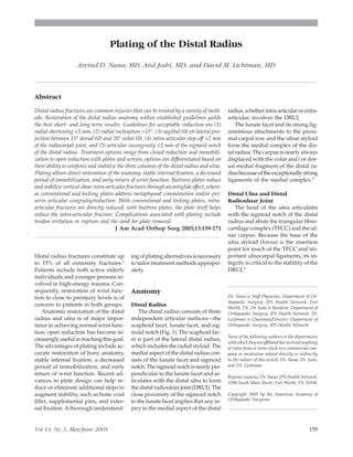
  • 1. Plating of the Distal Radius Arvind D. Nana, MD, Atul Joshi, MD, and David M. Lichtman, MD Abstract Distal radius fractures constitute up to 15% of all extremity fractures.1 Patients include both active elderly individuals and younger persons in- volved in high-energy trauma. Con- sequently, restoration of wrist func- tion to close to preinjury levels is of concern to patients in both groups. Anatomic restoration of the distal radius and ulna is of major impor- tance in achieving normal wrist func- tion; open reduction has become in- creasingly useful in reaching this goal. The advantages of plating include ac- curate restoration of bony anatomy, stable internal fixation, a decreased period of immobilization, and early return of wrist function. Recent ad- vances in plate design can help re- duce or eliminate additional steps to augment stability, such as bone void filler, supplemental pins, and exter- nal fixation. Athorough understand- ing of plating alternatives is necessary to tailor treatment methods appropri- ately. Anatomy Distal Radius The distal radius consists of three independent articular surfaces—the scaphoid facet, lunate facet, and sig- moid notch (Fig. 1). The scaphoid fac- et is part of the lateral distal radius, which includes the radial styloid. The medial aspect of the distal radius con- sists of the lunate facet and sigmoid notch.Thesigmoidnotchisnearlyper- pendicular to the lunate facet and ar- ticulates with the distal ulna to form the distal radioulnar joint (DRUJ). The close proximity of the sigmoid notch to the lunate facet implies that any in- jury to the medial aspect of the distal radius,whetherintra-articularorextra- articular, involves the DRUJ. The lunate facet and its strong lig- amentous attachments to the proxi- mal carpal row and the ulnar styloid form the medial complex of the dis- tal radius. The carpus is nearly always displaced with the volar and/or dor- sal medial fragment of the distal ra- diusbecauseoftheexceptionallystrong ligaments of the medial complex.2 Distal Ulna and Distal Radioulnar Joint The head of the ulna articulates with the sigmoid notch of the distal radius and abuts the triangular fibro- cartilage complex (TFCC) and the ul- nar carpus. Because the base of the ulna styloid (fovea) is the insertion point for much of the TFCC and im- portant ulnocarpal ligaments, its in- tegrity is critical to the stability of the DRUJ.3 Dr. Nana is Staff Physician, Department of Or- thopaedic Surgery, JPS Health Network, Fort Worth, TX. Dr. Joshi is Resident, Department of Orthopaedic Surgery, JPS Health Network. Dr. Lichtman is Chairman/Director, Department of Orthopaedic Surgery, JPS Health Network. None of the following authors or the departments with which they are affiliated has received anything of value from or owns stock in a commercial com- pany or institution related directly or indirectly to the subject of this article: Dr. Nana, Dr. Joshi, and Dr. Lichtman. Reprint requests: Dr. Nana, JPS Health Network, 1500 South Main Street, Fort Worth, TX 76104. Copyright 2005 by the American Academy of Orthopaedic Surgeons. Distal radius fractures are common injuries that can be treated by a variety of meth- ods. Restoration of the distal radius anatomy within established guidelines yields the best short- and long-term results. Guidelines for acceptable reduction are (1) radial shortening <5 mm, (2) radial inclination >15°, (3) sagittal tilt on lateral pro- jection between 15° dorsal tilt and 20° volar tilt, (4) intra-articular step-off <2 mm of the radiocarpal joint, and (5) articular incongruity <2 mm of the sigmoid notch of the distal radius. Treatment options range from closed reduction and immobili- zation to open reduction with plates and screws; options are differentiated based on their ability to reinforce and stabilize the three columns of the distal radius and ulna. Plating allows direct restoration of the anatomy, stable internal fixation, a decreased period of immobilization, and early return of wrist function. Buttress plates reduce and stabilize vertical shear intra-articular fractures through an antiglide effect, where- as conventional and locking plates address metaphyseal comminution and/or pre- serve articular congruity/reduction. With conventional and locking plates, intra- articular fractures are directly reduced; with buttress plates, the plate itself helps reduce the intra-articular fracture. Complications associated with plating include tendon irritation or rupture and the need for plate removal. J Am Acad Orthop Surg 2005;13:159-171 Vol 13, No 3, May/June 2005 159
  • 2. Columnar Classification of the Distal Radius and Ulna Conceptually, the distal radius and ulna may be divided into three col- umns based on the anatomy (Fig. 1, inset);thiscolumnarclassificationthen can be used to guide treatment plans. The distal radius is divided into the lateral and medial columns, which an- atomically correlate with the scaphoid facetandlunatefacet,respectively.The medial column of the distal radius is further subdivided into dorsal medi- al and volar medial columns. The lat- eral, dorsal medial, and volar medi- al columns correspond with Melone’s2 system for classifying intra-articular distal radius fractures. The ulnar col- umn represents the ulnar styloid and the TFCC. Pathophysiology In most activities of daily living, the dorsum of the distal radius is subject to tensile forces, whereas the volar surface is subject to compression. This is reflected in the bony architecture of the distal radius, with its strong volar buttressing cortex and thinner cancellous dorsal surface. When the wrist is subjected to a nonphysiolog- ic load, as in a dorsally directed com- pression force (eg, a fall on the out- stretched hand), predictable fracture patterns result. Melone2 aptly described the four major components of intra-articular distal radius fractures as radial sty- loid, dorsal medial, volar medial, and shaft fragments (Fig. 1). The transverse coronal split between the dorsal and volar medial distal radius fragments can be difficult to assess on plain ra- diographs.4 In many ways, the com- binations of displaced Melone com- ponentsreflectvariantsoffracturetypes andclassificationdescribedinthepast. For example, isolated radial styloid displacement represents a chauffeur’s fracture. Volar medial displacement describes Barton’s fracture; displace- ment of the dorsal medial fragment represents the common reversed Bar- ton’s or dorsal compression fracture. Displacementofallarticularfragments as a unit is comparable to the classic extra-articular Colles fracture (dorsal) or to Smith’s fracture (volar). Associated Injuries Associated injuries must be consid- ered in any comprehensive treatment plan for patients with distal radius and ulna fractures. Up to 68% of distal ra- dius fractures are associated with soft- tissue injuries, such as partial or com- plete tears of the TFCC, scapholunate ligament, and/or lunotriquetral lig- ament.5,6 Fractures involving the lu- nate facet (the medial aspect of the dis- tal radius) and fractures of the radial styloid cause the most intracarpal in- juries because of the strong ligamen- tous interconnections between the dis- tal radius and carpus.5 Intracarpal ligament disruption does not have to be repaired unless gross instability is noted on postreduction radiographs (eg, scapholunate diastasis). The 4- to 6-week immobilization necessary for distal radius healing is usually suf- ficient for ligament healing in nondis- placed injuries. Radiographic and clinical exami- nations of the injured wrist after sur- gical reduction are helpful in evalu- ating the integrity of the DRUJ and TFCC, both components of the ulnar column. TFCC tears are also common, particularly when the medial column of the distal radius and/or the ulnar styloid is intact. A fracture of the ul- nar styloid base and significant dis- placement (>2 mm) of an ulnar sty- loid fracture increase the risk of DRUJ instability.7 Treatment of the ulnar col- umn is focused on restoring DRUJ stability by closed, percutaneous, or open treatment of the ulnar styloid and the TFCC. Imaging Studies Posteroanterior (PA) and lateral ra- diographs are necessary for every pa- tient with a distal radius injury. The lateral view of the wrist places the an- terior surface of pisiform between the volar surface of the scaphoid (the scaphoid tuberosity) and the anteri- or surface of the capitate.8 Treatment options for distal radi- us fractures are based on the initial injury as well as on radiographs made after closed reduction. If the initial injury position is within acceptable guidelines9 (Table 1) for the patient’s functional requirements, any loss of reduction is usually insignificant as Figure 1 Distal radius anatomy. The fracture line between the volar medial and dorsal me- dial columns extends into the sigmoid notch and thus must also be evaluated on postreduc- tion radiographs. (Reproduced with permission from Trumble TE, Culp RW, Hanel DP, Geissler WB, Berger RA: Intra-articular fractures of the distal aspect of the radius. Instr Course Lect 1999;48:465-480.) Inset, Columnar classification of distal radius and ulna. Plating of the Distal Radius 160 Journal of the American Academy of Orthopaedic Surgeons
  • 3. long as the fracture is adequately im- mobilized and protected. Converse- ly, unacceptable initial displacement often means that loss of reduction af- ter nonsurgical treatment may require surgical intervention. The initial postinjury lateral view of the distal radius will demonstrate either the more common dorsal dis- placement or the less frequent volar displacement of the distal radius col- umns. The direction of initial displace- ment on the lateral radiograph gen- erally correlates with the side of both greatest cortical comminution and re- quired initial treatment. For example, if the lateral view shows dorsal dis- placement of the lunate facet, then the dorsal surface will have the greatest cortical fragmentation, and reduction will initially be focused dorsally. Postreduction or traction radio- graphs also are useful in assessing whether the distal radius fracture is intra-articular or extra-articular. The extent of cortical comminution is more easily visualized on postre- duction radiographs than on initial postinjury radiographs.11 All plain ra- diographs of distal radius fractures should include evaluation of radial inclination, radial length, ulnar vari- ance, radial tilt,9 articular step-off or gap, sigmoid notch step-off or gap, DRUJ subluxation/dislocation, and the presence and extent of displace- ment of ulnar styloid fractures (Fig. 2). Because of individual variability, appropriate radiographs of the unin- jured contralateral wrist are recom- mended to evaluate the patient’s nor- mal anatomy and corresponding radiologic measurements. Plain radiographs may be insuffi- cient to assess the extent of intra- articular and extra-articular fragmen- tation and injury to the DRUJ. Axial computed tomography (CT) scans with sagittal and coronal plane recon- structions can aid in visualizing die- punch fractures, volar rim fractures, and scaphoid facet fractures. Treatment of Distal Radius Fractures All patients with distal radius frac- tures warrant a trial of closed reduc- tion and plaster immobilization; ex- Table 1 Guidelines for Acceptable Reduction of Distal Radius Fractures* 1. Radial shortening <5 mm at the DRUJ compared with the contralateral side 2. Radial inclination on posteroanterior radiographs >15° 3. Sagittal tilt on the lateral projection between 15° dorsal tilt and 20° volar tilt 4. Intra-articular step-off or gap <2 mm of the radiocarpal joint10 5. Articular incongruity <2 mm of the sigmoid notch of the distal radius *These guidelines must be individualized to accommodate each patient’s functional activity levels and general medical status. Adapted with permission from Graham TJ: Surgical correction of malunited fractures of the distal radius. J Am Acad Orthop Surg 1997;5:270-281. Figure 2 The various angles to assess in distal radius fractures. A, Radial inclination (RI; normal, 22°). B, Radial length (RL; normal, 12 mm). C, Ulnar variance (UV; normal, 0 to −2 mm). D, Radial tilt (RT; normal, 11° volar). (Reproduced with permission from Graham TJ: Surgical correction of malunited fractures of the distal radius. J Am Acad Orthop Surg 1997; 5:270-281.) Arvind D. Nana, MD, et al Vol 13, No 3, May/June 2005 161
  • 4. ceptions include those with open injuries, those who cannot tolerate im- mobilization, and those with volar or dorsalverticalshearfractures(Barton’s fracture). Multiple trauma is also an indication for early surgical interven- tion. If the fracture can be reduced to within acceptable guidelines (Table 1) and maintained in satisfactory align- ment by plaster sugar-tong splint im- mobilization, it is considered to be sta- ble.Anunstablefractureischaracterized by inadequate reduction of a displaced fracture fragment or by the inability to maintain reduction after closed frac- turemanipulation.Dependingonfunc- tional expectations, surgical treatment may be considered for unstable frac- tures involving the medial column of the distal radius (Fig. 3). Determining short-term stability after closed reduction is difficult and is based largely on personal experi- ence in the treatment of distal radius fractures. If the surgeon thinks that a fracture is unstable and may present an unacceptable risk to the patient, then early open treatment with fix- ation should be considered. Success- ful closed treatment requires attention to detail by the physician as well as complete patient compliance with postreduction protocols. Normal wrist and forearm motion subjects the dorsal metaphysis of the distal radius to both tensile and com- pressive forces, but the volar surface transmits greater compressive forc- es.8 Restoration of these biomechan- ical relationships is necessary to es- tablish a stable reduction of the distal radius fracture. The first step toward a stable reduction is to create a sta- ble volar buttress. In distal radius fractures with dorsal comminution, volar integrity is reestablished by ac- curate cortical apposition of the large volar metaphyseal fragments. With comminuted volar metaphyseal frag- ments or inherently unstable volar vertical shear fractures (volar Barton fractures), stable fracture reduction can be achieved by placement of a volar buttress plate. Once volar sta- bilityisrestored,thedorsalmetaphys- eal fragments can be reduced against the stable volar buttress using exter- nal fixation, percutaneous pins, and/ or bone void filler (bone graft).8 Autograft, allograft, or other bone graft substitutes may be used to fill the metaphyseal defect created after reduction of distal radius fractures. Comminuted metaphyseal fragments are characterized by more compressed cancellous bone and a greater void af- ter reduction. Conversely, large meta- physeal fragments are associated with less compressed cancellous bone and thus a lower requirement for bone grafting. A bone void filler has sev- eral advantages, including mechan- ical support,12 providing osteoconduc- tive material for the bone defect, faster bone healing, and decreased incidence of loss of reduction.12-15 Hydroxyapa- tite cement without additional fixa- tion is inadequate for the treatment of distal radius fractures;16 it should be combined with external fixation to supplement any bone void filler. Volar buttress plate Medial column fracture of the distal radius Large dorsal metaphyseal fragments Unstable dorsal metaphyseal fragments Stable dorsal metaphyseal fragments or no dorsal fracture Small dorsal metaphyseal fragments Percutaneous pins and splint immobilization Splint immobi- lization Stable volar metaphyseal fragments after closed reduction* or no volar fracture Unstable volar metaphyseal fragments after closed reduction Conventional volar plate with dorsal percutaneous pins Volar plate with distal locking screws/pegs Small dorsal metaphyseal fragments Large dorsal metaphyseal fragments Unstable dorsal metaphyseal fragments Stable dorsal metaphyseal fragments Volar plate with distal locking screws/pegs Dorsal plate with distal locking screws/pegs Conventional volar plate with bone void filler and external fixation Dorsal buttress plate Dorsal bone void filler and neutralization external fixation Figure 3 Plating recommendations for fractures of the medial column of the distal radius. Treatment choice depends on surgeon pref- erence and experience. * = accurate cortical apposition of the volar metaphyseal fragments. Plating of the Distal Radius 162 Journal of the American Academy of Orthopaedic Surgeons
  • 5. Restoration of volar stability has important radiocarpal implications because the stronger and more impor- tant radiocarpal ligaments are at- tached to the volar surface. Volar in- tegrity therefore is critical because it facilitates adequate reduction of dor- sal metaphyseal fragments against a stable volar buttress and because it prevents possible volar radiocarpal instability.8 Plate Fixation Plate fixation is primarily indicat- ed for unstable fractures of the volar medial column of the distal radius; it is also helpful for combined volar and dorsal medial column injuries. Although some authors recommend dorsal plates for isolated dorsal me- dial column injuries of the distal ra- dius, other methods of treatment are available for these injuries. Volar plates with locking screws or pegs may be effective for extensive dorsal comminution. Distal radius plates are categorized by location of use and type of plate. Plates categorized by location may be used on the dorsal medial, volar me- dial, and radial styloid aspects of the distal radius. Certain plate designs address two areas simultaneously (ie, dorsal medial distal radius and radi- al styloid, or volar medial distal ra- dius and radial styloid). Smaller im- plants are also available to stabilize each area separately. The two principal types of distal radius plates are buttress plates and plates that can span metaphyseal comminution and/or maintain artic- ular congruity and reduction. Only the buttress plate, with its antiglide effect,reducesintra-articularfractures (eg, volar Smith or Barton fractures [Fig. 4] or dorsal Barton fracture). All other distal radius plates require di- rect reduction of intra-articular frac- tures; the plating serves to maintain the alignment. Plates that maintain alignment can be further subdivided into two types: conventional and locking. In conven- tional plate design, stability of the construct is ultimately achieved through apposition of the bone and plate by screw purchase in the bone. Conventional plates can be used dor- sally or volarly. The use of dual small conventional plates on the dorsal dis- tal radius also has been described.17 When cortical or cancellous screws are used in the distal holes of the but- tress plate, they are most effective if the opposite cortex is not comminut- ed. If the opposite cortex is commi- nuted, the surface could collapse be- cause of both axial forces across the wrist joint and toggle of the screws in the plate, which may result in loos- ening of the distal screws.18 Plates with locking distal screws or pegs support the subchondral bone and resist forces across the articula- tion that may displace the articular fragments (Figs. 5, 6, and 7). Locking screws or pegs offer numerous advan- tagesoverregularscrewsbecausetheir stability is achieved through plate de- sign. The screws or pegs become fixed- angle devices via threaded heads that engage the threaded distal screw holes of the plate for stability. New plate de- signs allow locking screw insertion in the most distal screw holes as well as in the proximal screw holes. Of his- torical note, the dorsal plate used by Gesensway et al19 employed a blade plate construct with multiple tines to achieve fixed-angle fixation. Locking screws or pegs support the subchondral bone without rely- ing on the purchase of the screws or pegs in bone, and they are indepen- dent of opposite cortex comminution. Accordingly, no bone void filler is re- quired to prevent collapse at the site of comminution. The distal screws or pegs—whethercortical,cancellous,or fixed-angle—serve to maintain reduc- tion of articular fragments, but they do not directly reduce the articular fracture. Distal Radius Plating Plating offers direct restoration of the distal radius through stable fix- ation. The stability of the construct en- sures more predictable healing of the fracture and thus shortens the peri- od of immobilization of the wrist. The principal advantage of distal radius platingistheearlyreturnofwristfunc- tion,animportantcriterionforpatients who expect wrist function after inju- rytoreturntopreinjurylevels.Conven- tional plates can be used for buttress and/or neutralization support of a distal radius fracture. Plates with lock- ing screws or pegs do not rely on an- atomic contour of the plate to obtain stability. Stabilization of the opposite cortex through locking screws or pegs significantly decreases or eliminates the need for further stabilization of the opposite cortex fragmentation. Figure 4 The antiglide effect of a buttress plate helps reduce a volar vertical shear frac- ture (volar Barton fracture) of the distal ra- dius. The most proximal screw is placed first; the penultimate distal screw (A) reduces the fracture (arrows) (B) before placement of the final distal screw (C). (Reproduced with per- mission from Jupiter JB: Complex articular fractures of the distal radius: Classification and management. J Am Acad Orthop Surg 1997; 5:119-129.) Arvind D. Nana, MD, et al Vol 13, No 3, May/June 2005 163
  • 6. Surgical Treatment Extra-articular Distal Radius Fractures These injuries are transverse or short oblique fractures proximal to the DRUJ and are usually amenable to closed treatment methods with sup- plementary percutaneous pin stabi- lization. For an unstable distal shaft fracture with minimal or no commi- nution that is either dorsally or vo- larly displaced, a volar plate is suf- ficient to achieve stable fixation. A locking volar plate does not offer any specific advantage over convention- al volar plating systems for this type of fracture. However, for an extra- articular fracture with dorsal or volar comminution, a locking volar plate may offer stability; it also restores length without the mobilization of extensor tendons that is required for dorsal plating. A dorsal plate for an extra-articular fracture with dorsal comminution is considered excessive treatmentwithoutsufficientbenefitbe- cause these fractures can be routine- ly pinned and stabilized in a closed fashion. Bone void filler and external fixation may be used for severely com- minuted extra-articular fractures. Intra-articular Distal Radius Fractures The concept of four-part displace- ment is useful in conceptualizing a treatment algorithm, especially with regard to the indications for distal ra- dius plating. When evaluating a pa- tient with a distal radius fracture, three distinct areas (columns) are as- sessed (Fig. 1, inset). The medial col- umn of the distal radius represents Melone’s dorsal and volar die-punch radius fragments; plates and/or bone void filler are particularly applicable for restoration of this column. The ra- dial styloid component—the lateral distal radius column—is then re- viewed to determine whether local- ized treatment is sufficient (eg, per- cutaneous pins or screws for the displaced radial styloid) or whether treatment should be combined with fixation of the medial column of the distal radius (eg, a volar or dorsal plate that also fixes the styloid). Fi- nally, the ulnar column, representing the TFCC and the ulnar styloid com- ponents, is addressed. Intra-articular Fractures of the Lateral Column The radial styloid fracture and its corresponding scaphoid facet are usually displaced proximally by ax- ial compression forces. Often the frag- ment is rotated in pronation, but this is not easily appreciated on routine radiographs. CT scan and dynamic fluoroscopy can help delineate this Figure 5 A, With conventional screws, stability is achieved through friction between the plate undersurface and the underlying bone (arrows) resulting from screw compression at the interface. This stability is augmented by bicortical screw purchase, which also decreases toggle between the screw and plate hole (represented by the rectangles). Friction at the in- terface must be higher than the axial forces to ensure absolute stability. Thus, anatomic con- touring of the conventional screw/plate system is needed to obtain a stable construct. B, In a locking screw system, the threaded screw head locks in the threaded screw hole of the plate to attain stability. Consequently, axial forces in the bone are transmitted to the plate rather than the screw, and no screw toggle can occur (arrows). Because stability with a locking screw does not require compression between the bone and the plate (rectangles), the periosteal blood supply under the plate is preserved. (Adapted with permission from Appenzeller A, Chris- tensen R, Frenk A, Gilbert S, Schavan R: The development of the distal femur LISS. Injury 2001;32[suppl 3]:5-25.) Figure 6 In the distal radius, the principal advantage of a plate with locking screws is sta- bility of the construct, even in the presence of segmental bone defect or comminution of cor- tex opposite the plate. Because the screws lock into the plate, the axial forces are transmitted to the plate. This diagram shows locking screws along the entire plate. F = axial force in the direction of the arrow. (Adapted courtesy of Synthes [USA], Paoli, PA.) Plating of the Distal Radius 164 Journal of the American Academy of Orthopaedic Surgeons
  • 7. rotational deformity. Isolated radial styloid fractures should raise con- cerns about concomitant intracarpal injury. Adisplaced radial styloid usu- ally can be reduced by closed means (ligamentotaxis by wrist flexion and ulnar deviation) and stabilized with two percutaneous pins. Reduction and pin placement are assessed intra- operatively with fluoroscopy or, if open reduction is used, under direct visualization (Figs. 8 and 9). Occa- sionally the radial styloid fragment must be manipulated into position, using the pins as joysticks, before se- curing the position by advancing the pins into the opposite cortex. The di- rection of the styloid pins is from ra- Figure 7 Anteroposterior (A) and lateral (B) radiographs of a distal radius injury. The fracture, which demonstrates volar and dorsal in- stability, is fixed with a volar locking plate (C and D). The dorsal instability is best seen in the postoperative lateral view (D), which shows a dorsal gap just proximal to the locking pegs. The locking pegs stabilize both the dorsal and volar articular fragments. In this patient, bone void filler was not used in the dorsal gap, and the distal ulna was not surgically stabilized. (Courtesy of Arvind D. Nana, MD, Fort Worth, TX.) Figure 8 Anteroposterior (A) and lateral (B) radiographs of a wrist at initial presentation. Anteroposterior (C) and lateral (D) radiographs of the wrist in longitudinal traction. The patient is immobilized in a plaster sugar-tong splint. (Courtesy of David M. Lichtman, MD, Fort Worth, TX.) Arvind D. Nana, MD, et al Vol 13, No 3, May/June 2005 165
  • 8. dial volar to ulnar dorsal. Other op- tions for styloid fixation include percutaneous lag screw fixation or a small buttress plate and pins.20 Plate fixation of the radial styloid is usually done in combination with stabilization of medial column inju- ries of the distal radius. Volar or dor- sal distal radius plates include radial styloid stabilization in their construct and therefore do not need separate ra- dial styloid plating or pinning. Cer- tain smaller implant designs are avail- able for separate plating of radial styloid fractures, but this is almost al- ways in combination with medial col- umn stabilization (whether dorsal or volar). Separate lateral and medial column plating of the distal radius is most mechanically stable when the plates are angled 50° to 90° to each other.11,17,20,21 Separate radial styloid plates are placed on the dorsal radial or most radial aspect of the distal ra- dius; this placement requires a low- profile design to avoid irritation of the brachioradialis tendon or dorsal ex- tensor tendons. Intra-articular Fractures of the Medial Column Unstable volar fractures are ame- nable to volar plate stabilization. If the fracture is isolated (ie, there is no dor- sal fragment instability), then a sim- ple buttress plate may be used to re- constitute the volar cortex. If the volar metaphysis is comminuted, bone void filler may be used to fill in the gap and also to support the distal ar- ticular surface. The integrity of the joint should not be violated by dis- secting the volar radiocarpal liga- ments. When necessary, the volar cor- tex can be hinged open like a book to inspect and realign volar intra- articular injuries. When dorsal fragment instability also is present, several options are available. If a volar buttress plate has been applied, then the dorsal frag- ment must be stabilized from a dor- sal approach. If the dorsal fragment is large, it can be fixed by one or (pref- erably) two percutaneous cross pins entering the dorsal medial fragment distally and aimed proximally and ra- dially (Figs. 8 and 9). As with the ra- dial styloid fragment, percutaneous pins can be used as joysticks to ma- nipulate the dorsal fragment into po- sition. The pins also may be placed into the fracture line (intrafocal Ka- pandji pinning [Fig. 10]), starting more perpendicular to the dorsal cor- tex, and then used to reduce the dor- sal fragment by moving the pins more nearly parallel to the dorsal cortex, with subsequent fixation into the volar cortex. If the dorsal medial fragment is comminuted, reducing and holding the area with pin fixation may be dif- ficult. The fracture can be opened dor- sally, usually through a longitudinal approach between the third and fourth dorsal extensor compartments. An approach ulnar to the fourth com- partment also may be used if the com- minution is limited to the dorsal ul- nar corner and semilunar notch. The floor of the third extensor compart- ment is incised longitudinally; then the fourth extensor compartment (sheath and tendons) is sharply ele- Figure 9 Same patient as in Figure 8. Anteroposterior (A) and lateral (B) radiographs made 1 week after injury. The laterally displaced radial styloid fragment and volarly displaced volar medial fragment (arrows) are evident. Instability of these fragments is easily demon- strated by their inability to maintain initial closed reduction. Anteroposterior (C) and lateral (D) postoperative radiographs show that the volar medial fragment is stabilized with a volar plate. The radial styloid is fixed with two percutaneous pins; the dorsal cortex with its large dorsal fragments is stabilized with a dorsal percutaneous pin. The ulnar styloid also was stabilized because of the fracture through its base, marked displacement (>2 mm), and clinical instability of the DRUJ after stabilization of the distal radius. (Courtesy of David M. Lichtman, MD, Fort Worth, TX.) Plating of the Distal Radius 166 Journal of the American Academy of Orthopaedic Surgeons
  • 9. vated off the dorsal distal radius. Bone grafting of the dorsal metaphy- sis is now possible. If the joint needs to be viewed, the dorsal capsule can be incised transversely. Reduction un- der direct visualization usually can be done, and bone void filler is often used to stabilize and buttress the dor- sal cortex. If necessary, a transverse capsular incision may be used to ex- plore the radiocarpal joint and reduce intra-articular or intracarpal injuries. Some surgeons use low-profile dor- sal plates at this point to maintain ar- ticular reduction and stability; others apply an external fixator for 3 to 4 weeks to avoid use of a dorsal plate. An alternative for treating both dorsal and volar instability is to use a plate with locking screws or pegs to transfix both volar and dorsal cor- tices of the medial column of the dis- tal radius (Fig. 7). Satisfactory results have also been achieved using lock- ing screw–plate combinations from the dorsal side.22-24 Although dorsal plates do permit adequate healing of the fracture, their secondary effects—particularly on surrounding soft-tissue structures— render them a less appealing option. Disadvantages of dorsal plates in- clude the need for mobilization of ex- tensor tendons to achieve proper plate placement, possible tendon ir- ritation or rupture because of a prom- inent plate or screws, and the possi- bility of additional surgery to remove the symptomatic dorsal plate. In isolated unstable fractures of the dorsal medial column, the decision to treat with pin fixation or with open bone grafting plus neutralization ex- ternal fixation depends on the size of the fragments. Treatment is the same as when combined with volar insta- bility: percutaneous pins for large fragments or open reduction, bone void filler, and external fixation for comminuted fractures (small frag- ments). Once again, low-profile dor- sal plates can be applied instead of an external fixator, but locking screws or pegs are not necessary in this in- stance. A lateral column injury of the dis- tal radius is usually treated concom- itantly or after stabilization of the me- dial column. As noted, if the fracture pattern permits, the styloid can be fixed with either a volar or dorsal plate. In most instances, the radial sty- loid can be fixed separately using two percutaneous pins, as described for isolatedradialstyloidfractures.Aper- cutaneous lag screw or a small dorsal radial buttress plate are also options. Surgical Approach Volar Radial Approach The volar radial (Henry) approach is utilitarian; it can expose the entire volar radial surface up to the DRUJ (Fig. 11, A). This exposure uses the in- terval between the flexor carpi radi- alis (FCR) and the radial artery or goes through the floor of the FCR ten- don sheath. The pronator quadratus muscle is elevated in a subperiosteal fashion to visualize the volar distal radius.25 Orbay26 and Orbay and Fernandez27 also describe the release of the first extensor compartment and the insertion of the brachioradialis off the lateral distal radius to permit ac- cess to the dorsal surface and to fa- cilitate the reduction of the lateral col- umn of the distal radius (radial styloid). Release of the volar capsule or the ligamentous attachments on the volar rim may lead to volar ra- diocarpal instability and should be avoided. Carpal tunnel release through the distal extension of this approach is not recommended because of potential injury to the volar cutaneous branch of the median nerve. If carpal tunnel release is indicated, a separate stan- dard or a mini carpal tunnel incision is recommended. Prophylactic carpal tunnel release is not routinely per- formed21,28,29 unless acute carpal tun- nel symptoms are present or unless the distal fragment is significantly dorsally displaced for a prolonged period of time. Volar Ulnar Approach The volar ulnar approach offers limited exposure to the radial column and is indicated for injuries to the vo- lar aspect of the medial column and the DRUJ (Fig. 11, B). The interval be- tween the flexor carpi ulnaris and the Figure 10 With intrafocal Kapandji pinning, the pin is placed through the fracture site and maneuvered to elevate the fragment. Once adequate reduction is achieved, the pin is then driven through the opposite cortex to achieve stability. This technique can be used to restore radial inclination (A) or volar tilt (B). (Adapted with permission from Palmer AK: Fractures of the distal radius, in Green DP [ed]: Operative Hand Surgery. New York, NY: Churchill Liv- ingstone, 1993, pp 929-971.) Arvind D. Nana, MD, et al Vol 13, No 3, May/June 2005 167
  • 10. finger flexor tendons is used for this approach. While retracting the flexor tendons radially and protecting the ulnar nerve and artery, the pronator quadratus muscle is mobilized off the distal ulna. This exposure has the ad- vantage of permitting distal extension to complete a carpal tunnel release. Postoperative Management After surgery, immobilization is de- sirable to facilitate soft-tissue heal- ing and resolution of swelling. Plas- ter splinting is preferable to the use of circumferential or bivalved casts because use of casts may lead to po- tential complications such as finger edema and compartment syndrome. Postsurgical follow-up includes suture removal at 10 to 14 days and serial radiographs for the first 2 weeks if deemed necessary. Arm el- evation and finger range of motion are important components of early rehabilitation. Conversion to a cir- cumferential cast may be considered at 2 weeks; however, complete wrist immobilization beyond the fourth postoperative week usually is not desirable because it negates the ad- vantage of early restoration of wrist stability with plating. During the pe- riod of immobilization, therapy is focused on finger motion; once im- mobilization is discontinued, ther- apy expands to include wrist motion (flexion, extension, ulnar deviation, and radial deviation) and forearm rotation (pronation, supination). A removable Velcro wrist splint then can be provided for support and comfort while the patient engages in gentle activities of daily living and active range-of-motion exercises. With plating of extra-articular frac- tures, or for fractures with well-fixed intra-articular fragments, immobili- zation can be discontinued as early as 2 weeks after surgery as long as the incision is sufficiently healed and ede- ma has decreased to allow wrist mo- tion. For more complex intra-articular fractures treated with plating, immo- bilization for at least 4 weeks is nec- essary to permit healing of the intra- articular fragments and to allow wrist motion without risk of displacement of the intra-articular fragments. For 6 to 8 weeks, splints can be worn at night, and only light physical thera- py is recommended. Strenuous push- ing, pulling, twisting, or lifting should be avoided for the first 3 months to facilitate optimal bone healing. Figure 11 A, The volar radial approach uses the interval between the flexor carpi radialis tendon and the radial artery. The pronator quadra- tus is elevated sharply, starting at its insertion on the distal radius. B, Top detail, the volar ulnar approach can be extended distally to release the median nerve from the carpal tunnel. Bottom detail, at the level of the distal radius, the flexor tendons and the median nerve are re- tracted radially to expose the volar medial distal radius. (Adapted with permission from Fernandez DL, Jupiter JB: Fractures of the Distal Radius. New York, NY: Springer, 1996, pp 67-102.) Plating of the Distal Radius 168 Journal of the American Academy of Orthopaedic Surgeons
  • 11. Complications The most common complication of plate fixation in the distal radius is tendon injury; 25% of patients eval- uated reported everything from ten- don irritation to frank rupture.17,24,30 With the volar plate, tendon injuries have included rupture of the flexor pollicis longus,22,31,32 FCR tenosyno- vitis,28 and dorsal extensor tendon in- volvement as a result of protruding screws.28,33 Dao et al34 reported radi- al artery pseudoaneurysm secondary to volar distal radius plate design. Median nerve dysfunction (ie, pares- thesia, carpal tunnel syndrome, or re- flex sympathetic dystrophy) is asso- ciated with volar plating, but routine prophylactic carpal tunnel release in this scenario is not considered stan- dard treatment.21,28,29 Adverse effects are more common with dorsal plat- ing than with volar plating. As with dorsal plates, tendon- related complications requiring plate removal also occur after volar plat- ing; in one study, 13 of 73 patients (18%) required plate removal after volar plating.32 Tendon irritation is thought to be caused by prominent or sharp plate edges,17,23,30 prominent or loose screws,35 and cellular reac- tion to the titanium metal of the plates.34,36 Extensor tendon rupture can be caused by screws that are loose or backing out,30,33,37 by sharp edges created by cutting of the plate,17 and by prominent design of the distal as- pect of the dorsal plate.36,38,39 Many authors have reported a high inci- dence (up to 30%) of dorsal plate re- moval secondary to tendon inflam- mation or rupture.17,23,30,34-36 To prevent tendon injury, some recom- mend that a portion of the extensor retinaculum be interposed between the plate and the tendon or tendon sheath,21,30,40 or that the dorsal plates be routinely removed.23,37,40 Tendon rupture has been reported as early as 8 weeks and as late as 7 months af- ter surgery.30,38 Plate breakage is an- other reason for plate removal.37 Loss of reduction also can be a problem and is seen more frequently with the use of conventional plates when the opposite cortex is not fur- ther augmented with pins, bone void filler, or external fixation.24,34,41,42 Outcomes Functional outcomes of distal radius fractures depend on multiple factors, including radial shortening, radial in- clination, sagittal tilt, intra-articular step-off or gap, and articular congru- ity of the sigmoid notch (Table 1). Al- though intra-articular step-off >2 mm has been shown to lead to radio- graphic osteoarthritis, this does not necessarily correlate with poor func- tional outcome.10 Radial shortening >5 mm, however, may have a marked impact on outcome because it can af- fect both the DRUJ and the radiocar- pal joint.9,10,43 Outcome studies for each type of dorsal plate design (conventional, dual, and locking) have been per- formed, but the average follow-up is only 12 to 19 months for population groups ranging from 21 to 73 patients. Two studies of conventional plate de- sign demonstrated 72% to 95% good- to-excellent functional results, with a reported complication rate of 20% to 23%. The largest study evaluated dual dorsal plating in the treatment of dis- tal radius fractures; however, even with 97% good-to-excellent results, the incidence of complications was 21%.17,24,30 Of the dorsal plates with locking screws or pegs, the Synthes pi plate (Paoli, PA) demonstrated a 57% good- to-excellent outcome and a 23% com- plication rate.39 Subsequent reports on the pi plate show good-to- excellent functional results of 56% to 68%.21,44 All of the studies on dorsal plating, regardless of design, include the complication of plate removal as a secondary procedure.21,39,44 The rea- son for plate removal is usually ex- tensor tenosynovitis or rupture. Keating et al42 examined the results of the volar buttress plate in 79 pa- tients and found that functional re- covery associated with malunion was significantly less than in those with good anatomic restoration. Malunion was the most frequent complication (28%) of all complications reported (40.5%).42 Orbay26 and Orbay and Fernan- dez27 examined the use of the volar plate with locking pegs for the treat- ment of dorsally displaced fractures of the distal radius; they reported 100% good-to-excellent results. The authors attributed the success of the treatment plan to stable internal fix- ation and preservation of dorsal soft tissues, which facilitate early fracture healing; decreased need for bone grafting; and low incidence of tendon injury. In their study, 9 of 29 patients (31%) had preoperative median nerve symptoms and also underwent car- pal tunnel release. One patient had extensor tendon irritation secondary to a long peg that necessitated hard- ware removal, but all other plates were left in place.26,27 No study has a follow-up >19 months; thus, conclu- sive long-term recommendations for distal radius plating cannot be made based on these reports. Summary Improved restoration of the anatom- ic relationship in unstable distal ra- dius fractures can lead to improved, early functional outcome; the trend is to achieve this goal through open reduction and internal fixation. Ben- efits of plate fixation include direct vi- sualization of the fracture, stable in- ternal fixation, a shortened period of immobilization, and early return of extremity function. Recent plate de- signs include locking screws or pegs that maintain articular reduction as well as span and stabilize the com- minution of the opposite cortex. The outcome of plating distal ra- dius fractures cannot be compared to Arvind D. Nana, MD, et al Vol 13, No 3, May/June 2005 169
  • 12. other surgical options because of a lack of long-term follow-up and the wide variation of indications for plat- ing in published studies. The three- column system for distal radius and ulna fractures is a simple and prac- tical approach to understanding and treating these common, yet some- times complex, injuries. The risks and benefits of plating must be balanced for each patient and weighed with complete understanding of the patho- logic anatomy and physiology of each specific fracture pattern. The OKO video ″Intra- articular Distal Radius Fractures,″ by Christopher H. Allan, MD, is avail- able at http://www5.aaos.org/oko/ jaaos/main.cfm. References 1. Sanders WE: Distal radius fractures, in Manske PR (ed): Hand Surgery Update. Rosemont, IL:AmericanAcademy of Or- thopaedic Surgeons, 1996, pp 117-123. 2. Melone CP Jr: Articular fractures of the distal radius. Orthop Clin North Am 1984;15:217-236. 3. Palmer AK: The distal radioulnar joint. Orthop Clin North Am 1984;15:321-335. 4. Andersen DJ, Blair WF, Steyers CM, Adams BD, El-Khouri GY, Brandser EA: Classification of distal radius frac- tures: An analysis of interobserver reli- ability and intraobserver reproducibil- ity. J Hand Surg [Am] 1996;21:574-582. 5. Geissler WB, Freeland AE, Savoie FH, McIntyre LW, Whipple TL: Intracarpal soft-tissue lesions associated with an intra-articular fracture of the distal end of the radius. J Bone Joint Surg Am 1996; 78:357-365. 6. Richards RS, Bennett JD, Roth JH, Milne K Jr: Arthroscopic diagnosis of intra-articular soft tissue injuries asso- ciated with distal radius fractures. J Hand Surg [Am] 1997;22:772-776. 7. May NM, Lawton JN, Blazar PE: Ulnar styloid fractures associated with distal radius fractures: Incidence and implica- tions for distal radioulnar joint instabil- ity. J Hand Surg [Am] 2002;27:965-971. 8. Nana AD, Lichtman DM: Distal-third forearm fractures. Available at http:// www.emedicine.com/orthoped/topic 79.htm. Accessed April 27, 2005. 9. Graham TJ: Surgical correction of malunited fractures of the distal radius. J Am Acad Orthop Surg 1997;5:270-281. 10. Knirk JL, Jupiter JB: Intra-articular frac- tures of the distal end of the radius in young adults. J Bone Joint Surg Am 1986; 68:647-659. 11. Rikli DA, Regazzoni P: Fractures of the distal end of the radius treated by inter- nal fixation and early function. J Bone Joint Surg Br 1996;78:588-592. 12. Fernandez DL, Geissler WB: Treatment of displaced articular fractures of the ra- dius. J Hand Surg [Am] 1999;24:102-107. 13. Leung KS, Shen WY, Leung PC, Kinnin- month AWG, Chang JCW, Chan GPY: Ligamentotaxis and bone grafting for comminuted fractures of the distal ra- dius. J Bone Joint Surg Br 1989;71:838-842. 14. Putnam MD, Fischer MD: Treatment of unstable distal radius fractures: Meth- ods and comparison of external distrac- tion and ORIF versus external distrac- tion-ORIF neutralization. J Hand Surg [Am] 1997;22:238-251. 15. Trumble TE, Schmitt SR, Vedder NB: Fac- tors affecting functional outcome of dis- placed intra-articular distal radius frac- tures. J Hand Surg [Am] 1994;19:325-340. 16. Jeyam M,Andrew JG, Muir LT, McGov- ern A: Controlled trial of distal radial fractures treated with a resorbable bone mineral substitute. J Hand Surg [Br] 2002;27:146-149. 17. Jakob M, Rikli DA, Regazzoni P: Frac- tures of the distal radius treated by in- ternal fixation and early function. J Bone Joint Surg Br 2000;82:340-344. 18. Small Fragment Locking Compression Plate (LCP): Technique Guide. Paoli, PA: Synthes (USA), 2002, pp 1-20. 19. Gesensway D, Putnam MD, Mente PL, Lewis JL: Design and biomechanics of a plate for the distal radius. J Hand Surg [Am] 1995;20:1021-1027. 20. Swigart CR, Wolfe SW: Limited incision open techniques for distal radius frac- ture management. Orthop Clin North Am 2001;32:317-327. 21. Hahnloser D, Platz A, Amgwerd M, Trentz O: Internal fixation of distal ra- dius fractures with dorsal dislocation: π-plate or two ¼ tube plates?Aprospec- tive randomized study. J Trauma 1999; 47:760-765. 22. Fuller DJ: The Ellis plate operation for Smith’s fracture. J Bone Joint Surg Br 1973;55:173-178. 23. Fitoussi F, Ip WY, Chow SP: Treatment of displaced intra-articular fractures of the distal end of the radius with plates. J Bone Joint Surg Am 1997;79:1303-1312. 24. Finsen V, Aasheim T: Initial experience with the Forte plate for dorsally dis- placed distal radius fractures. Injury 2000;31:445-448. 25. Trumble TE, Culp RW, Hanel DP, Gei- ssler WB, Berger RA: Intra-articular fractures of the distal aspect of the ra- dius. Instr Course Lect 1999;48:465-480. 26. Orbay JL: The treatment of unstable distal radius fractures with volar fixa- tion. Hand Surg 2000;5:103-112. 27. Orbay JL, Fernandez DL: Volar fixation for dorsally displaced fractures of the distal radius: A preliminary report. J Hand Surg [Am] 2002;27:205-215. 28. Jupiter JB, Fernandez DL, Toh C-L, Fell- man T, Ring D: Operative treatment of volar intra-articular fractures of the dis- tal end of the radius. J Bone Joint Surg Am 1996;78:1817-1828. 29. Schneeberger AG, Ip WY, Poon TL, Chow SP: Open reduction and plate fix- ation of displaced AO type C3 fractures of the distal radius: Restoration of artic- ular congruity in eighteen cases. J Orthop Trauma 2001;15:350-357. 30. CarterPR,FrederickHA,LaseterGF:Open reduction and internal fixation of unsta- bledistalradiusfractureswithalow-profile plate:Amulticenter study of 73 fractures. J Hand Surg [Am] 1998;23:300-307. 31. Bell JSP, Wollstein R, Citron ND: Rup- ture of flexor pollicis longus tendon. J Bone Joint Surg Br 1998;80:225-226. 32. Nunley JA, Rowan PR: Delayed rupture of the flexor pollicis longus tendon af- ter inappropriate placement of the π plate on the volar surface of the distal radius. J Hand Surg [Am] 1999;24:1279-1280. 33. Wong-Chung J, Quinlan W: Rupture of extensor pollicis longus following fixa- tion of a distal radius fracture. Injury 1989;20:375-376. 34. Dao KD, Venn-Watson E, Shin AY: Ra- dial artery pseudoaneurysm complica- tion from use of AO/ASIF volar distal radius plate: A case report. J Hand Surg [Am] 2001;26:448-453. 35. Schnur DP, Chang B: Extensor tendon rupture after internal fixation of a dis- tal radius fracture using a dorsally placed AO/ASIF titanium Pi plate. Ann Plast Surg 2000;44:564-566. 36. Lowry KJ, Gainor BJ, Hoskins JS: Exten- sor tendon rupture secondary to the AO/ASIF titanium distal radius plate Plating of the Distal Radius 170 Journal of the American Academy of Orthopaedic Surgeons
  • 13. without associated plate failure: A case report. Am J Orthop 2000;29:789-791. 37. Kambouroglou GK, Axelrod TS: Com- plications of the AO/ASIF titanium dis- tal radius plate system (π plate) in in- ternal fixation of the distal radius:Abrief report. J Hand Surg [Am] 1998;23:737-741. 38. Lucas GL, Fejfar ST: Complication in internal fixation of the distal radius. J Hand Surg [Am] 1998;23:1117. 39. Ring D, Jupiter JB, Brennwald J, Buchler U, Hastings H: Prospective multicenter trial of a plate for dorsal fix- ation of distal radius fractures. J Hand Surg [Am] 1997;22:777-784. 40. Peine R, Rikli DA, Hoffmann R, Duda G, Regazzoni P: Comparison of three dif- ferent plating techniques for the dorsum of the distal radius: A biomechanical study. J Hand Surg [Am] 2000;25:29-33. 41. Hove LM, Nilsen PT, Furnes O, Oulie HE, Solheim E, MolsterAO: Open reduc- tion and internal fixation of displaced in- traarticular fractures of the distal radi- us. Acta Orthop Scand 1997;68:59-63. 42. Keating JF, Court-Brown CM, Mc- Queen MM: Internal fixation of volar- displaced distal radial fractures. J Bone Joint Surg Br 1994;76:401-405. 43. Catalono LW III, Cole RJ, Gelberman RH, Evanoff BA, Gilula LA, Borelli J Jr: Displaced intra-articular fractures of the distal aspect of the radius. J Bone Joint Surg Am 1997;79:1290-1302. 44. Campbell DA: Open reduction and in- ternal fixation of intra-articular and un- stable fractures of the distal radius us- ing the AO distal radius plate. J Hand Surg [Br] 2000;25:528-534. Arvind D. Nana, MD, et al Vol 13, No 3, May/June 2005 171